Pediatrics Final (new content)
1/106
There's no tags or description
Looks like no tags are added yet.
Name | Mastery | Learn | Test | Matching | Spaced |
|---|
No study sessions yet.
107 Terms
Normal ureter and bladder anatomy
With contraction of the bladder during urination, the uretero-vesicular junction (UVJ) closes, ensuring a uni-directional flow of urine from the ureters into the bladder
Vesicoureteral reflux
the retrograde passage of urine at the uterovesicular junction (UVI) into the ureters. Urine refluxes from the bladder into the ureters when the bladder contracts due to incompetent UVJ
Diagnosis of VCUR
A history of urinary tract infections causing febrile episodes raises suspicion for VCUR, a voiding cystourethrogram will show reflux and hydronephrosis
voiding cystourethrogram
the urethra is catheterized and the bladder is filled with contrast material, images are taken of the bladder full and with voiding, if refluxing from the bladder is occurring the contrast will be seen in the ureter and possibly the kidney as shown in the VCUG image
signs of VCUG with reflux
Kidney- filled with contrast, ureter that is engorged from refluxing urine, bladder- filled with contrast
Manifestations of VCUR
reflux and stasis of urine, hydronephrosis, more severe urinary tract infections, scarring of the kidney tissue, kidney injury (complication- UTI)
Signs of a UTI in infants and young children
Fever, V/D, abdominal/flank pain, dysuria, abdominal distention, lethargy, irritability, apnea in newborns
hydronephrosis
stretching/swelling of the renal pelvis due to chronic reflux and obstruction of urine flow which can lead to kidney tissue damage, sometimes discovered during fetal ultrasound
Grades of Hydronephrosis
1 is the least severe, and 5 is the most severe
medical treatment of VCUR
- prophylactic, low dose antibiotic (usually PCN or sulfa medication) to prevent urinary tract infection until child grows out of the anatomical defect
- Frequent voiding to avoid a full bladder that will aggrevate refluxing
- increased fluid intake to encourage frequent voiding and reduce risk of infection
- urine culture is obtained if there are signs and symptoms of infection
parent education for VCUR
- S/S of UTI, report suspicion to PCP
- rationale for low-dose prophylactic antibiotics
- education of required testing
- female children: front-to-back cleansing after toiletings, most common infecting organism in UTI is E. Coli\
- encourage fluid intake and frequent voiding
- uncircumcised males: routine hygiene to reduce risk of UTI
- Follow up: urine analysis/culture, VCUG
procedural treatment for VCUR: Surgical
ureteral re-implantation, performed if medical rx fails, open or laparoscopic approach used, ureters are removed from bladder and re-implanted at an appropriate angle and depth through the bladder wall to prevent reflux
procedural treatment for VCUR: STING
subureteral trans-urethral injection, endoscopic injection, trans-urethral injection of a bulking agent performed into the submucosa of the UVJ, bulking agent enhances competence of junction closure during bladder contraction, performed via cystoscopy as shown in the picture, child with be NPO prior to this procedure in preparation for sedation or anesthesia
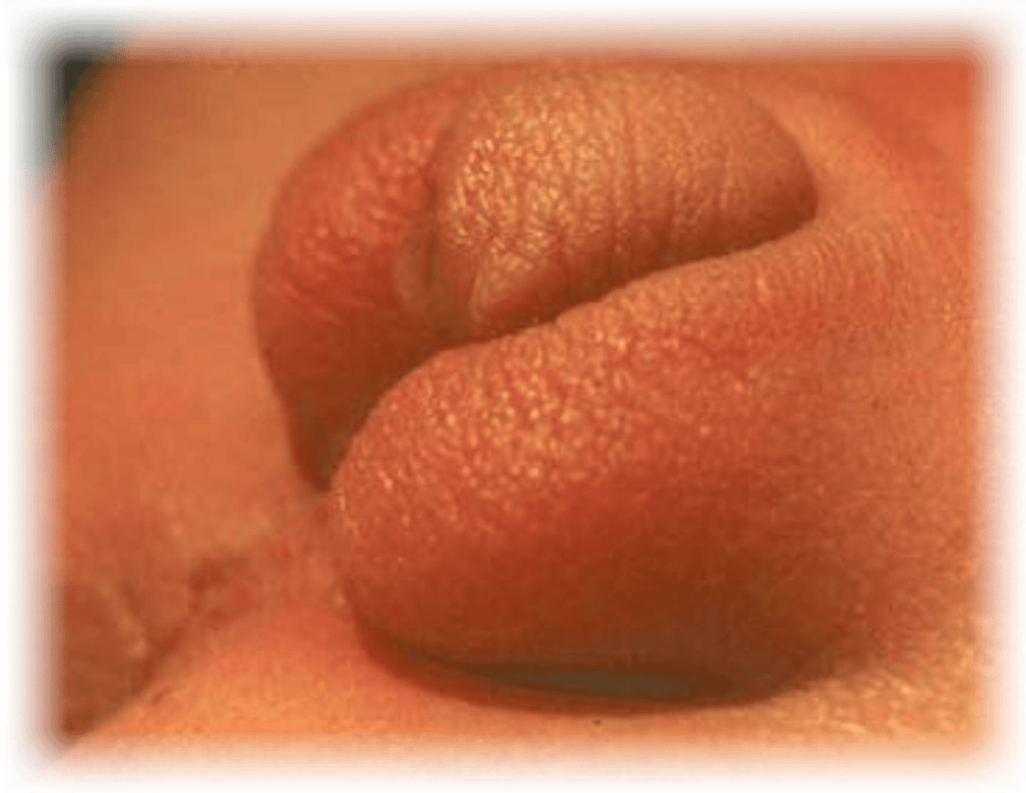
Hypospadias
congenital anomaly where the urethral opening is on the ventral surface of the penis, genetic predisposition
Hypospadias associated risks
mis-directed urine flow, infertility due to abnormal semen flow, erectile dysfunction, abnormal appearance of penis
Characteristic appearance of Hypospadias
small prepuce, chordee, severity variable, urethral opening can occur at any location along ventral surface, even close to the scrotom, bifid scrotom
Hypospadias: small prepuce
small or no foreskin noted
Hypospadias: chordee
a curvature of the penis may be present
Hypospadias: degree of severity variable
degree of curvature and distance of urethral opening from penis tip along the ventral surface can vary
Bifid scrotum
a cleft or split in the midline of the scrotum, appearing as two separate scrotal halves

treatment for Hypospadias
no circumcision is done, surgical correction: performed at 6 to 12 months of age before toliet training initiated
Post-op care for ureteral re-implantation and hypospadias repair
routine pain management, foley/stent care- ensure tube patency and monitor output hourly, administer antibiotic until foley/stent removal to prevent infection, administer oxybutynin
oxybutynin
anti-spasmodic of smooth muscle (bladder), be aware of extended release versus immediate release dosage forms for this medication
Similarities between Hypospadias and VCUR
congenital abnormalities, variable degrees of severity, post op care includes pain management, foley care, and anti-spasmodic therapy
differences between Hypospadias and VCUR
VCUR can be treated without surgery for mild forms, VCUR can lead to infection and organ damage, VCUR requires imaging to diagnose, timing of surgery for hypospadias is typically earlier in life
Acute Glomerulonephritis (AGN)
Immuno-pathogenic condition usually following an infection, Group A beta-hemolytic streptococcal infection (acute poststreptococcal glomerulonephritis- APSGN), secondary causes can include: SLE and sickle cell disease, occurs suddenly about 10-21 days following infection, typically seen in the school-aged child, greater incidence in males, usually lasts 2-4 weeks
glomerofiltration rate (GFR)
in normal GFR physiology, blood is filtered as it flows through Bowman's capsule. Urine is created from the filtration that occurs across the glomerular membrane.
Patho of AGN
immune complexes deposit in basement membrane of glomeruli, glomerular edema and trauma result from the immune complex deposition, from edema and trauma- decreased glomerular filtration rate and hematuria occurs, decreased GFR leads to excessive accumulation of water and sodium, expanded plasma and interstitial fluid (hypovolemia) occurs from the retention of water and sodium
Outward effects of AGN
tea-colored urine and periorbital edema
Manifestations of AGN
decreased UOP, elevated specific gravity from concentrated urine (over 1.015), tea-colored urine from hematuria, peri-orbital and peripheral edema, anorexia and vomitting (due to retention of wastes from decreased GFR), irritability and lethargy from edema, hypertension ad headache from hypervolemia, proteinuria and hematuria from glomeruli trauma, serum lab values (elevated BUN and Cr, ASO titer is evidence of recent strep infection)
Medical care for AGN
antibiotic if active infection present, diuretic therapy for hypervolemia: furosemide, anti-hypertensive agent, low Na+ diet and restricted fluid intake, possible K restriction (oligouria)
Nursing care for AGN
monitor fluid balance (strict I and O, daily weight, VS with BP Q4h), monitor BUN and Cr results-potential complication of renal failure, monitor urine for blood/protein and track BP as outcome measures for disease improvement, implement seizure precautions; potential complication of hypertensive encephalopathy, monitor breath sounds- potential risk for pulmonary edema/effusion
Nephrotic syndrome
an increase in permeability of the glomeruli to plasma proteins caused by an auto-immune response
Idiopathic Nephrotic syndrome
most common type of nephrotic syndrome, typically effects children 2-6 years of age, illness course can be weeks to months/relapses
Secondary Nephrotic syndrome
Caused by a condition that damages the glomeruli such as AGN
congenital Nephrotic syndrome
hereditary disorder characterized by protein loss, edema and intravascular volume contraction
edema caused by protein loss in urine because albumin is a key factor in holding onto water in the blood
3 types of pressure in the bowman's capsule
colloid osmotic pressure, hydrostatic blood pressure, hydrostatic capsule pressure. The pushing and pulling enforces homeostasis of the blood volume as it passes through the Bowman's capsule
colloid osmotic pressure (COP)
created by plasma proteins which PULLS fluid INTO the vascular space
hydrostatic blood pressure
created by blood pressure which PUSHES fluid OUT OF the vascular space
hydrostatic capsule pressure
created by Bowman's Capsule that PUSHES against the blood hydrostatic pressure
Patho of Nephrotic Syndrome
Loss of serum proteins into the urine leading to reduced colloidal osmotic pressure of the intravascular space (PULL) resulting in leakage of fluid into body tissue and intravascularly hypovolemic, decreased renal blood flow leading to: release of renin resulting in vasoconstriction and increased secretion of ADH and aldosterone leading to Na+ and fluid rentention
manifestations of Nephrotic Syndrome due to loss of serum protein
hypo-albuminemia (norm is 3.5-5.5), significant proteinuria causing frothy urine, hyperlipidemia from low serum albumin
manifestations of Nephrotic Syndrome due to loss of COP and intravascular fluid leak into tissue
Anasarca- significant generalized edema, fatigue and poor appetite from severe edema, increased specific gravity (over 1.015), weight gain
manifestations of Nephrotic Syndrome due to decreased intravascular volume and hypovolemia
decreased UOP, low or normal BP, elevated BUN from dehydration, elevated Cr
effects of Nephrotic Syndrome
anasarca- severe tissue edema from hypoalbuminemia, frothy urine from high urine protein level
Nephrotic Syndrome and immunosupressive treatment primary
teroid therapy: extended course of weeks to months required, early responders have best prognosis for recovery
- side effects: growth suppression, weight gain, hyperglycemia, immunosuppression
Nephrotic Syndrome and immunosupressive treatment secondary
Cyclophosphamide-immunosuppressant agent/chemo, cyclosporine- DMARD, immunosuppressive agent, used in non-responders
Supportive care for Nephrotic Syndrome
albumin infusion and diuretic
- albumin pulls fluid from the tissue into the vascular space followed by diuresis with the diuretic
- diuretics such as: furosemide, spironolactone, metolazone
Fluid and salt restriction
short term complications of Nephrotic Syndrome
- immunosuppression from medications
- infections due to immunosuppression and interstitial fluid accumulation: (peritonitis, pneumonia, cellulitis)
- thromboembolism from combination of hypovolemia, reduced mobility, and hyperlipidemia
- Skin breakdown from anasarca, reduced mobility, poor nutrition
- potential relapse of nephrotic syndrome
long term complications of Nephrotic Syndrome
renal failure and renal insufficiency
Nursing implications of Nephrotic Syndrome
- monitor fluid balance: strict I's and O's and daily weight
- monitor urine protein, BUN, or Cr for signs of improvement from therapy
- monitor VS and BP for signs of hypovolemia or infection
- monitor abnormal girth for improvement in edema
- monitor respiratory status for pulmonary complications
- monitor skin integrity and implement preventative skin care
- promote infection control measures due to immunosuppressive therapy
- promote nutrition with salt and fluid restriction
- encourage ambulation to prevent thromboembolism and/or SCDs
- educate family on all of the above
renal biopsy: care before biopsy
- NPO prior for 4-6 hours if patient is sedated for procedure
- check coagulation studies prior for bleeding risk
renal biopsy: care after biopsy
renal tissue is highly vascular; therefore care focuses on prevenetion of and monitoring for bleeding:
- monitor VS, UOP, and hematocrit immediately after procedure
- bedrest x 24 hours
- no heavy lifting for 5-7 days
- lay supine on back roll to keep pressure on needle aspiration site immediately after procedure
Types of GU dysfunction
- Motility disorders: GERD, Hirchsprung
- Obstructive disorders: plyoric stenosis, intussception
- Inflammatory disorders: appendicitis
GERD
Inadequate muscle tone of the sphincter located at the entrance of the stomach, resulting in a reflux of stomach contents
Manifestations of GERD
Upper respiratory manifestations, hoarseness, irritability, cyanotic spells (b/c of upper resp effects), vomiting, crying with feeding, Failure to thrive
Is GERD normal in peds?
some reflux is normal, especially in infants 4-12 months, there more prone to reflux b/c sphincter is not mature
Children at risk for GERD
- children with a neurological condition (high or low muscle tone)
- obese children
- preterm infants
- children with a chronic lung disease- increased abdominal pressure from coughing and hyperinflation
- children with tracheoesophageal fistula- poor GI motility
- any condition that creates increased pressure on the abdomen
how to diagnose GERD
pH probe or barium swallow x-ray
pH probe
gold standard test, 24 hours monitoring for reflux, inserted via nares into lower esophagus, sensors along the probe register level/duration of acid reflux, involved and expensive but the most reliable
Barium Swallow x-ray
shows GI anatomy, child drinks barium contrast material followed by x-ray, reflux of barium can be seen, opague- seen on radiography, can be done outpatient
Potential complications of GERD
- Esophagitis from acid: chronic blood loss, food refusal
- Baretts esophagus: inflammation and scarring, cancer risk
- Chronic infections from high level reflux: aspiration pneumonia, sinusitis, otitis media
- Failure to Thrive: repeated emesis, food refusal, developmental delay
- Acute Life Threatening Event: aspiration, death
Non-medical treatment for GERD
- Anti-reflux positioning
- Thicken feeding: 1 tsp to 1 tbsp rice cereal
- avoid bedtime feeding
- avoid caffeine
- Position prone with head elevated 30 min after feeding- infants must be monitored if sleeping
- decrease volume of feedings and increase frequency
- avoid tight clothing
- avoid rigorous play after feedings
Naso-jejunal tube feeding and GERD
feeding into the jejunum prevents reflux since the stomach is bypassed, can't do a large volume, small volume but continous
GERD medications
- H2-receptor antagonists (reduce gastric acid): Ranitidine, cimetidine
- Proton pump inhibitors (reduce gastric acid secretion and increase LES tone, and heal): omeprazole, lansoprazole, pantoprazole (IV only)
- Motility agent (move food through GI tract): metoclopramide
- Sucralfate is given to coat the esophagus if esophagitis is present
Surgical intervention for GERD
Nissen fundoplication with or without G tube placement (fundus wrapped tightly around lower espohagus to prevent reflux)
complications of Nissen Fundoplication
Dumping syndrome, loosening of the surgical wrap may lead to GERD reoccurence, Gas-bloat syndrome
Dumping syndrome
Gastric contents empty too quickly into duodenum leading to discomfort, tachycardia, and diaphoresis. Giving feedings more slowly may help.
Gas-bloat syndrome
inability to burp or vomit d/t the tight surgical wrap, if the patient has a G-tube then venting the tube can provide release of gas or allow regurgitation of the stomach contents
care for enteral tubes
elevate HOB for feedings, provide non-nutritive sucking for infants with pacifier, use equipment specifically for enteral feeding (IV tubing/pumps are not used for feeding b/c that can lead to serious errors), verify tube placement prior to use: each tube is different in placement verification, use clean technique: wear clean gloves
NGT
nose to stomach, inserted by nurse, placement check by x-ray or air auscultation or aspiration of stomach contents, feeding by gravity or pump and continous or bolus
NJT
nose to jejunum, inserted by nurse, placement checked by x ray or length of tubing exposed, feeding technique pump only continous only
GT
abdomen surface to stomach, inserted by surgeon, placement checked by tube appearing intact or aspiration of stomach contents, feeding technique through gravity or pump and continous or bolus
JT
abdomen surface to jejunum, surgeon, inserted by surgeon, placement checked by tube appears intact, feeding by pump only and continous only
Hirchsprung disease patho
lack of ganglion cells in a section of the bowel leads to un-opposed sympathetic stimulation leads to constricted bowel in aganglionic section of bowel causing blockage of stool proximal to aganglionic bowel
Hirchsprung disease
congenital agonglionic megacolon, lack of ganglion cells of the intestinal tract, typically involves the rectum and distal colon, greater risk in males, increased risk in Trisomy 21
Hirchsprung manifestations
abdominal distention, vomiting- may be bilious, constipation, meconium ileus (thick and sticky, blocks intestine), ribbon-like stool, foul-smelling stool, dilated proximal colon on x-ray
Diagnostic testing for Hirchsprung
- Rectal biopsy: tissue specimen taken to evaluate presence or absence of ganglion cells
- Barium enema: used to evaluate location of the "transition zone"- the area between normal bowel and aganglionic bowel
treatment for Hirchsprung
surgery with the goal of removing the aganglionic section of the intestinal tract to allow normal stooling
types of surgery for Hirchsprung
1. pull through procedure- aganglionic section is resected and the normal intestine is pulled through and anastomosed to the rectum
2. two step procedure: the aganglionic section is resected and a diverting ostomy is created, re-anastomosis to the rectum is performed at a later date after inflammation resolves
Preop care for Hirchsprung surgery
NPO, serial abdominal circumference to monitor for enterocolitis, TPN and lipids for nutrition, cleanse bowel- saline enemas, IV antibiotics- prevent postop infection, neomycin enema- reduce intestinal flora to prevent postop infection
postop care for Hirchsprung surgery
TPN, IV antibiotics, NGT to low intermittent suction, for pull thorugh frequent diaper changes and protective skin barrier d/t loose stool as well as rectal dilations to prevent rectal strictures at the anastomomosis site, ostomy care for 2 step procedure
Complications of Hirchsprung
Enterocolitis, stool incontinence (leakage of stool can occur after surgery), Short bowel syndrome, anastomatic strictures
enterocolitis
acute bowel inflammation that can lead to perforation, peritonitis, and shock. Manifestations include fever, abd distention, and tenderness, explosive diarrhea. Can occur before or after surgery
Short bowel syndrome
decreased intestinal surface area due to significant removal of diseased bowel. Malabsorption of nutrients, diarrhea, bacterial overgrowth, and dysmotility of the intestines can occur. Can occur after surgery
Anastomotic strictures
can occur after surgery, rectal dilation may be required
Pyloric stenosis
pyloric sphincter hypertrophies causing a narrow outlet from the stomach, develops shortly after birth, risk factors: male, first born child, and caucasian
Pyloric stenosis manifestations
- Palpable olive shaped mass over abdomen
- non-bilious and projectile vomiting
- Failure to thrive
- dehydration and electrolyte imbalance
- metabolic alkalosis from repeated emesis ( serum pH over 7.45)
dehydration and electrolyte imbalance s/s
irritability, weakness, lethargy, sunken fontanel and lack of tears, poor urine output, dry mucus membranes
Diagnostic testing for pyloric stenosis
ultrasound shows thickened pylorus
pyloric stenosis preop care
monitor electrolytes and venous pH, correct dehydration, electrolyte imbalance, and metabolic alkalosis with IV fluids, insert NG tube to low intermittent suction.
pyloric stenosis operative procedure
laparoscopic pyloromyotomy, incision through the pylorus muscle
pyloric stenosis post op care
control incision pain, monitor vitals, monitor Is and Os, small feedings, incision monitoring and care
intussusception
Telescoping of the bowel and mesentery leads to the following series of events: (lymphatic and venous obstruction, edema and ischemia of bowel wall, bleeding and increased mucous in the bowel), unknown cause, increased incidence in young children over 5 years of age, male incidence greater then females
intussusception manifestations
currant jelly stool d/t intestinal bleeding and mucous production, heme and stool, intermittent and crampy abdominal pain, bilious vomitting, sausage-shaped abdominal mass
intussusception diagnostics
ultrasound
intussusception complications
bowel perforation and peritonitis
bowel perforation conditions of risk
Appendicitis, intussception, enterocolitis, abdominal trauma, GI ulcer
bowel perforation manifestations
fever, abdominal pain and distention, nausea and emesis, elevated WBC, signs of sepsis
bowel perforation interventions
NPO with IVF, NGT to LIS, pain control, monitor GI distention/ VS/ bowel sounds/ lab results, antibiotics