Cavity Preparation Terminology and Concepts
1/79
There's no tags or description
Looks like no tags are added yet.
Name | Mastery | Learn | Test | Matching | Spaced |
|---|
No study sessions yet.
80 Terms
primary etiologic factors
agent (bacteria)
environment (diet)
host (tooth)
additional etiologic factors
time (demin and remin)
fluoride
saliva
social and demographic factors
primary organism in caries initiation
streptococcus mutans
bacteria that initiates root surface lesions
actinomyces
what bacteria is still considered the primary organsim in root caries?
strep mutans
bacteria that contributes to caries progression
lactobacillus acidoophilus
common sites of origin
pit and fissure
smooth enamel
root surfaces
teeth must be both ___ and ___ for caries detection
clean
dry
what surface is most susceptible to caries
root surface
what smooth surface has the highest caries incidence amongst smooth surfaces?
proximal surfaces
what is the most likely area to exhibit caires?
pit and fissure enamel surfaces
how to detect pit and fissure lesions
visual inspection (best)
bitewing radiograph
tactile exam
how to detect proximal surface lesions
bitewing radiograph (most accurate in posterior teeth)
periapical radiograph/ visual/ fiber optic transillumination (anterior teeth)
how to detect smooth surface lesions
visual
who created the common caries and cavity preparation classification system used in dentistry?
GV Black
is GV Black's classification and preparation system still used today?
yes
class 1 lesion
caries affecting pit and fissure, on occlusal, buccal, and lingual surfaces of posterior teeth, and lingual of anterior teeth
class 2 lesion
caries affecting proximal surfaces of molars and premolars
class 3 lesion
caries affecting proximal surfaces of central incisors, lateral incisors, and cuspids NOT involving the incisal edge
class 4 lesion
caries affecting proximal INCLUDING incisal edges of anterior teeth
class 5
caries affecting gingival 1/3 of facial or lingual surfaces of anterior or posterior teeth
class 6
caries affecting cusp tips of molars, premolars, and cuspids
2 other types of classification systems
number of surfaces involved
type of surface involved
number of surfaces involved classification
simple = 1 surface
compound = 2 surfaces
complex = 3+ surfaces
type of surfaces involved classification
smooth surface = sides of teeth
pit and fissure = occlusal surfaces, grooves, and pits
tooth preparation
mechanical alteration of a tooth to receive a restorative material which will return the tooth to proper form, function, and esthetics
what significantly increases restoration longevity?
achieving proper outline form
preparation walls can be
external or internal (no connection to prep)
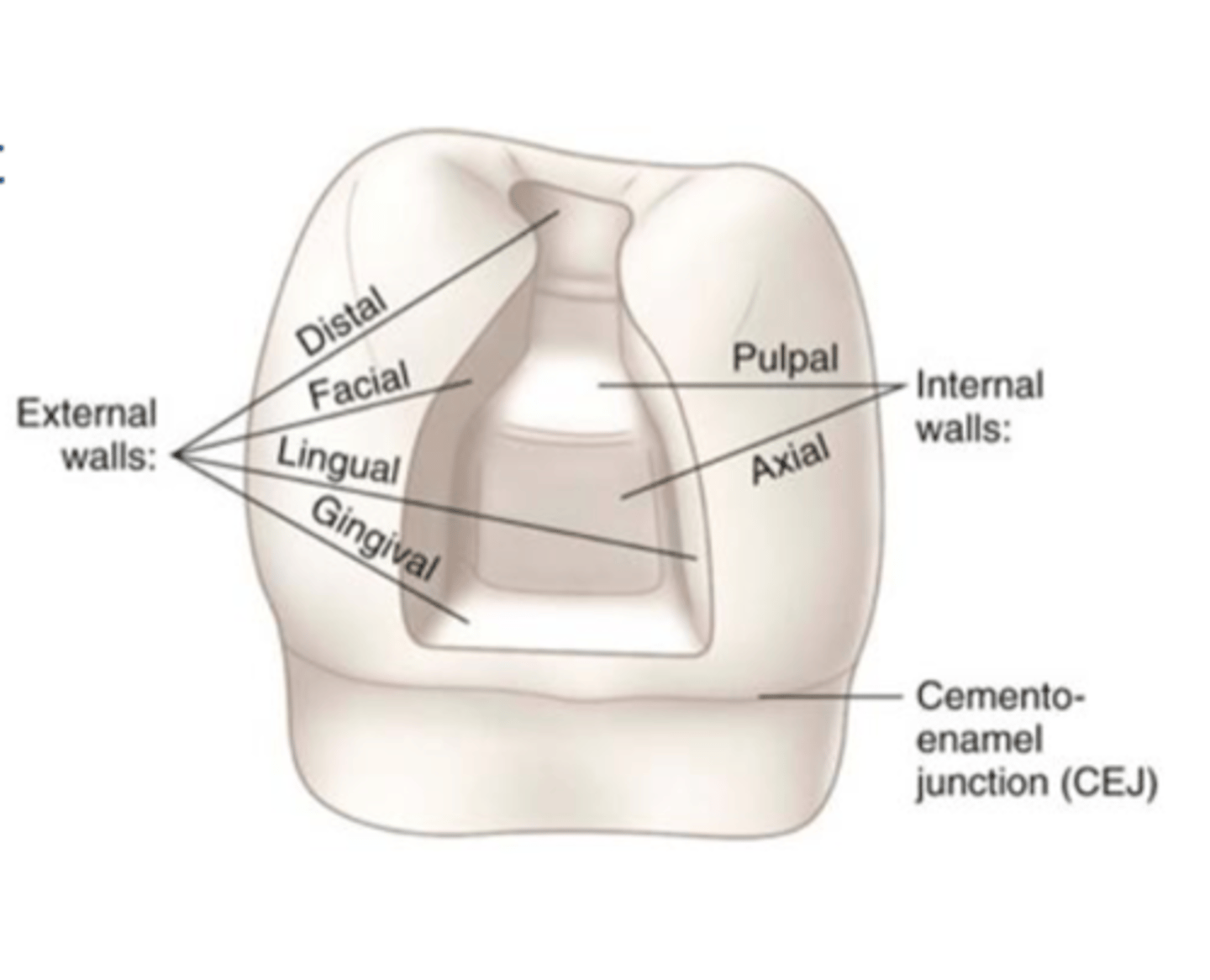
external preparation walls
buccal, distal, mesial, lingual, gingival
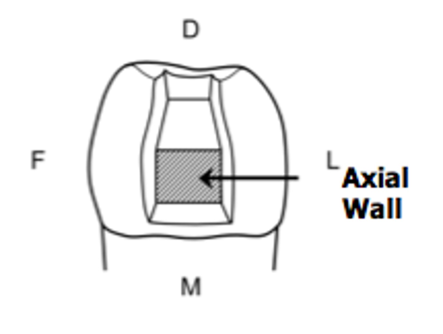
internal preparation walls
axial and pulpal
axial internal wall
parallels long axis of tooth, vertical plane of the tooth adjacent to pulp
pulpal internal wall
perpendicular to long axis of tooth closest to the pulp in the horizontal plane
when are the terms floors/ seats used instead of walls?
when the wall exists perpendicular to the long axis of the tooth
pulpal floor
gingival seat
line angles
junction of 2 walls; named based on the two walls
ex. lingual-gingival line angle
axial-pulpal line angle
point angles
junction of 3 walls; named based on the 3 walls
ex. axial-facial-gingival point angle
margin
junction of a cavity wall + external tooth surface
cavosurface margin
margins follow the entire preparation circumferentially
cavosurface angle
angle of tooth structure formed by the junction of a prepared wall + external surface of tooth (90º)
steps for a cavity preparation
1. outline form
2. resistance and retention form
3. convenience form
4. removal of remaining carious dentin
5. finishing enamel walls
6. cleansing the preparation
step 1 outline form
process of placing the cavity margins (cavosurface) in the tooth
should be visualized BEFORE any tooth reduction... measure twice, cut once
factors influencing outline form
1. location of the carious lesion
2. size of the carious lesion!!!
3. tooth anatomy
4. type of restorative material
5. esthetics
6. positioning of adjacent structures
7. functional requirements
8. retentive factors
step 1
outline form
what is the primary determinant in outline form?
lateral spread of decay in dentinal layer
aka SIZE of lesion (lateral)
what aspect of carious lesion size is not considered for outline form
lesion depth
final outline is not established until
carious dentin and overlying enamel have been removed
unsupported enamel
enamel not supported by dentin
when is the final outline form established?
when you have a DEJ free of carious tooth structure and no unsupported enamel
step 2
resistance and retention form
resistance form
shape/ placement of the preparation walls that enable both the restoration and the tooth to withstand, without fracture, masticatory forces delivered principally in the long axis of the tooth
resistance form 4 requirements
1. flat floors perpendicular to masticatory forces
2. restriction of preparation extensions to allow strong cuspal/ marginal ridges (conserve unaffected tooth) (keep marginal ridges)
3. inclusion of weakened tooth structure in the preparation design to prevent tooth fracture (cap a compromised cusp)
4. consideration of restorative materials: amalgam should have 90 degree cavosurface angle
retention form
shape/ form of preparation that resists displacement of the restoration from tipping/lifting forces
___ considers what is holding the material in the cavity preparation?
retention form
retention form 7 requirements
1. dovetails
2. convergent walls
3. grooves / slots
4. pins (large preps only)
5. frictional resistance of walls (nearly parallel, vertical or minimally tapered)
6. acid etch with bonding systems
7. mutually divergent rounded areas*
what type of retention does acid etch help with?
composite resin micromechanical retention
retention divots
divergent rounded area
purpose of dovetails in retention form
proper position maintains width and strength of marginal ridges
step 3 convenience form
shape/ form of preparation that provides the easiest way to operate/ restore the tooth
inadequate convenience form
prevents proper instrumentation
3 factors that influence convenience form
1. extension of the preparation
2. changing the direction of approach
3. changing the instrumentation utilized
why is preservation of unaffected structure crucial?
it helps resistance form
step 4
removal of remaining carious dentin
when is the remaining carious dentin/ infectious structure removed?
after basic cavity design has been completed
incipient lesion
dentin penetration by carious process 0.5mm or less
extensive lesion
advanced dentin penetration by the carious process
what is the ideal preparation depth?
0.5mm inside the DEJ after steps I, II and III have been completed
what step does not apply with incipient lesions?
step 4, removal of remaining carious dentin
technique for removing carious tooth structure and why
remove it ONLY from the affected area to avoid pulpal involvement and maintain resistance form
step 5
finishing enamel walls
what do you smooth and refine to finish enamel walls (step 5)?
walls of cavity prep + cavosurface angles
3 purposes of finishing preparation walls
1. good seal between restorative material + tooth
2. good marginal junction
3. max strength to at margin
why is a good marginal junction important?
it is less noticeable to the patient and facilitates cleansing (less plaque retentive)
factors to consider during wall finishing 4
- direction of enamel rods
- dentinal support for enamel rods
- restorative material that will be used
- location of preparation margins
when does the angulation of enamel rods vary?
varies with location of tooth structure and between primary vs. permanent teeth
fissured surfaces enamel rods (occlusal surfaces)
rods converge from DEJ to surface in areas of pits and grooves
rods diverge from DEJ to surface in areas of cusps and ridges
smooth surfaces enamel rods (proximal, facial, lingual)
- perpendicular to long axis of the tooth in the middle 1/3
- incline occlusally in the occlusal 1/3
- incline gingivally in the gingival 1/3
for proper enamel support, we want to finish our enamel walls so that enamel rods that form cavosurface have their inner ends...
resting on sound dentin
full length rods from DEJ to cavosurface are preferred
where do full length enamel rods receive support from?
DEJ + shorter rods w/ inner end resting on dentin
step 6
cleansing the preparation
cleansing the preparation entails
1. removing all debris from cavity preparation
2. dry and inspect for any remaining weakened structure (carious dentin and unsupported enamel)
caries on mesial aspect of secnd premolar is classified as what?
smooth surface caries