Biopsychology
1/111
There's no tags or description
Looks like no tags are added yet.
Name | Mastery | Learn | Test | Matching | Spaced |
|---|
No study sessions yet.
112 Terms
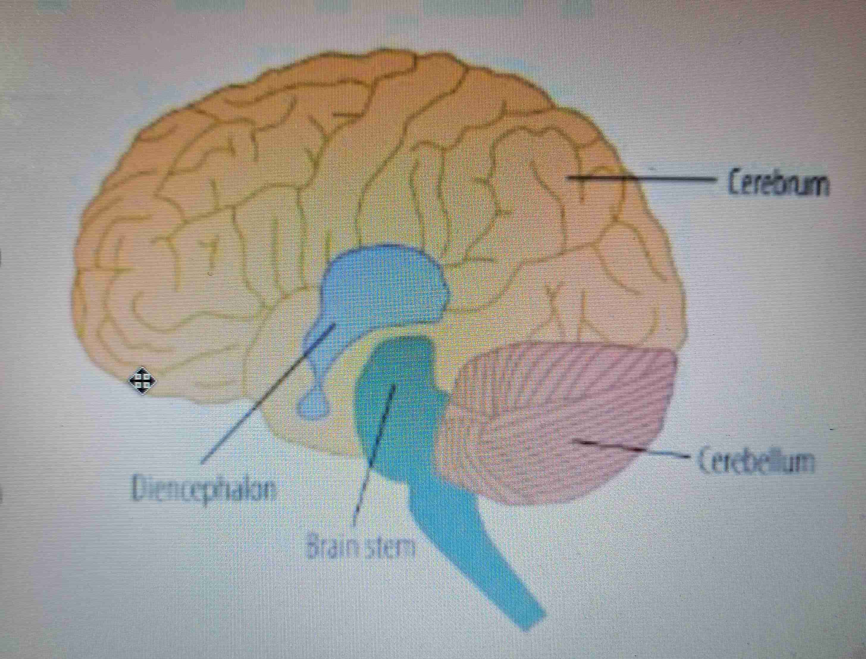
Which four parts is the brain divided into?
cerebrum, cerebellum, diencephalon and brain stem
Which four lobes is the cerebrum divided into?
frontal, temporal, parietal and occipital
Frontal lobe function
thought and production of speech
occipital lobe function
processes visual info.
temporal lobe function
processes auditory info.
parietal lobe function
processes sensory info. and plays an important role in spatial navigation
cerebellum function
motor skills and balance
which two structures is the diencephalon split into
the thalamus and the hypothalamus
thalamus function
relays nerve impulses from senses to appropriate parts of the brain where they can be processed
hypothalamus function
homeostasis and controls the release of hormones from the pituitary gland
brain stem function
regulates automatic functions
which NS is split into the Sympathetic NS and Parasympathetic NS
autonomic
somatic NS function
responsible for voluntary actions. maintains communication between CNS AND PNS using 12 pairs of cranial nerves and 31 pairs of spinal nerves
autonomic NS function
important in regulating actions over which you have no conscious control (like homeostasis)
sympathetic NS function
prepares the body for fight or flight via the use of the stimulating transmitter, noradrenaline
parasympathetic NS function
returns the body to its resting state after the fight or flight response via the use of the neurotransmitter, acetylcholine
evaluate theories of the “fight or flight” response
gender bias (androcentrism) - Early research into stress used only male animals and found the “fight or flight” response. Later research showed that females often respond with “tend and befriend” — showing that gender differences were overlooked. This is beta bias.
If our understanding of 'normal' behaviour comes from research involving all-male samples, then any behaviour that deviates from this standard is classed as 'abnormal' or 'inferior'.
This leads to female behaviour being misunderstood and even pathologised (taken as a sign of illness).
Von Dawans found that men can also respond with friendly and cooperative behaviour (e.g during 9/11)
sensory neurons (what they look like and function)
they are attached to a receptor cell and they are responsible for carrying impulses to CNS
relay neurons (what they look like and function)
they don't have a myelin sheath and they allow sensory and motor neurons to communicate. They lie within the CNS
motor neurons (what they look like and function)
they control muscles and they release neurotransmitters that bind to the receptors on muscles to trigger a muscle contraction.
Muscle relaxation is caused by inhibition of the motor neuron
dendrites function
receive signals from other neurons
axon function
a long fibre that carries impulses also known as an action potential
myelin sheath function
insulates the axon so that impulses travel faster
synaptic vesicles function and location (in neuron)
these are located at the end of the axon and they contain neurotransmitters
the action potential reaches vesicles and causes them to release neurotransmitters through a process called exocytosis
axon terminal function
connects the neuron to others using a process called synaptic transmission
when does excitation in the postsynaptic neuron occur?
receptor stimulation results in an increase in the positive charge of the postsynaptic neuron and increases the likelihood of the neuron passing on the impulse
when does inhibition occur in the postsynaptic neuron?
receptor stimulation results in the increase in the negative charge of the postsynaptic neuron and decreases the likelihood of the neuron passing on the impulse
what is ‘re-uptake’ in synaptic transmission?
the remaining neurotransmitters after transmission are broken down and reabsorbed by the presynaptic neuron and stored for later use
types of neurotransmitters
Dopamine - attention, learning
Acetylcholine - memory, learning
Noradrenaline - ‘fight or flight’
Serotonin - emotion, sleeping
GABA - fear, anxiety
what is summation?
the addition of EPSPs and IPSPs
how is a synapse unidirectional?
receptors only on postsynaptic neuron and neurotransmitters only released from presynaptic
what is spatial summation?
several presynaptic neurons can combine to cause an action potential in a postsynaptic neuron
what is temporal summation?
several action potentials produce synaptic transmissions in order to produce a postsynaptic action potential
what is divergence? (synaptic transmission)
impulses from one presynaptic neuron can be passed to several postsynaptic neurons
the entire process of synaptic transmission
action potential → axon terminal + vesicles → exocytosis → diffuse and bind to specific receptors → summation → reuptake (An ATV Enters DuBai So Rapidly)
As the action potential reaches the synaptic vesicles and the axon terminal, it causes the vesicles to release neurotransmitters as part of a process called exocytosis. These neurotransmitters diffuse across the synaptic gap (down a concentration gradient) where they bind to SPECIFIC receptors on the dendrite of the post-synaptic neuron.
Depending on the neurotransmitter, there is either an excitatory or inhibitory effect on the firing of the next action potential. The effects are then terminated by a process called re-uptake, where the remaining neurotransmitters are broken down and reabsorbed by the presynaptic neuron.
what is something that stimulates ‘fight or flight’ called?
a stressor
fight or flight process (6 steps)
person enters a stressful situation
the amygdala sends a distress signal to the hypothalamus
hypothalamus activates SAM pathway
the sympathetic NS stimulates the adrenal medulla
adrenal medulla secretes adrenaline + noradrenaline into bloodstream
physiological changes occur to prepare body
why might females have a “tend and befriend” response instead? what causes it?
this might be due to a need to protect offspring from predators.
females have oestrogen, which increases oxytocin levels, resulting in a reduced stress response
pituitary gland function
it stimulates the release of hormones from other glands
what is the pituitary gland divided into?
anterior and posterior lobes
what does the anterior lobe in the pituitary gland release? what is this hormone for?
ACTH, which stimulates the adrenal cortex and the release of cortisol. cortisol manages long-term stress
what does the posterior lobe in the pituitary gland release? what is this hormone for?
oxytocin - responsible for uterus contractions during childbirth
what does the pineal gland release? what is this hormone for?
melatonin - responsible for biological rhythms
what does the thyroid gland release? what is this hormone for?
thyroxine - responsible for regulating metabolism
what is hyperthyroidism?
too much thyroxine (skinny)
what is hypothyroidism?
too little thyroxine (fat)
what is the adrenal gland divided into? what do they do?
adrenal medulla - releases adrenaline and noradrenaline which are used in fight or flight
adrenal cortex - releases cortisol which stimulates glucose release
what do the testes release? what is this hormone for?
testosterone - responsible for development of secondary male sex characteristics during puberty
what do the ovaries release? what is this hormone for?
oestrogen - involved in the regulation of the menstrual cycle and development of secondary female sexual characteristics
what are the SAM and HPA systems?
SAM pathway - immediate shock response
HPA axis - countershock response
where is the motor centre located and what does it do?
it is located in the frontal lobe and is responsible for voluntary movements by sending signals to muscles
where is the somatosensory centre located and what does it do?
it is located in the parietal lobe and receieves incoming sensory info from the skin to produce sensations.
where is the visual centre located, what does it do and how does it work?
it is located in the occipital lobe and processes colour, shape and movement.
processing starts in the retina →optic nerve → thalamus → visual cortex
where is the auditory centre located, what does it do and how does it work?
it is located in the temporal lobe and it processes acoustic information like volume, tempo and pitch.
pathways begin at the cochlea (soundwaves converted to nerve impulses) and then the brain stem (basic decoding e.g. intensity/duration) before reaching the auditory cortex where a response is initiated.
where is broca's area and what does it do?
it is located in the left frontal lobe and is responsible for speech production
where is wernicke's area and what does it do?
it is located in the left temporal lobe and is responsible for language comprehension
support for localisation of function from case studies
Phineas Gage suffered damage to the frontal lobe and as a result his mood + personality changed.
indicates that frontal lobe is responsible for these
support for localisation of function from brain scanning studies
Peterson used brain scans to show how Wernicke's area was active during a listening task and how Broca's was active during a reading task.
brain scans are an objective and scientific way of showing localisation of function
support for localisation of function from neurosurgery procedures
Dougherty showed that procedures like the cingulotomy is successful with OCD patients. this involves damaging the cingulate gyrus
evidence against localisation of function due to no localisation of higher order functions
Lashley showed that learning was not localised to one specific area.
this suggests that higher order functions are dealt with more holistically.
evidence against localisation of function in the plasticity of the brain
stroke victimes can recover lost abilities over time.
this suggests that localisation of function is not set and determined
what is hemispheric lateralisation?
the idea that the brain’s two halves are functionally different and that each hemisphere has functional specialisations.
what is each side of the brain responsible for?
left - language
right - visual motor tasks
how are the two hemispheres connected?
the corpus callosum are nerve fibres which facilitate interhemispheric communication
what were the dynamics of the hemispheric lateralisation study?
Sperry and Gazzaniga investigated patients that had undergone a commissurotomy (they had lost interhemispheric communication)
they conducted 3 different tests:
describe what is seen
tactile test (sensory)
drawing task
which hemisphere of the brain processes the left visual field?
right side (and vice versa)
what was found in the “describe what you see” task of the hemispheric lateralisation experiment?
when the picture was processed by the right hemisphere, patients were unable to describe the picture.
this demonstrated the superiority of the left hemisphere in terms of language
what was found in the tactile test of the hemispheric lateralisation experiment?
when an object was placed in their left hand (processed right hemisphere), they were unable to describe what they felt.
however they could identify it by selecting a similar object from a series of alternate objects
what was found in the drawing task of the hemispheric lateralisation experiment?
their left hand (controlled by the right hemisphere) would consistently draw better pictures (even though the participants were all right-handed)
evaluation of sperry and gazzaniga’s hemispheric lateralisation experiment.
highly controlled + scientific
causation could be established between stimuli and responses
small sample size
low population validity makes the findings less generalisable
what is brain plasticity?
the brain’s ability to change and adapt because of experience
what is functional recovery?
the transfer of functions from a damaged area → undamaged area
study for brain plasticity
Maguire et al. used MRI scans to analyse the sizes of the hippocampus in London taxi drivers and a control group.
Results:
increased grey matter was found in the posterior hippocampus of the taxi drivers
A positive correlation was found netween time spent as a taxi driver and volume of the posterior hippocampus
problem with Maguire’s taxi driver study
It only recorded snapshot data, meaning that it only recorded data of that specific time:
Doesn’t show us the hippocampi grew over time
Maybe people with bigger hippocampi become taxi drivers - not the other way around
what is neuronal pruning
the removal of redundant or otherwise unnecessary synaptic connections. it occurs between childhood and adulthood.
what are the mechanisms for recovery?
axonal sprouting
recruitment of homologous areas
neuronal unmasking
what is axonal sprouting?
the growth of new nerves which connect to undamaged nerves to form new pathways
what is recruitment of homologous areas?
when an area on the opposite hemisphere compensates if an area is damaged on one side
what is neuronal unmasking?
this is when secondary neural pathways are activated to enable functioning to continue.
what are the three possible views on how stem cell implantation into the brain can repair it?
they directly replace dead/dying cells
they secrete growth factors in order to ‘rescue’ the injured cells
an uninjured brain site is linked to the damaged region via a neural network
study on stem cells
Taijiri et al. provided stem cells to rats after brain trauma and found development of new neurons in the area of injury
study on neuron activity (plasticity)
Davidson et al. compared the neuronal activity of Tibetan monks with controls as they meditated
Bigger gamma waves were found in monks
study on recruitment of homologous areas
Danelli et al. examined a patient who had his left hemisphere removed at the age of 2. By the age of 17, his language performance was practically the same as the control group (after an intense rehabilitation programme)
evalutation of Danielli’s study on recruitment of homologous areas
Strengths:
Application - intense rehabilitation programmes help → get used
Brain scans v. objective
Weaknesses:
Can't be generalised - esp. to old people (plasticity declines with age)
Correlational data, not causational (how do we know the rehab programme really led to the recovery?)
4 ways to evaluate the ways of studying the brain
Spatial resolution (smallest detectable feature)
Temporal resolution (speed of the scanner’s detectors to detect change)
Invasive vs. non invasive (insertion)
Causation (between activity and function)
how does an fMRI scan work
They detect changes in blood oxygenation that occurs due to activity in specific parts of the brain that are active during certain processes that are completing physical demands (therefore demonstrates localisation).
strengths of fMRI scans
safe - no radiation like PET scans
high spatial resolution (detail) compared to EEGs + ERPs
non-invasive
weaknesses of fMRI scans
expensive
poor temporal resolution compared to EEGs + ERPs
only measures blood flow, not neuronal activity, making it difficult to tell which kind of activity is being measured. Therefore causation is problematic.
how does an EEG work
measures electrical activity via electrodes attached to the scalp.
provides a general state of the brain’s activity to indicate neurological issues.
strengths of EEGs
useful in clinical diagnosis
high temporal resolution compared to fMRI
non-invasive
weaknesses of EEGs
can’t detect activity in deep region of the brain. Only superficial regions → poor spatial resolution (compared to fMRI) → problematic causation
how do ERPs work
measures electrical activity in brain with electrodes on scalp like EEGs, but with these, a stimulus is presented many times and an average response is graphed (‘averaging’).
strengths of ERPs
more specific than EEGs
higher temporal resolution, compared to fMRI
non-invasive
cheaper than fMRI scans
weaknesses of ERPs
poor spatial resolution compared to fMRI as only superficial regions
some stimuli are unable to be presented multiple times
e.g. startling sounds like a scream or loud bang. These will lose their psychological impact over time due to habituation
time consuming for both researchers and participants
how do post mortems work
areas of damage in a person’s brain can be studied after their death and related to differences in their behaviour prior to their death
strengths of post mortems
patient is dead, so they won't have discomfort
fundamental in understanding the brain and localisation of function
weaknesses of post mortems
difficult to acquire informed consent
acquiring retrospective consent is difficult as family members may not reflect the participants true wishes
post-mortems may be conducted on individuals with impaired cognitive capacity - they may not have understood when they agreed to do the procedure
brain could have been affected during death, affecting results
length of the circadian rhythm and what resets it
The circadian rhythm lasts 24 hours and is reset by light.
how does the circadian rhythm work
Light is detected by the eye, which sends messages (related to brightness) to the suprachiasmatic nuclei (SCN)
The SCN then uses this info. to coordinate activity of the entire circadian rhythm
Sleeping and wakefulness are determined by homeostasis
what is an endogenous pacemaker
our body’s internal biological clocks that regulate our rhythms (e.g. SCN)