Comprehensive Eye and Ear Anatomy, Functions, and Disorders
 Call Kai
 Learn
Learn Practice Test
 Spaced Repetition
 Match
Match Flashcards
 Knowt Play
1/97

There's no tags or description
Looks like no tags are added yet.
Name | Mastery | Learn | Test | Matching | Spaced | Call with Kai | Chat |
|---|
No analytics yet
Send a link to your students to track their progress
98 Terms
Auditory
Pertaining to sense of hearing
Acoustic
Pertaining to sound or hearing
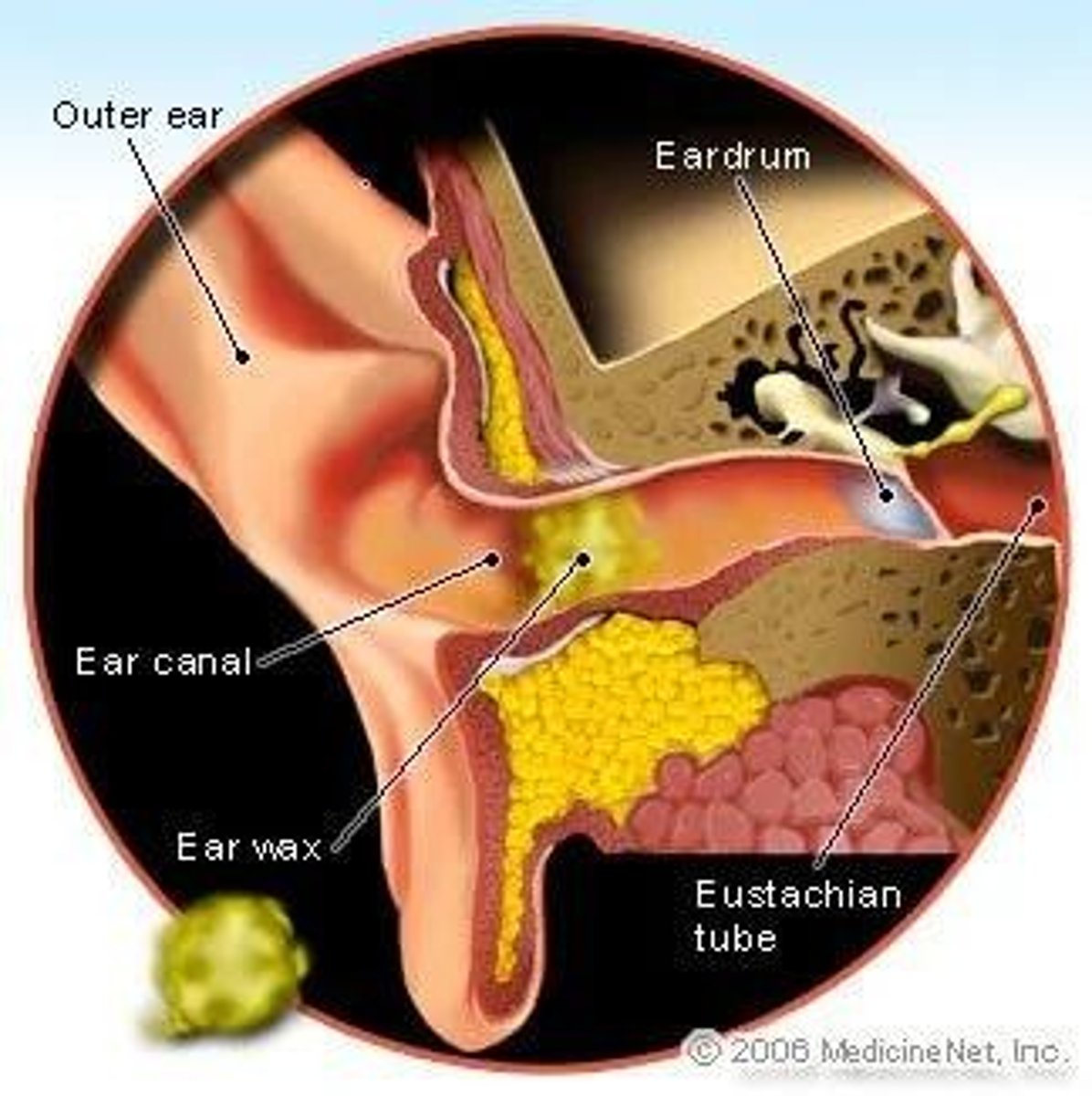
Cerumen
Earwax secreted by ceruminous glands lining auditory canal
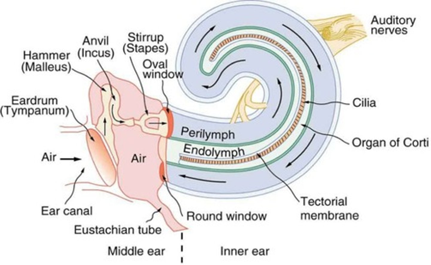
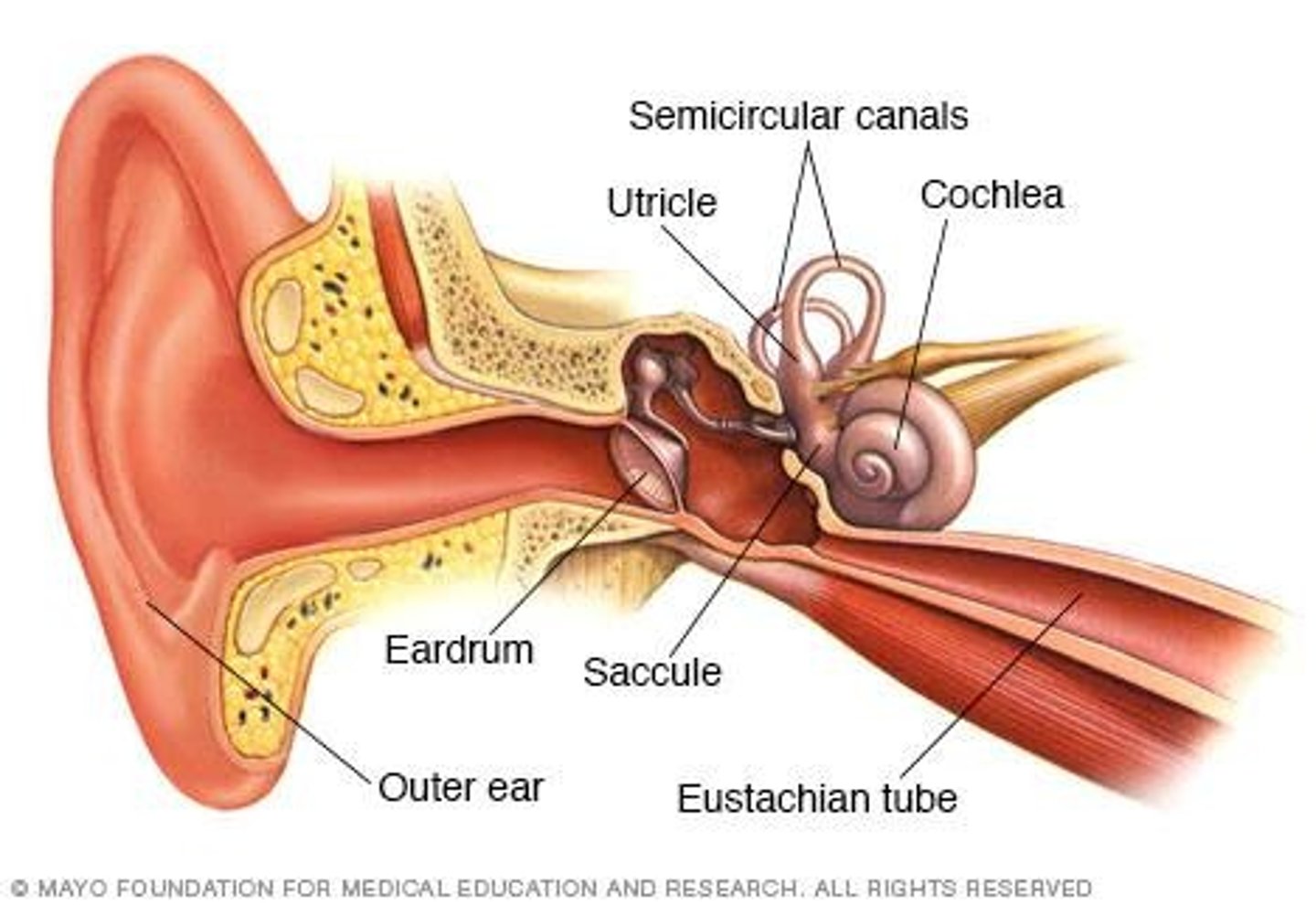
Tympanic Membrane
Eardrum located between outer and middle ear
Mastoid process
Temporal bone containing hollow air space surrounding the middle ear
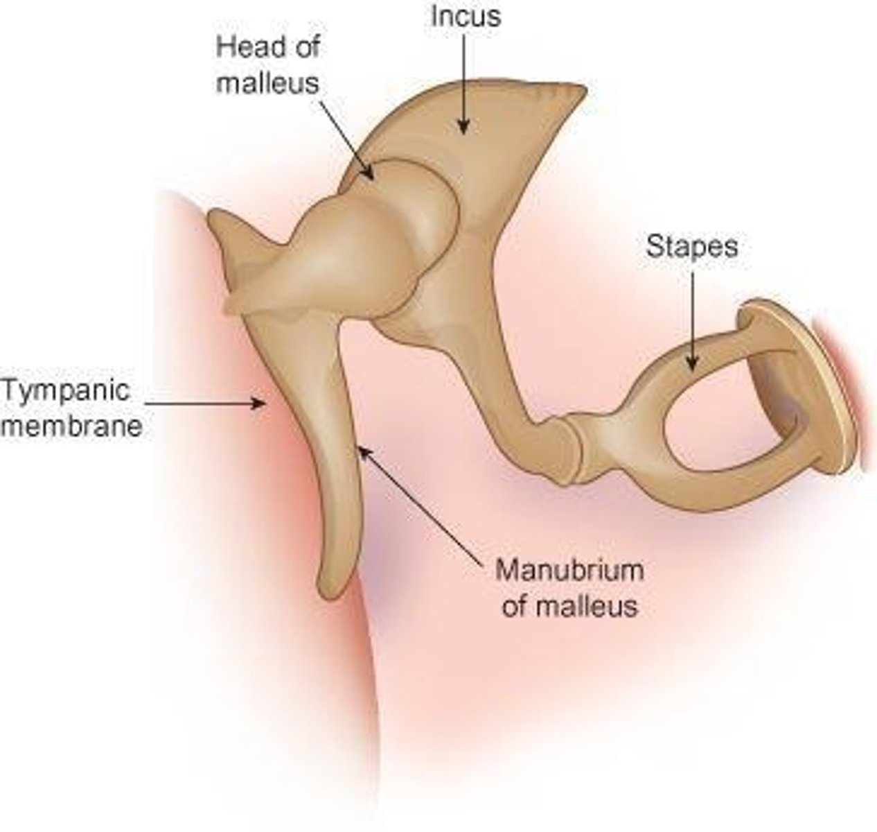
Auditory Ossicles
Transmit sound waves from eardrum to inner ear by vibration
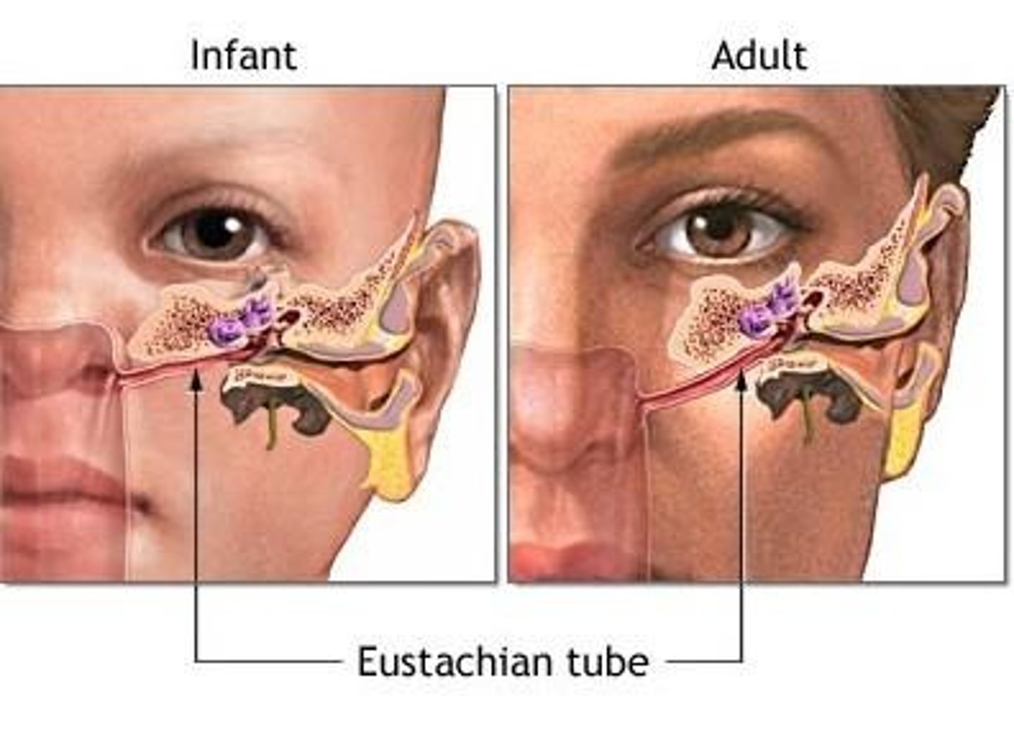
Eustachian Tubes
Lead from middle ear to nasal cavity and throat
Inner Ear
Contains sensory receptors for hearing and balance
Oval window
Membrane separating the middle ear from the inner ear
Cochlea
Snail-shaped structure where sound vibrations are converted into nerve impulses
Organ of Corti
Relays vibrations to auditory nerve fibers
Semicircular canals
Contain endolymph and sensitive hair cells that assist with equilibrium
Acoustic nerves
Transmit information to the brain
Air Conduction
Process of sound waves entering the ear through pinna
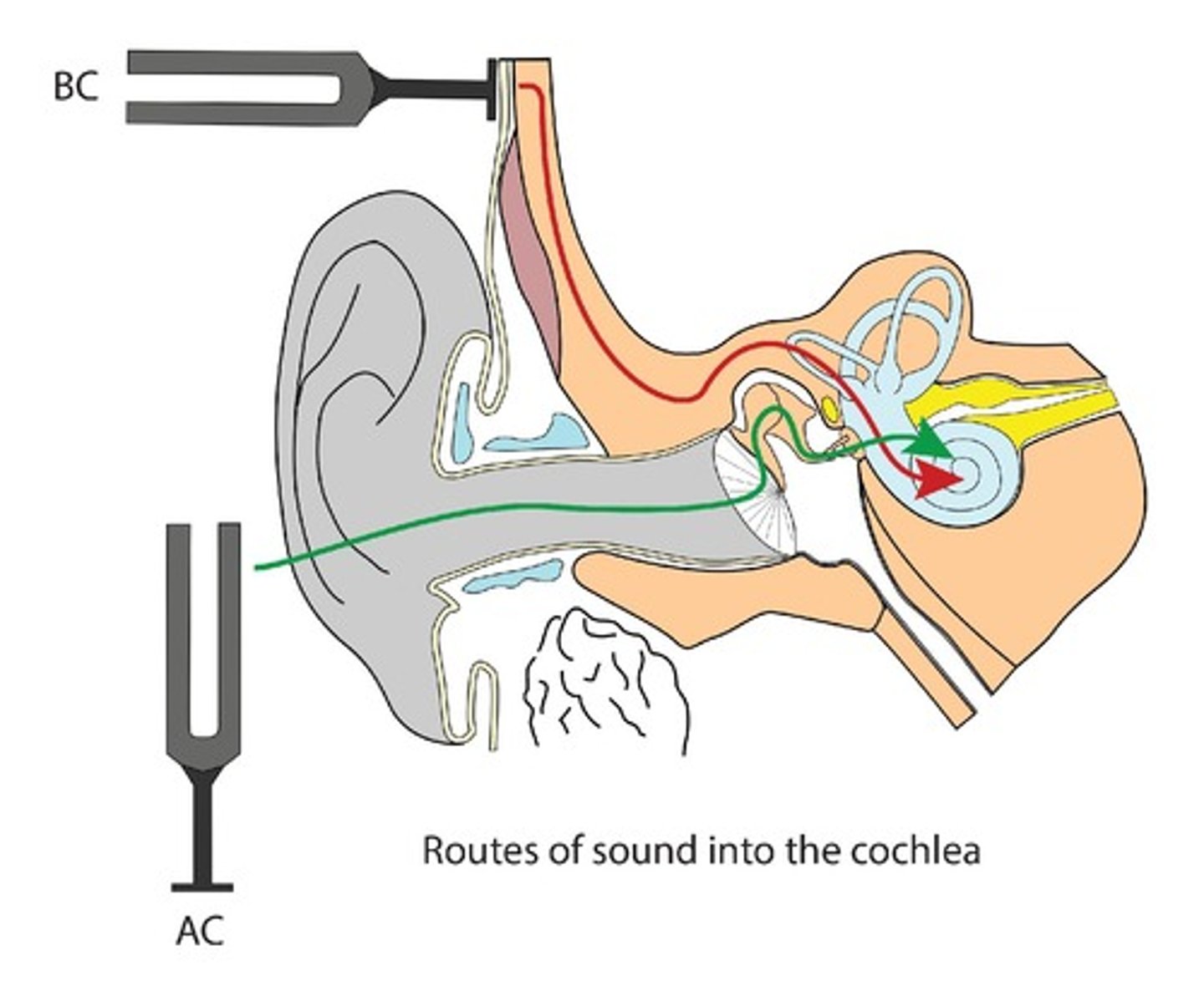
Bone Conduction
Occurs as eardrum vibrates causing auditory ossicles to vibrate
Sensorineural Conduction
Occurs when sound vibrations reach inner ear
Audiologist
Specializes in measurement of hearing function and rehabilitation of hearing impairments
Speech-language pathologist
Assists patients with problems related to swallowing, speech, and communication disorders
Otalgia
Pain in the ear
Otorrhea
Any discharge from the ear
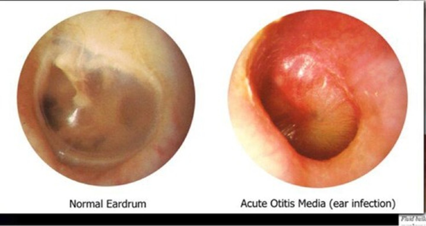
Otitis Media
Inflammation of middle ear
Vertigo
Sense of dizziness and loss of balance
Tinnitus
Ringing, buzzing, or roaring sound in one or both ears
Hearing Loss
Complete or partial loss of ability to hear
Presbycusis
Gradual loss of sensorineural hearing related to aging
Conductive hearing loss
Sound waves are prevented from passing air to fluid-filled inner ear
Sensorineural hearing loss
Due to damage to auditory nerve or hair cells in inner ear
Audiometry
Use of audiometer to measure hearing acuity
Otoscope
Instrument used to examine external ear canal
Tympanometry
Use of air pressure in ear canal for testing disorders of middle ear
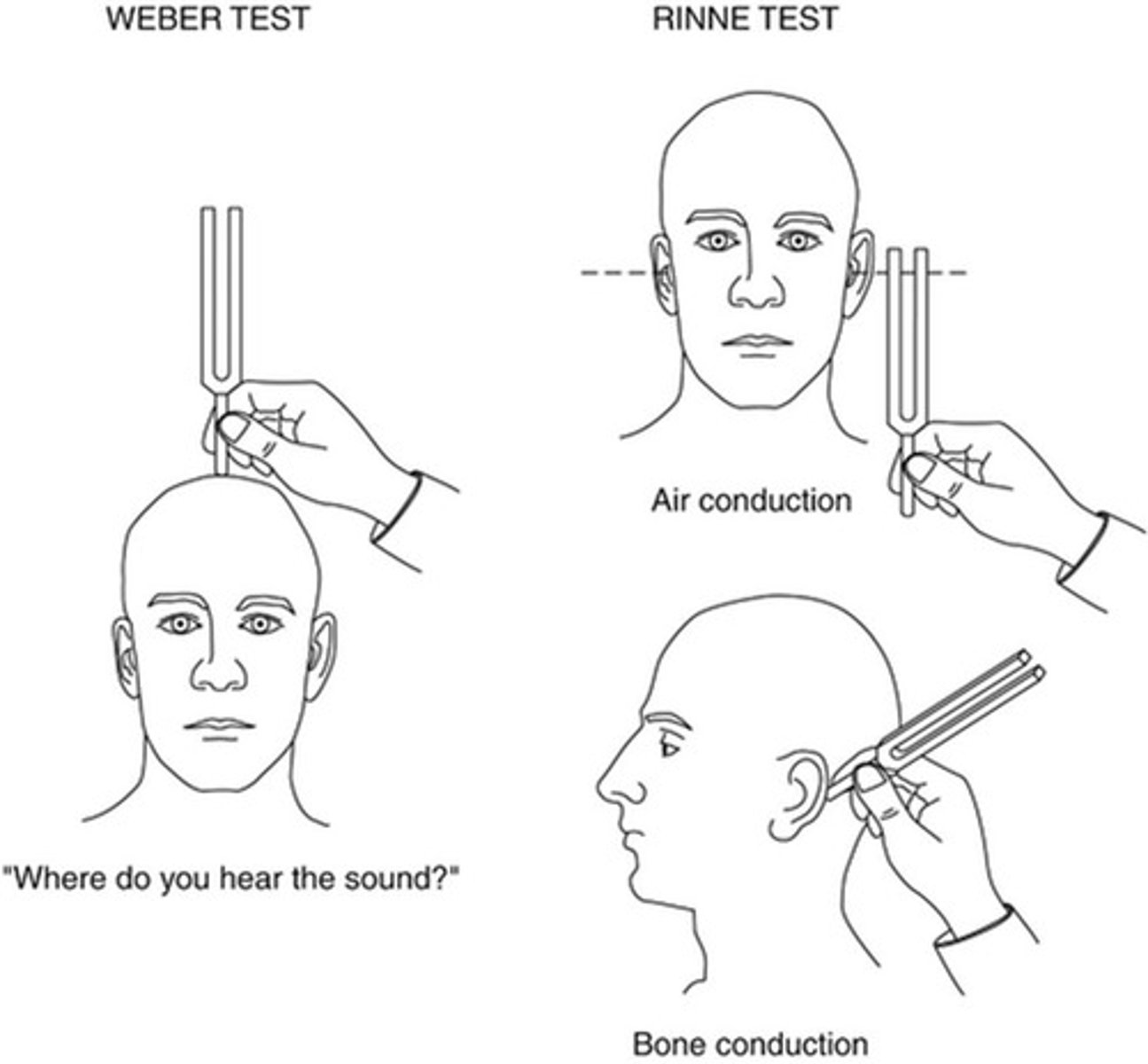
Weber and Rinne tests
Use of tuning fork to distinguish between conductive and sensorineural hearing losses
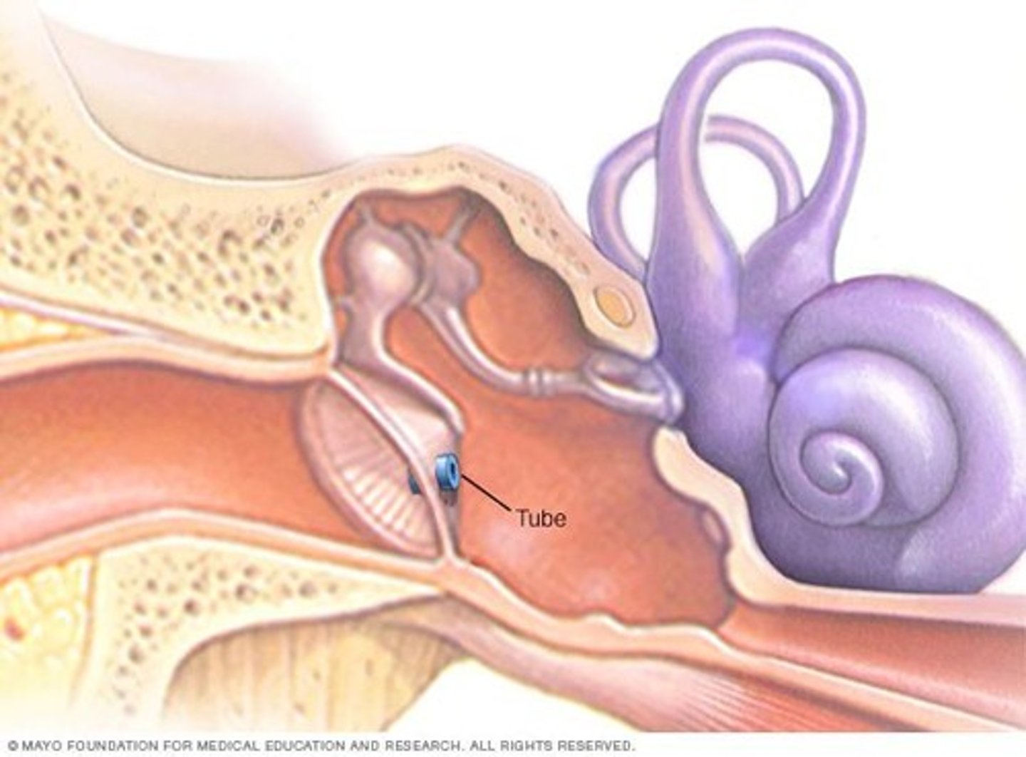
Tympanostomy Tubes
Ventilating tubes inserted into the eardrum for drainage and pressure relief
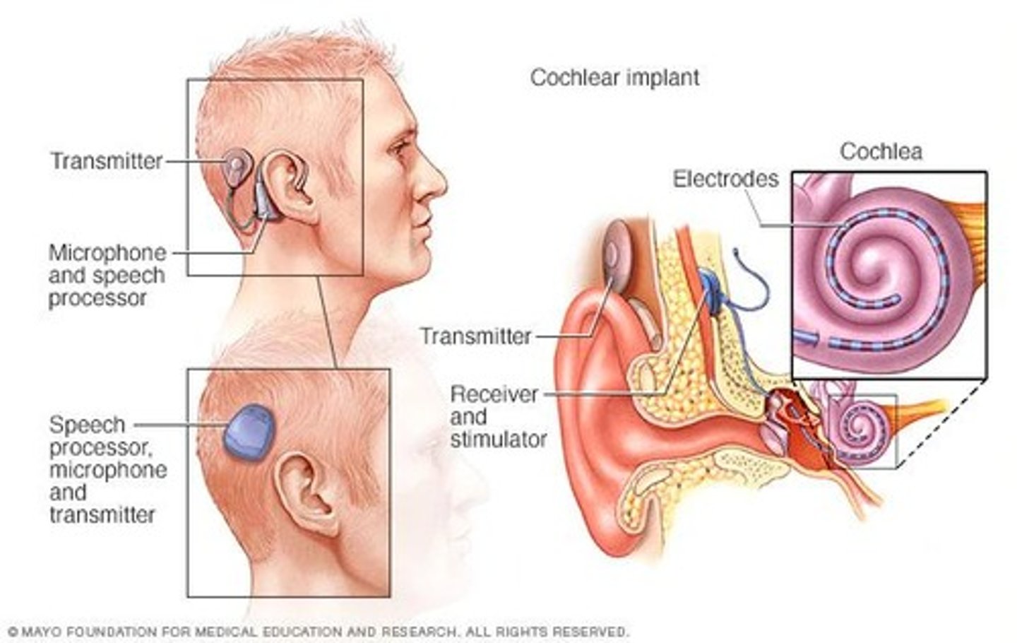
Cochlear implant
Bypasses damaged portion of ear and directly stimulates auditory nerves
Hearing aids
Electronic devices worn to correct hearing loss
Analog hearing aid
Uses microphone to detect/amplify sounds
Digital hearing aid
Computer chip converts incoming sound into code that is filtered before being amplified
Adnexa of the Eyes
The structures outside the eyeball, including the orbit, eye muscles, eyelids, eyelashes, conjunctiva, and lacrimal apparatus.
Bony Orbit
Cavity of skull that contains and protects the eyeball and associated muscles, blood vessels, and nerves; also known as the eye socket.
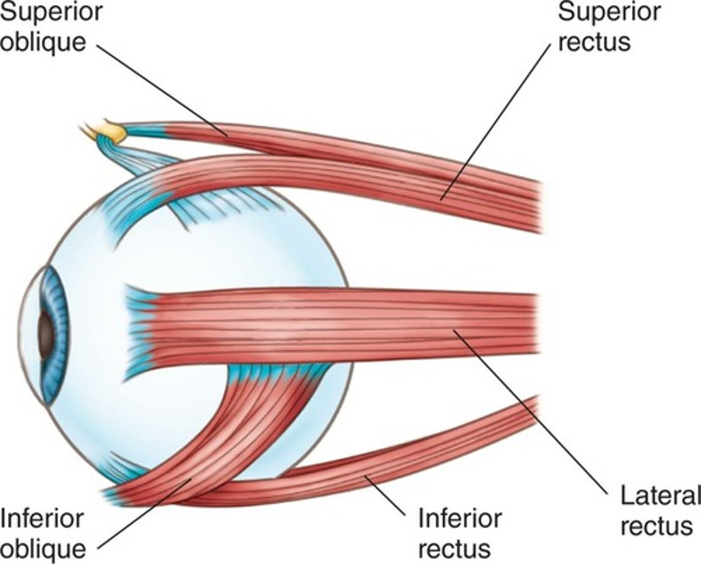
Muscles of Eye
Six major eye muscles (three pairs): superior and inferior oblique muscles, superior and inferior rectus muscles, lateral and medial rectus muscles.
Binocular vision
Coordination of eye muscles allowing for depth perception.
Depth perception
Ability to see things in three dimensions.
Eyelids, Eyebrows, and Eyelashes
Provide protection from foreign matter, excessive light, and injuries.
Cilia
Small hair making up eyebrows/eyelashes.
Conjunctiva
Transparent mucous membrane lining underside of the eyelid protecting the exposed surface of the eyeball.
Lacrimal Apparatus
Structures producing, storing, and removing tears.
Lacrimal glands
Located on underside of upper eyelid just above outer corner of each eye.
Lacrimal fluid
Maintains moisture on the anterior surface of eyeball.
Lacrimal canal
Ducts at the inner corner of each eye that collect tears emptying them into lacrimal sacs.
Lacrimal sac
Enlargement of the upper portion of the lacrimal duct.
Lacrimal duct
Passageway draining excess tears into nose.
Ocular
Pertaining to the eye.
Extraocular
Outside the eyeball.
Intraocular
Within the eyeball.
Sclera
White of the eye that maintains shape and protects delicate inner layers of tissue.
Choroid
Opaque middle layer of eyeball that provides blood supply to the entire eye.
Retina
Innermost layer lining posterior segment of the eye that receives nerve impulses and transmits them to the brain via optic nerve.
Anterior Segment of Eyeball
Front one-third of eyeball located behind the cornea and in front of the iris.
Aqueous humor
Helps maintain shape and nourishes intraocular structures.
Intraocular pressure
Measures fluid pressure inside the eye which is regulated by the rate at which aqueous humor enters/leaves the eye.
Posterior Segment of Eyeball
Remaining two-thirds of the eyeball located behind the iris and in front of the ligaments holding the lens in place.
Ciliary Body
Set of muscles and suspensory ligaments that adjust thickness of the lens and refine the focus of light rays on retina.
Iris
Colorful circular structure surrounding the pupil that controls the amount of light entering through the pupil.
Cornea
Transparent outer surface of eye covering iris and pupil that focuses light rays entering the eye.
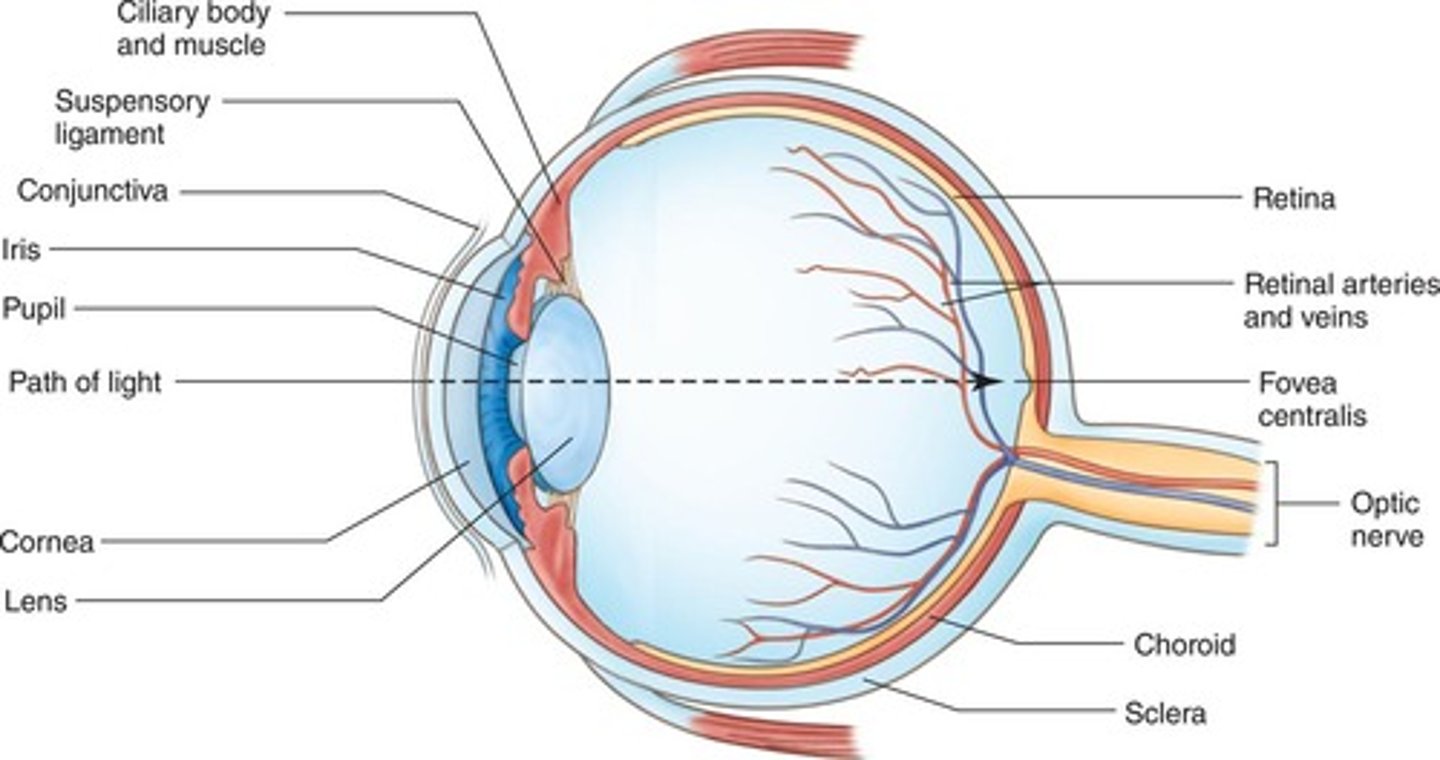
Pupil
Black circular opening in the center of the iris permitting light to enter the eye.
Lens
Clear, flexible, curved structure contained within clear capsule located behind the iris and pupil that focuses images on the retina.
Accommodation
The process whereby the eyes make adjustments for seeing objects at various distances.
Convergence
Simultaneous inward movement of the eyes toward each other to maintain single binocular vision as an object comes nearer.
Emmetropia
Normal relationship between the refractive power of the eye and the shape of the eye that enables light rays to focus correctly on the retina.
Refraction
Ability of the lens to bend light rays so they focus on the retina.
Visual Acuity
Ability to distinguish object details and shape at a distance.
Ophthalmologist
Specializes in diagnosing/treating diseases/disorders of eyes.
Optometrist
Provides diagnosis of eye diseases, measuring accuracy of vision determining need for corrective lenses.
Optician
Health care practitioner who designs, fits, and dispenses lenses for vision correction.
Conjunctivitis
Inflammation of conjunctiva usually due to infection or allergy; also known as Pinkeye.
Xerophthalmia
Dry eye due to decreased production of tears by tear glands.
Corneal abrasion
Injury to outer layers of cornea.
Diabetic retinopathy
Damage to retina as complication of uncontrolled diabetes.
Cataract
Loss of transparency of lens causing progressive loss of visual clarity.
Floaters
Particles of cellular debris in vitreous humor casting shadows on the retina.
Miosis
Contraction of pupil in response to light.
Mydriasis
Dilation of pupil.
Nystagmus
Involuntary, constant, rhythmic movement of eyeball.
Retinal detachment
Separation of some or all of the retina from the choroid.
Glaucoma
Group of diseases characterized by increased intraocular pressure resulting in damage to the retinal nerve fibers and optic nerve.
Macular Degeneration
Progressive condition causing damage to the macula, resulting in the loss of central vision.
Photophobia
Excessive sensitivity to light.
Strabismus
Improper eye alignment due to the inability of eye muscles to coordinate.
Presbyopia
Decline of near vision related to aging.
Astigmatism
Lack of focus due to uneven curvature of cornea.
Hyperopia
Farsightedness; light rays focus beyond the retina.
Myopia
Nearsightedness; light rays focus in front of the retina.
Blindness
Inability to see; legal blindness = best-corrected vision of 20/200.
Snellen chart
Measures visual acuity, recorded as fraction with 20/20 representing normal vision.
Slit-lamp ophthalmoscopy
Focusing the beam of light onto parts of the eye to permit examination of structures at front of the eye.
Tonometry
Measurement of intraocular pressure; abnormally high pressure may indicate glaucoma.
Corneal transplant
Surgical replacement of scarred/diseased cornea with clear corneal tissue from donor.
L A S I K
Treats vision conditions caused by the shape of cornea.
Retinopexy
Reattaches retinal detachment.