Diagnostic Imaging- Ultrasound Spleen, Liver, and Pancreas
1/137
There's no tags or description
Looks like no tags are added yet.
Name | Mastery | Learn | Test | Matching | Spaced |
|---|
No study sessions yet.
138 Terms
spleen
ID organ
lateral and VD
large breed canine spleens can be seen on both _____ and _____ images
abnormal- enlarged
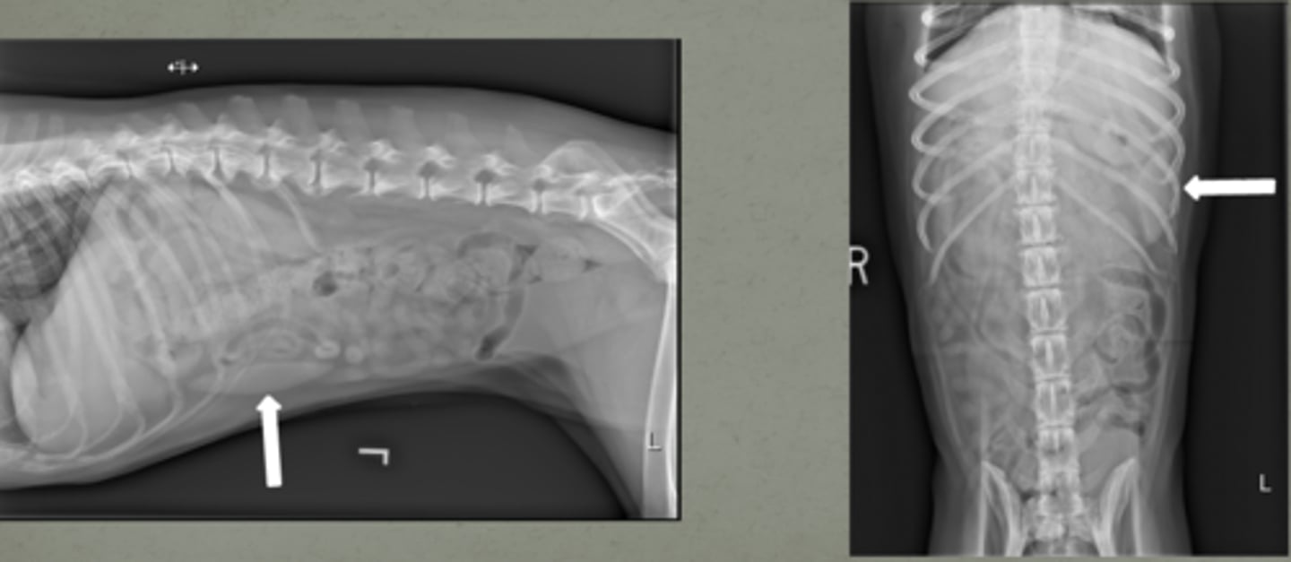
is this a normal or abnormal spleen?
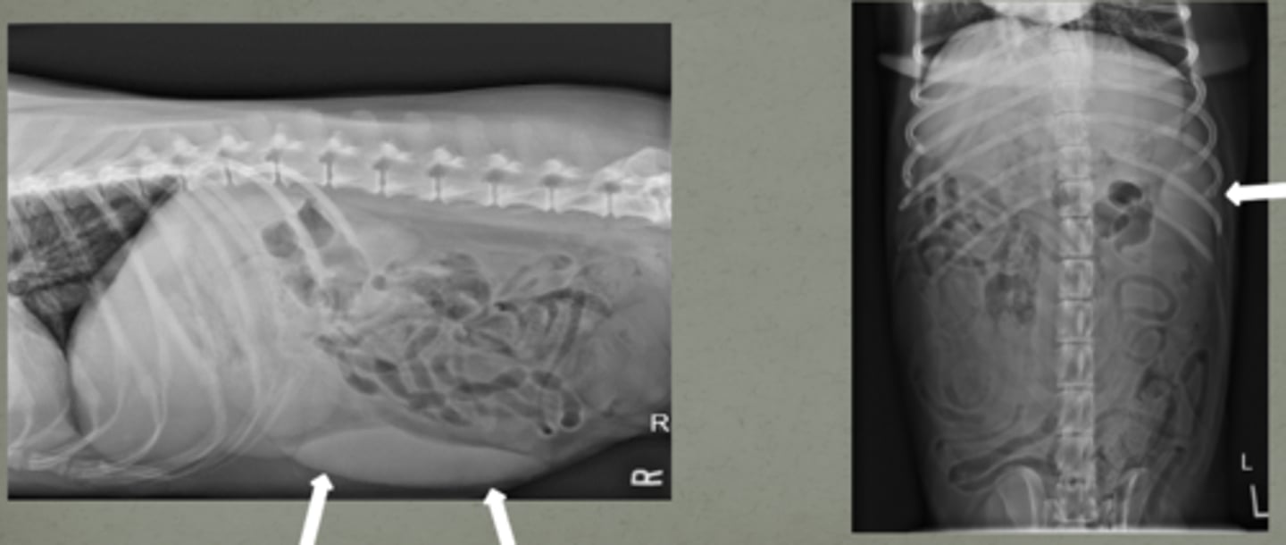
-left lateral
-lateral
in feline patients, the spleen will normally only be seen along the ________ body wall and typically not seen on _______ views
-splenomegaly
-splenic mass
-traumatic injury
-neoplasia
-anemia
-abdominal pain
what are spleen indications for scanning?
7.5-12
what MHz probe do you use for an ultrasound of the spleen for small animals?
transrectal- 5
transabdominal- 3-5
for ultrasound of the spleen for the horse, what MHz do you use transrectally? transabdominally?
cranial
the fundus of the stomach is located _______ to the spleen
caudal and deep
left kidney is _____ and ______ to spleen
caudal and medial
the left lobe of the pancreas is located ______ and ______ to the spleen
splenic
the left lobe of the pancreas is adjacent to the ______ vein
German Shepherd dogs
what breed of dog will have a larger spleen?
extends beyond the costal arch some crossing over to the right cranial to mid abdomen, some extending to the urinary bladder
where does the spleen extend in large dogs?
left cranial
cat spleens are usually in the ________ abdomen
<1 cm
normal spleen thickness for a cat?
-gastrosplenic
-left cranial
the head of the spleen is fixed by the ________ ligament and located in the ________ abdomen
body and tail
the _______ and ______ of the spleen are more mobile
-right
-left lateral
the body and tail of the spleen may cross over to the
_______ side or along the ____ ____ body wall
...
this is the spleen, just know what it looks like!!
well-defined
in normal US of the spleen, the capsule is usually ________
-homogenous
-less coarse
the spleen has ______ parenchyma with uniform finely stippled pattern less/more coarse than the liver
liver and renal cortex
the spleen is more hypoechoic than what two structures in the dog?
isoechoic or hypoechoic
in cats, the spleen can be _________ or ________ to the renal cortex (echogenicity-wise)
smooth
it is important to look for _______ borders of the spleen to detect masses
left- liver
right- spleen, b/c hyperechoic
which is the liver and which is the spleen based off of echogenicity?
splenic
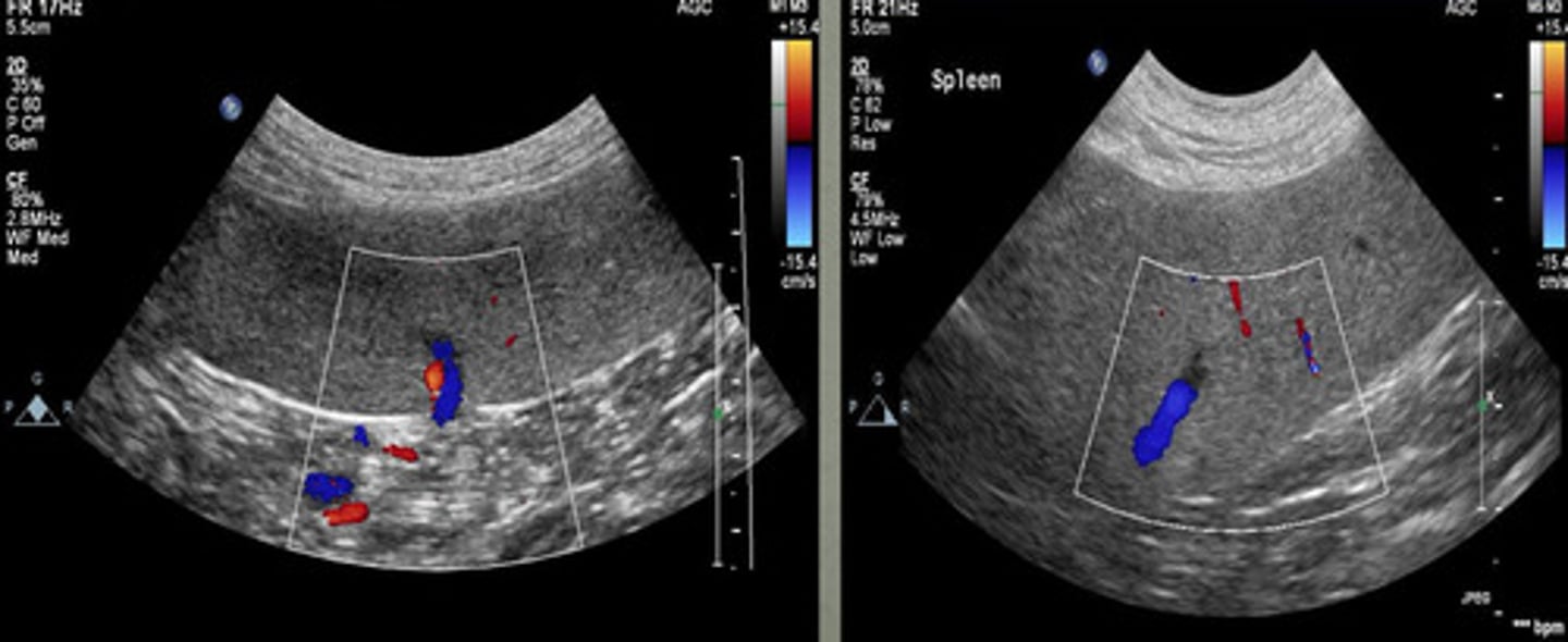
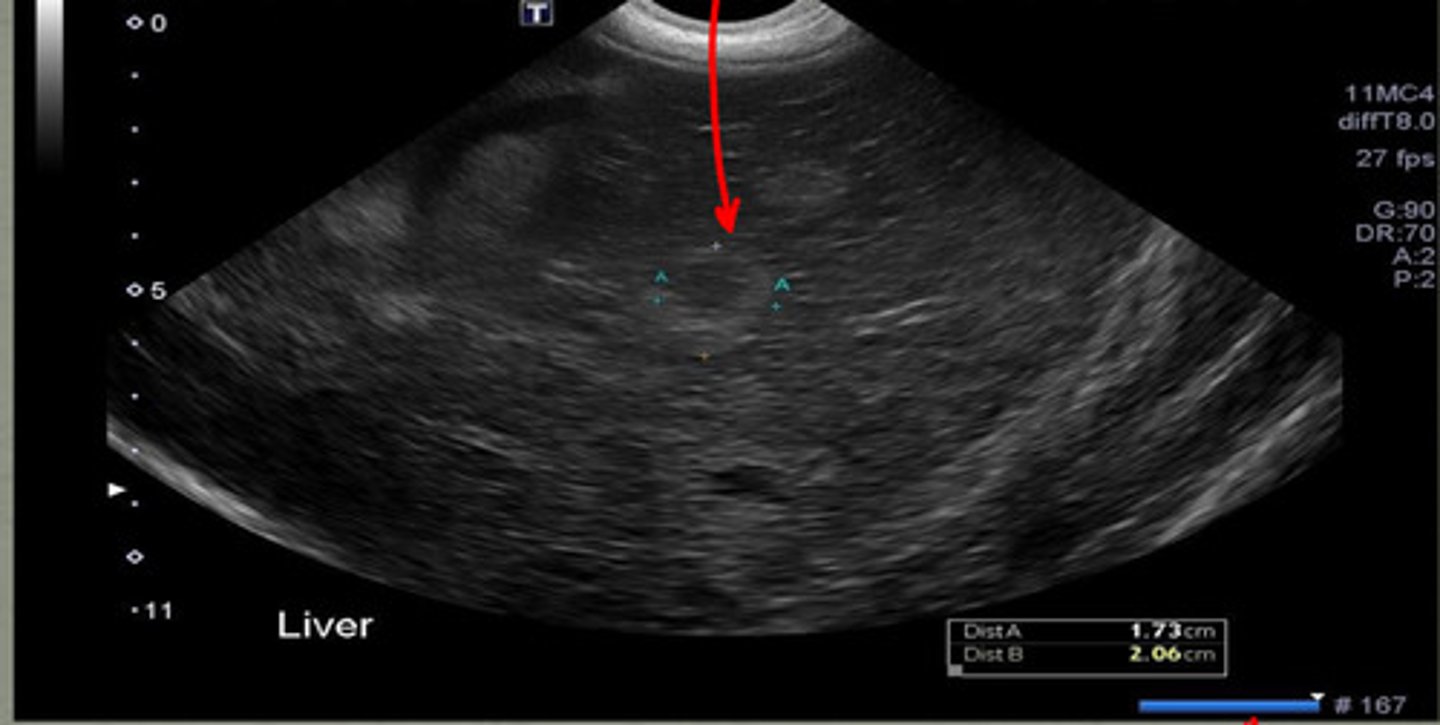
the _______ artery is not often detected without color doppler
-blue
-larger
-red
when using color doppler in ultrasound, veins appear _______ in color and are larger OR smaller than corresponding arteries depicted in ______ color?
color dopler
what type of tool is being used in this ultrasound to see the different vessels?
shadowing, hyperechoic
surrounding fat around the spleen causes _______ and appears what echogenicity?
yes, from surrounding fat
is this shadowing near the spleen normal?
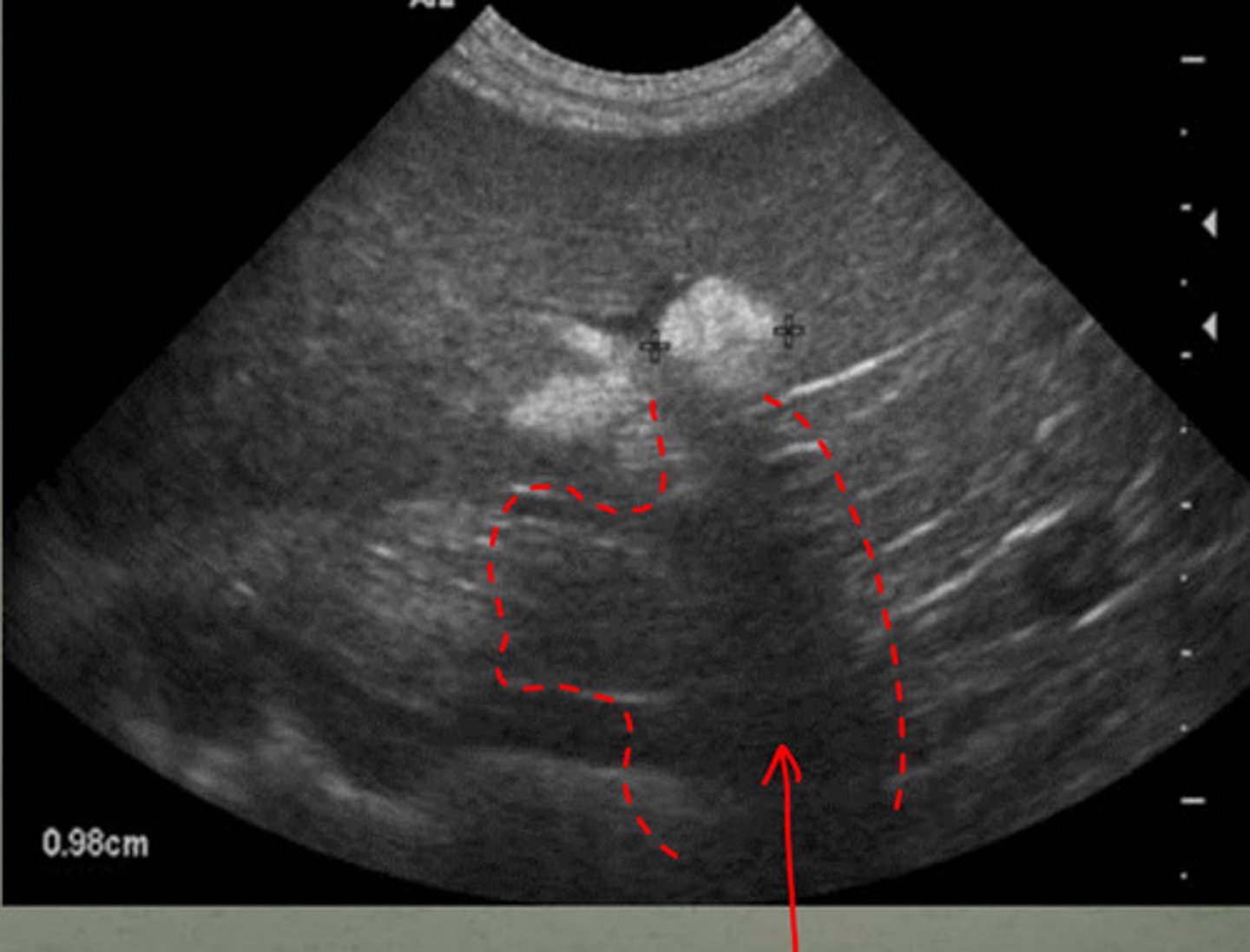
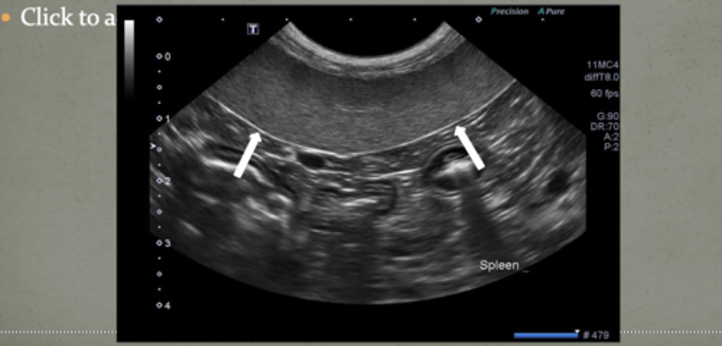
shadowing underneath the mass
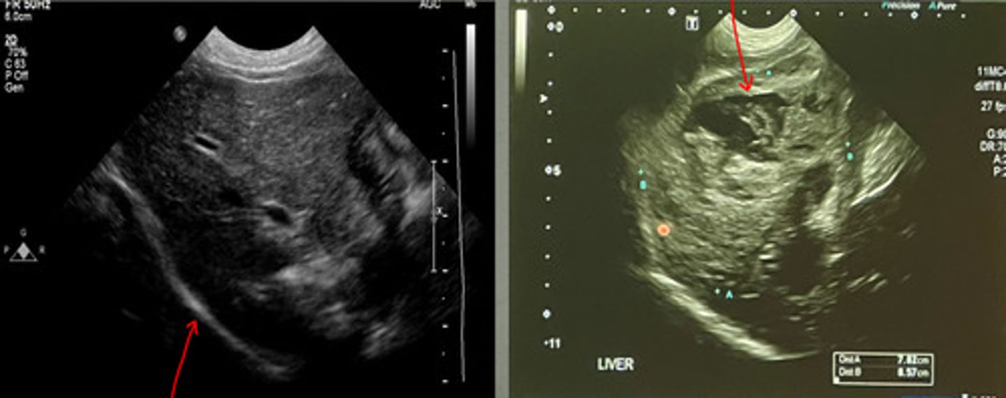
if there is a hyperechoic mass in the spleen, what artifact will occur due to it?
shadowing due to the splenic mass
what are the arrows indicating? (there is a mass in this spleen)
1. compare echogenicity of liver and renal cortex
2. classify lesion as focal or diffuse
3. focal lesions easier to recognize
4. uniform pattern/echotexture
what four things should you evaluate when ultrasounding the spleen?
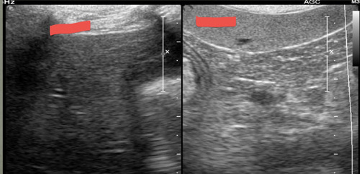
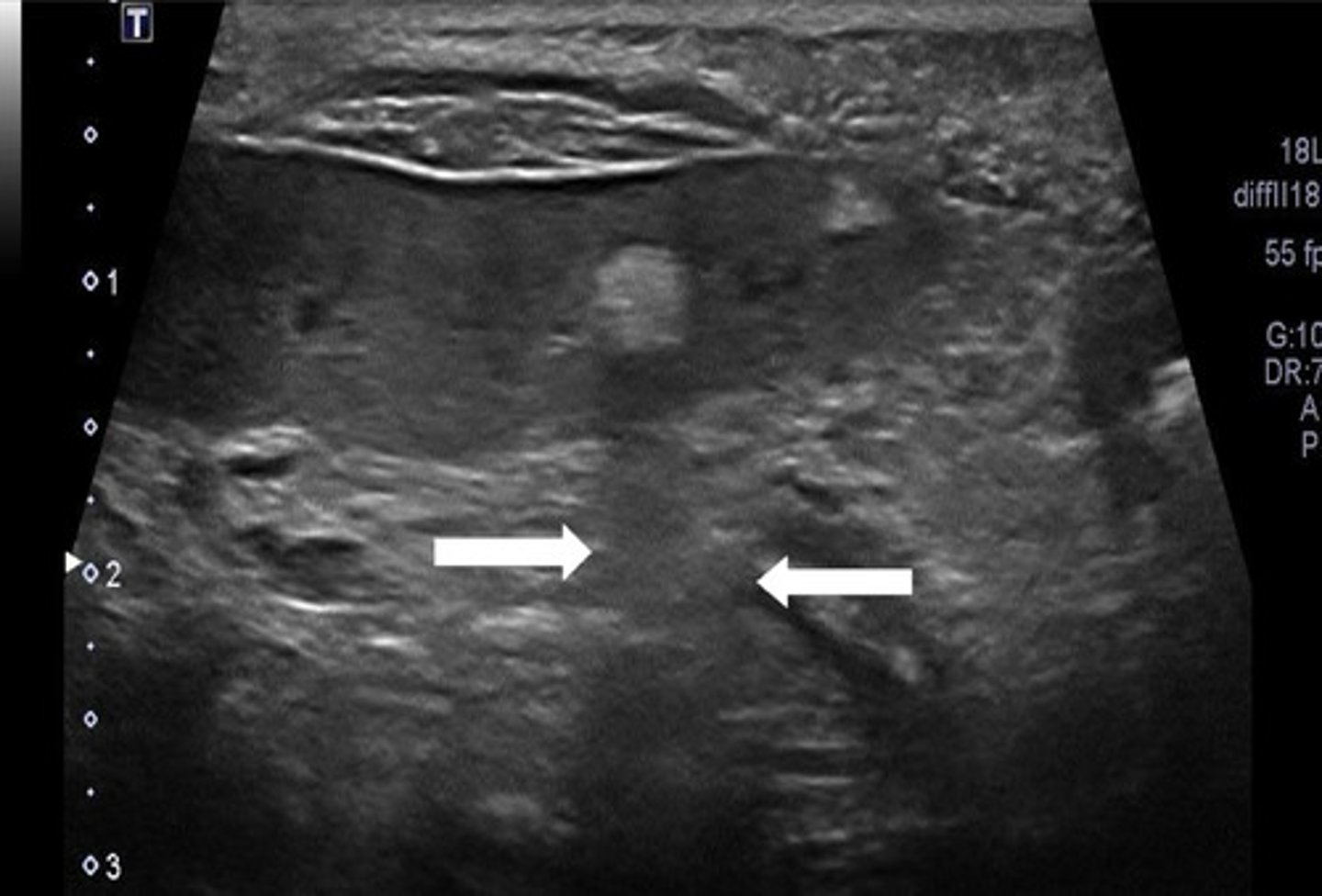
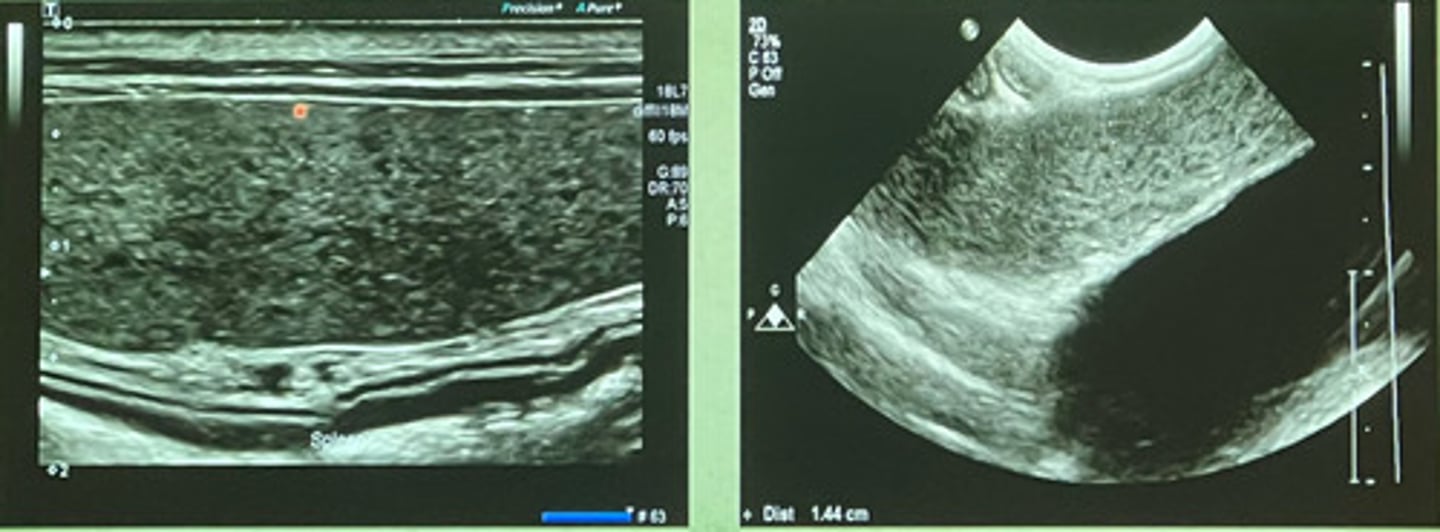
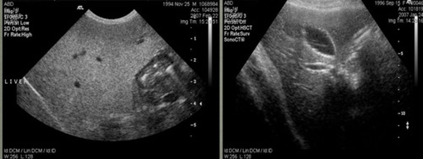
focal
are these ultrasound images of the spleen focal or diffuse?
diffuse
are these ultrasound images of the spleen focal or diffuse?
-clinical signs/history
-blood work indications
-hepatomegaly/mass
-tumor staging
-suspected diaphragmatic hernia
-liver biopsy/following response to therapy
what are some liver indications for ultrasound?
5-7.5 MHz
what frequency should you ultrasound the liver at?
-resolution
-penetration
high frequency = better ________
lower frequency = better ________
yes, to see deeper parts of the liver can switch to lower frequency or vice versa
is it okay to switch between different frequency probes when evaluating the liver?
-body conformation
-liver size
-GI gas
visualization of the liver is dependent on what 3 things?
subcostal
in small breed dogs with normal liver size, typically can be scanned from a ________ approach
intercostal
in large breed dogs with a small liver or ingesta in the stomach requires an ______ approach
hypoechoic
the liver is what echogenecity compared to the spleen?
uniform, coarse echogenic pattern and smooth sharp border
the normal liver appears how in regards to the echogenic pattern and border?
-obesity
-type of transducer
what two factors affect echogenicity of the liver?
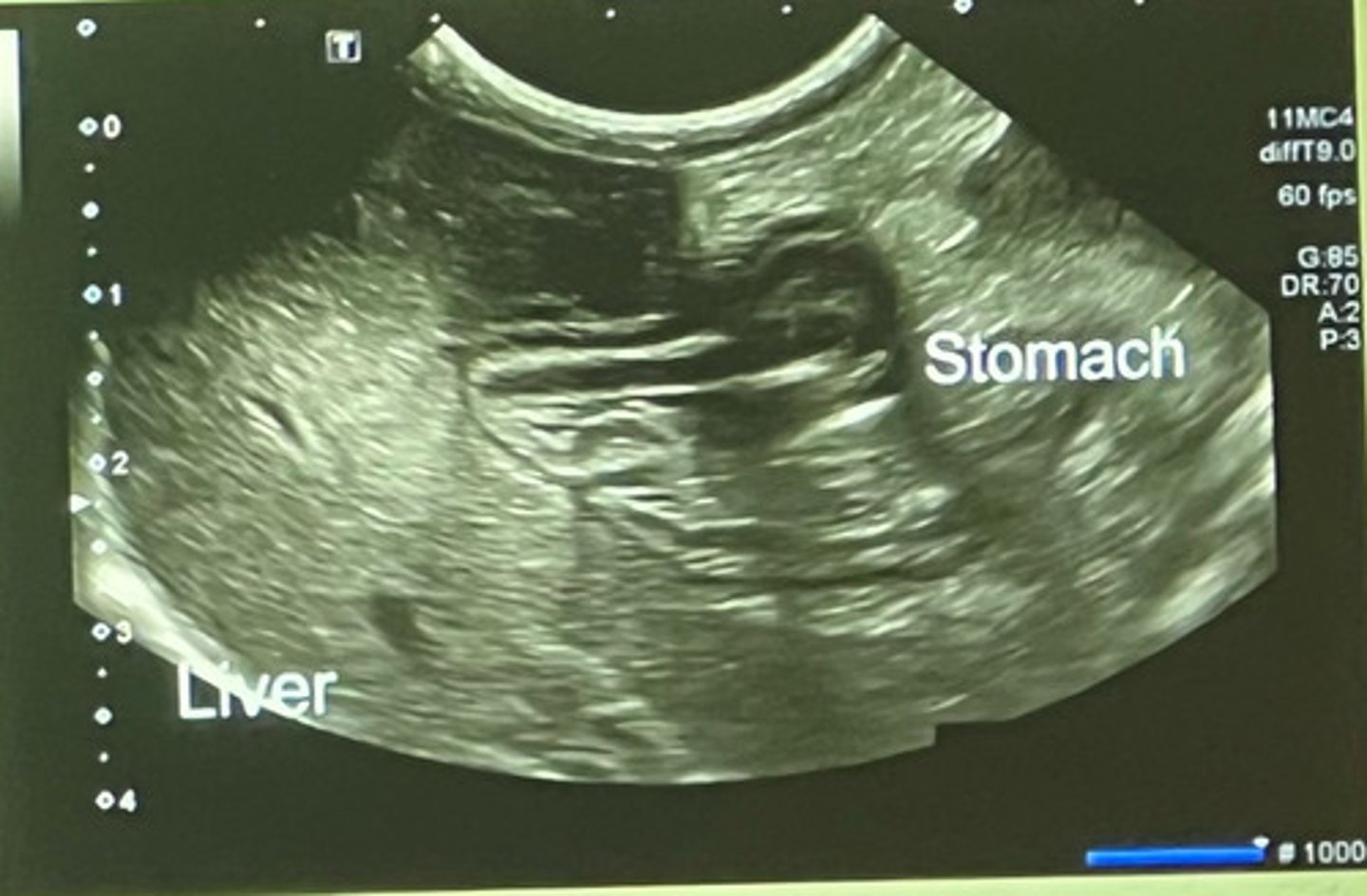
at the gastric fundus (or the spleen in some dogs)
where is the left caudal border of the liver located?
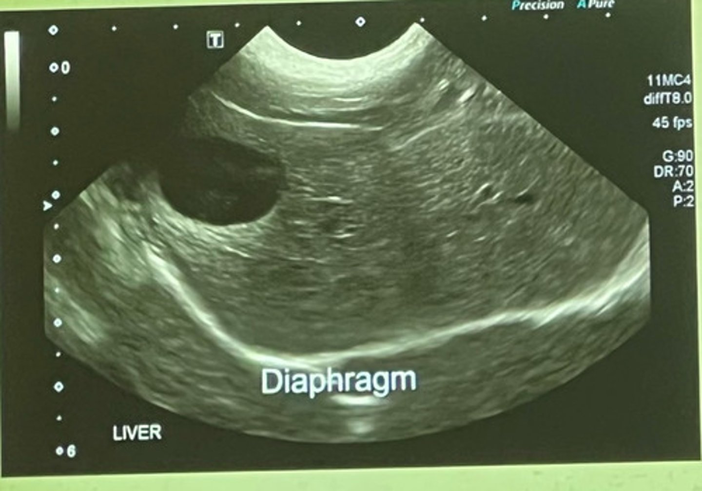
at the liver/diaphragm interface
where is the cranial border of the liver located?
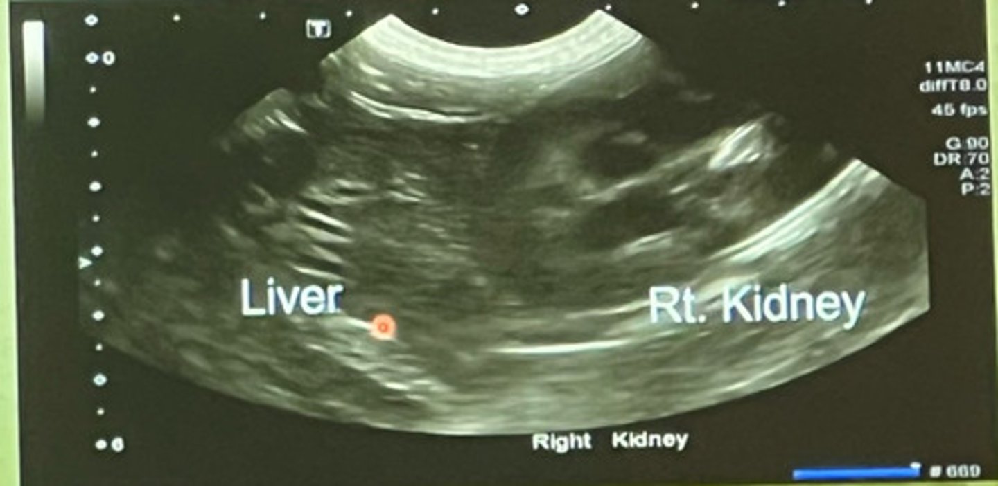
right kidney surrounded by caudate process of caudate lobe of the liver
where is the right caudal border of the liver located?
cranial border of liver
what part of the liver is this showing?
left caudal border of the liver
what part of the liver is this showing?
right caudal border of the liver
what part of the liver is this showing?
rounded and not sharp
the borders of the liver should be what shape?
intercostal
where are the normal borders located of the liver in large breed dogs?
beyond the costal arch
where do normal borders of the liver in small breed dogs extend to?
parallel
the stomach axis is ______ to the ribs
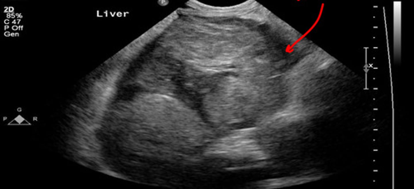
right- there is a mass on the ventral side
which of these US images of the liver is abnormal?
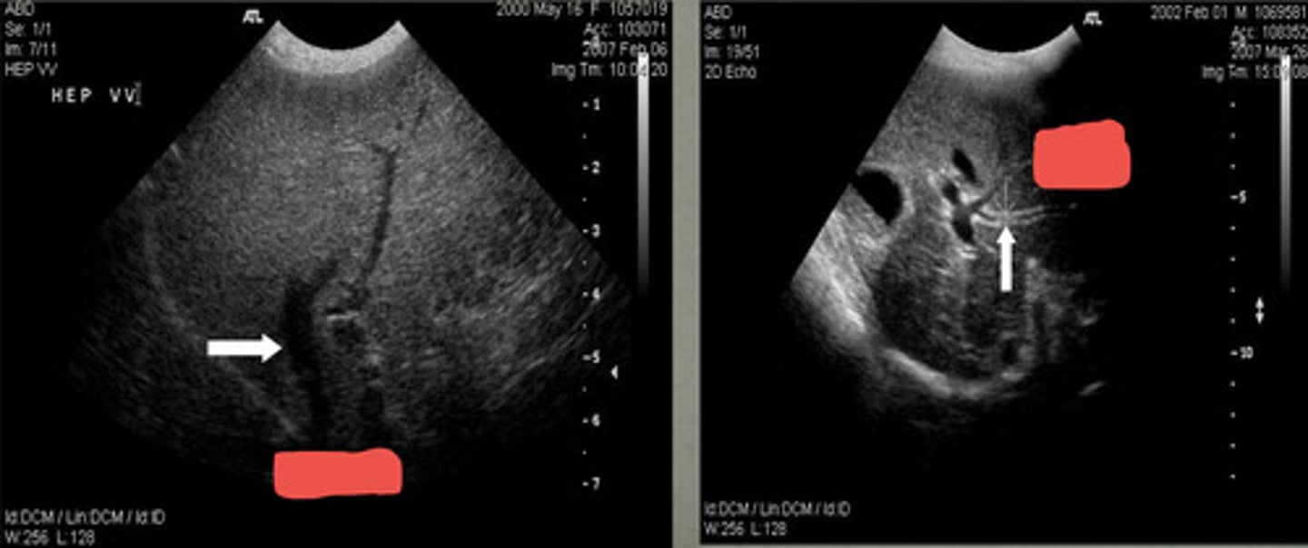
diaphragm
what is the red arrow pointing to in the left image?

right cranial dorsolateral aspect
the dorsal vessels of the liver are best viewed from what aspect of the abdomen?
dorsal and left
the aorta is located where?
no!
is the aorta compressible or pulsatile?
ventral and right
the caudal vena cava is located where in respect to the aorta?
yes
is the caudal vena cava compressible?
more ventral and left
the portal vein is located where in comparison to the caudal vena cava?
dorsal
the portal vein is ________ to the common bile duct
portal veins
the _____ veins have the most echogenic walls
at the diaphragm
hepatic veins join the caudal vena cava where?
anechoic
the hepatic veins are what echogenicity?
hepatic veins
do portal veins or hepatic veins have less delineated walls?
hyperechoic
portal veins have a more _______ border than the hepatic veins
left- hepatic vein
right- portal vein
which US shows the portal vein and which is hepatic vein?
hepatic
______ arteries are not detected on 2D images and only found with color flow Doppler
arteries
with color doppler, _______ are the small vascular structures depicted with red color
-cranially
-ribs
with a small liver on rads, the stomach axis will shift _______ and is not parallel with the ____
caudally
with hepatomegaly rads, the stomach axis shifts ______ and is not parallel with the ribs
right image
which US image shows the normal liver?
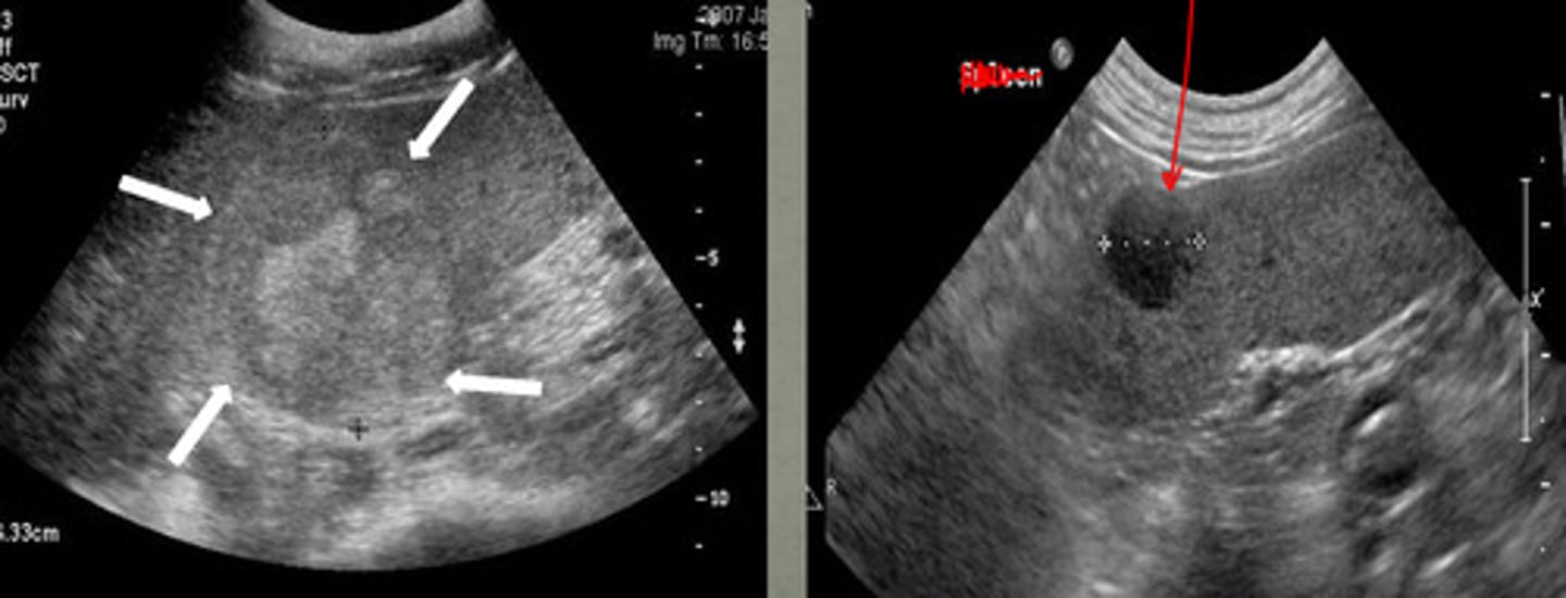
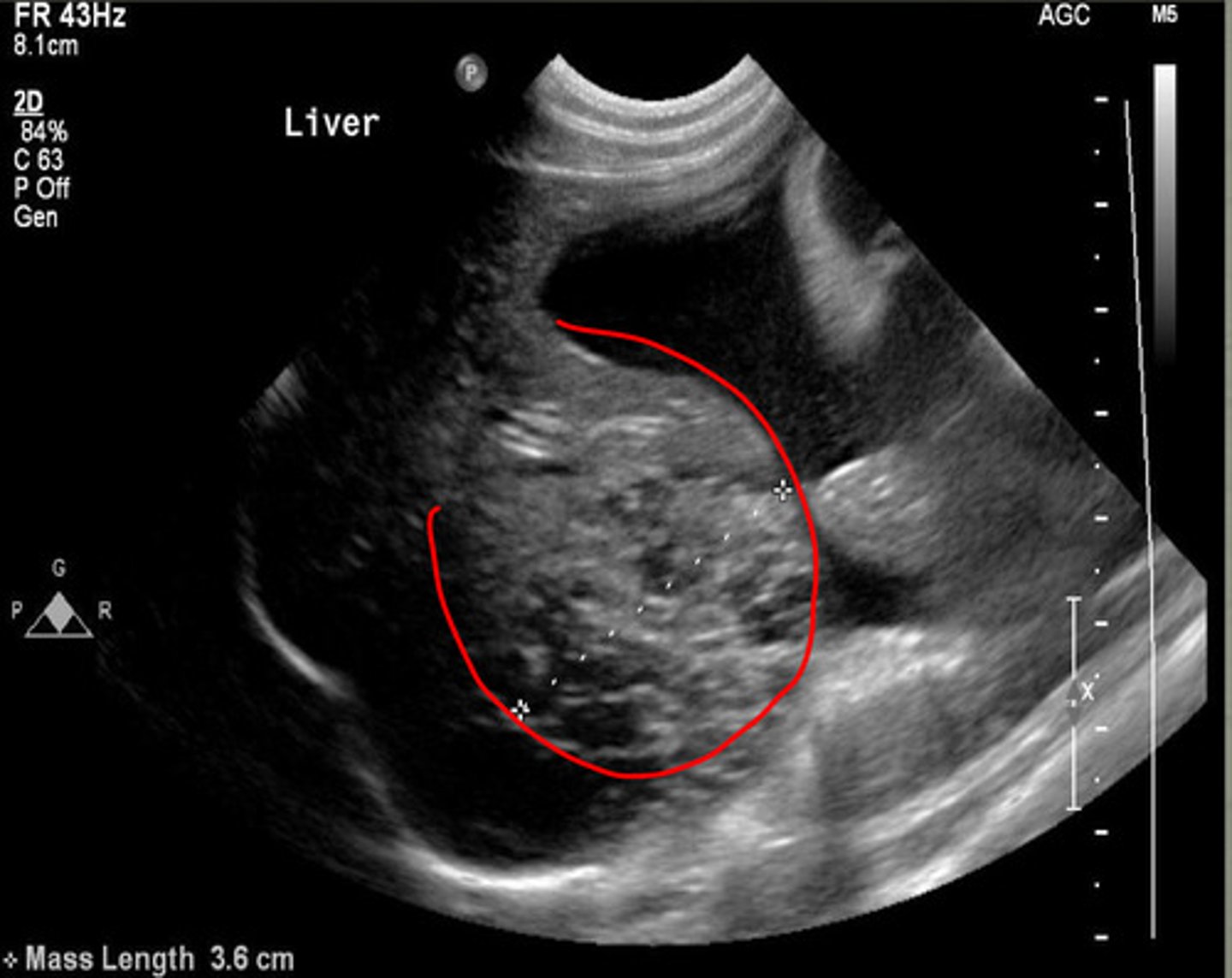
focal mass
what focal change does this show in the liver?
nodular lesions
what is the red arrow indicating from this ultrasound of the liver?
abnormal
is this a normal or abnormal liver US?
echogenicity- anechoic
shape- tear drop
how does the gallbladder appear on ultrasound? (echogenicity and shape)
the right side of the liver
where is the gallbladder located in respect to the liver?
1
the wall of the gallbladder is usually less than ____ mm in cats and dogs
anechoic
normal bile is what echogenicity?
biliary sludge or concretions
what is the echogenic material in the gallbladder?
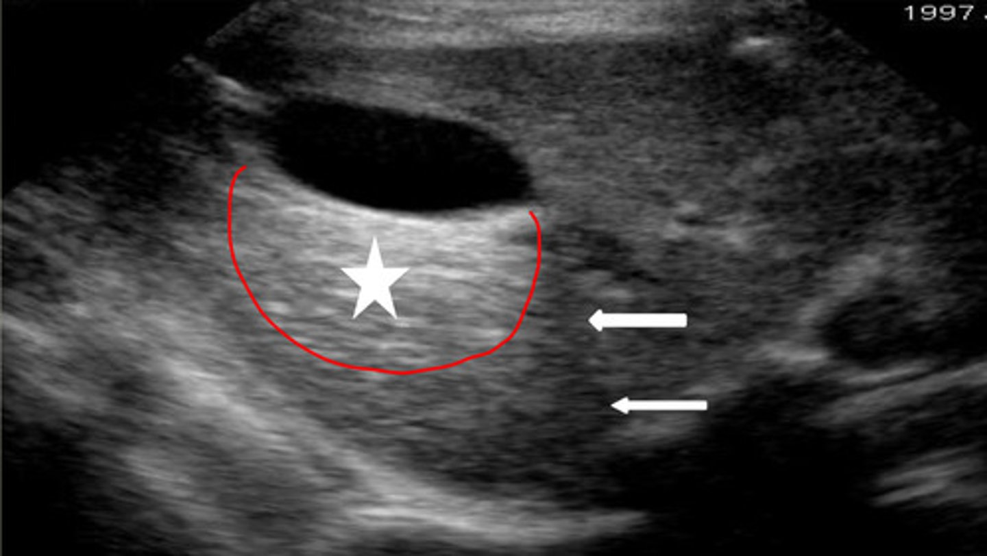
gallbladder
if this US image was taken on the right side of the liver, what structure would this be?

distal enhancement
what artifact is the red circle indicating underneath the gallbladder?
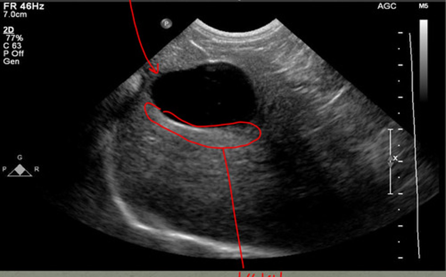
-distal acoustic enhancement
-refraction
what are the two artifacts associated with the gallbladder?
increases echogenicity in liver parenchyma deep to gallbladder
what does distal acoustic enhancement do?
bending artifact at edges of gallbladder, seen at curved surfaces
what is refraction artifact?
star- distal enhancement
arrow- refraction
does the star or arrows show distal enhancement or refraction?
common
the cystic duct joins the hepatic bile duct to become the _______ bile duct
common bile duct
what duct enters the duodenal wall?
portal vein
the common bile duct runs parallel to what vein?
intrahepatic biliary ducts
which biliary ducts are not normally visualized?
1
the wall of the common bile duct should not be greater than ____ mm
no
enlarged intrahepatic biliary ducts have _____ blood flow
color Doppler
what tool can be used to help distinguish an enlarged intrahepatic biliary duct from a blood vessel
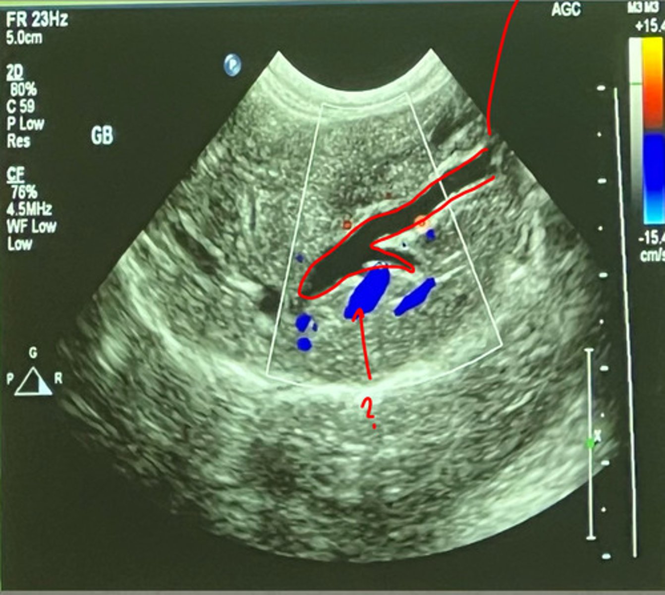
intrahepatic biliary duct
what duct is being shown with the red outline?
meals
gallbladder size can vary after what event?
yes
in cats, can the gallbladder neck be compressed with the US probe?
cats
in what species can the gallbladder have various shapes and may be duplicated?