ana 1 review (hard questions)
 Call Kai
 Learn
Learn Practice Test
 Spaced Repetition
 Match
Match Flashcards
 Knowt Play
1/105

There's no tags or description
Looks like no tags are added yet.
Name | Mastery | Learn | Test | Matching | Spaced | Call with Kai | Chat |
|---|
No analytics yet
Send a link to your students to track their progress
106 Terms
what are the most common chemical elemnts in living organisms and what general percentage of body mass
O - 65%
C - 18.5%
H - 9.5%
N - 3.3%
Ca - 1.5%
what is the difference between
molecule vs compound
inorganic molecule vs organic
molecule - 2 or more atoms, O2, N2
compound - molecule of 2 or more different elemnts
inorganic - without C-H covalent bonds
organic - with C-H covalent bonds
why is carbon a good molecule for life
diverse bonds by bonding with 4 other atoms
length, breanching, double bonds, rings
organic molecules are not soluble in water
c-h chains serve as compact energy stores
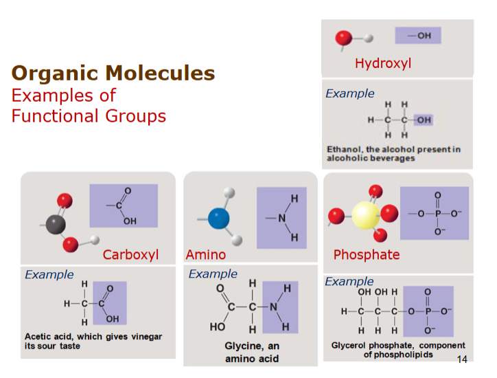
what are some functional groups
hydroxyl
carboxyl
amino
phosphate
4 major biolgoical molecules?
carbohydrates
nucleic acids
lipids
proteins
what percentage of our bodies are water/fluid, what part of that fluid is ICF and ECF?
55-60% of bodies are fluid
2/3 of which is ICF
1/3 of which is ECF
80% intersitital fluid
what are the chemical/physical properties of water and why
polar covalent bonds
reactant in many biochemical reactions
interact with ionic bonds
hydrogen bonds
lubricant
high heat capacity(takes long time to change temp)
evaporative cooling (Absorb heat)
describe types of carbohydrates and function
monosaccharides
disaccharides - 2 monosaccharides - glycosidic bond(form water, create bond between 2 hydroxyl)
polysaccharides - many
starch, glycogen, cellulose
function
provide energy
store energy
component in other molecules - deoxyribose
structural component - cellulose
describe types of lipids, bonds and saturated vs unsaturated
hydrophobic, mostly CH, little O
glycerol binds to fatty acid with dehydration (H + OH → H2O)
creates ester linakge
saturated fatty acid - no double bonds - straight - SOLID
unsaturated - one or more - BENT, liquid
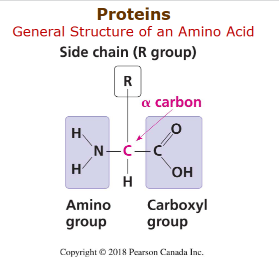
what is general structure of a protein, how do they form/connect, what are the functions?
amino group, side chain/R group, carboxyl group.
form with peptide bond, between OH and H, form H2O
connect C with N
can be primary, secondary, tertiary, or quaternary
functions
enzymatic
defensive - antibodies
storage -casein, ovalbumin
transport - hemoglobin
hormonal - insulin
receptor
movement - actin/myosin
structural - keratin
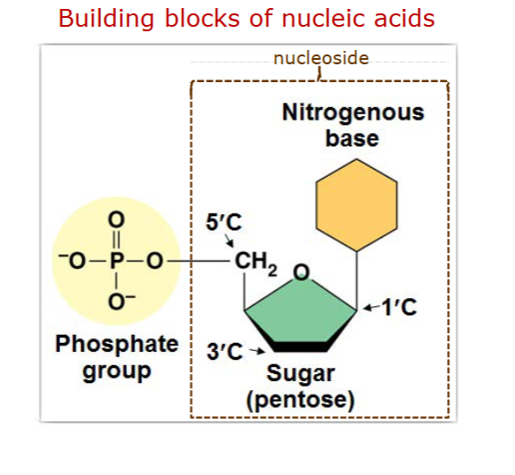
what is the general structure of a nucleotide?
nitrogenous base,
pyrimidines C T U
purines A G
phosphate group,
connected to nucleotide with phosphodiester bonds
sugar - ribose/deoxyribose
DNA - contain genetic info
RNA - carries copes if othat into tocytoplasm for protein synthesis
ATP - nucleotide as energy
what are the properties of life
Metabolism
anabollism - synthesis, energy used
catabolism - breakdown, energy released
responsiveness to environemnt
cellular organization
exchange of materials with envrionment
energy processing, movement
reperoduction, development, growth
what are the levels of structural organization of the body
chemical level
atoms, molecules
cellular level
tissue level - similar types of cells
organ lelvel - different types of tissues
organ system level - organs working together
organismal - many organ systems
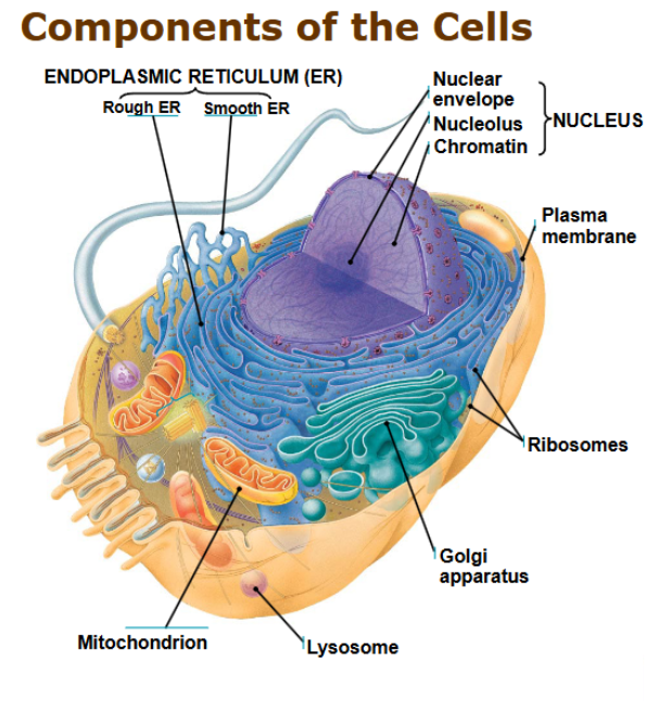
components of the cells and their functions?
endoplasmic reticulum
smooth - builds fats, stores calcium
rough - ribosomes, makes/folds/checks proteins
cell membrane - selective barrier
nucleus - home of DNA
ribosomes -
golgi apparatus - ships and receives proteins
lysosomes/vacuoles - contain enzymes, fuse together to digest
mitochondrion - cellular respiration
what are the cellular processes involved in the development of the human body
cell division
cell growth
cell differentation - specialization
nerve cells, muscle cells, RBC, WBC
describe the structure of a phospholipid
hydrophilic head - phosphate and glycerol
hydrophobic tail - saturated and unsaturated fatty acid
why is the cell membrane a fluid mosaic?
contains a collage of proteins, each with diversity of structures and functions
fluid - not static (unsaturated fatty acids and cholesterol)
what are the mmebrane transport processes
passive processes - conc gradient
simple diffusion
osmosis - water via aquaporins/bilayer
faciltated diffusion - membrane protein
active processes - against conc gradient
active transport - membrane protein
bulk transport - vesicle
phagocytosis - enfulgment of particles
pinocytosis - enfulgment of drops of ECF
receptor mediated endocytosis - specific substances
exocytosis - remove
hypertonic vs hypotonic
hypertonic - higher solute concentration than a cell - take water out (love water)
hypotonic - lower solute concentraton than cell - push water in (hates water)
what can epithelial tissue be classified in
can be
squamous - plate like
cuboidal
columnar
also be
simple
stratified
pseudostratified
what are some examples of epithelial tissue and their classification
simple squamous - easy transport of materials, capillaries and lungs, digestive cells
squamous stratified - epidermis, lines mouth, vagina
cuboidal simple - thyroid
cuboidal stratified - sweat glands and secretions
simple columnar - absorption of nutrients in intestine
pseudostratified - secrete and propel mucus in airways/trachea
what are neuron tissue cells examples
microglial cells
oligodendrocyte
neuron
astrocyte
what is connective tissue and its classification
tissue that supports binds or protects other tissues/organs
originates from mesenchymal cells(stem cells) → fibroblasts
can be
proper
loose (protein fibers spaced far apart)
areolar
adipose
reticular
dense
regular
irregular
supportive
cartilage
hyaline
fibrocartilage
elastic
bone
fluid
blood
lymph
what does the extracellular matrix of connective tissue contain (what protein fibers)
collagen - thick and strong
elastic - flexible
reticular fibers - thin, supporting mesh
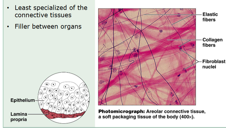
describe loose areolar (connectice tissue proper)
least specialized
filler between organs
elastic, collagen fibers
fibroblast nuclei
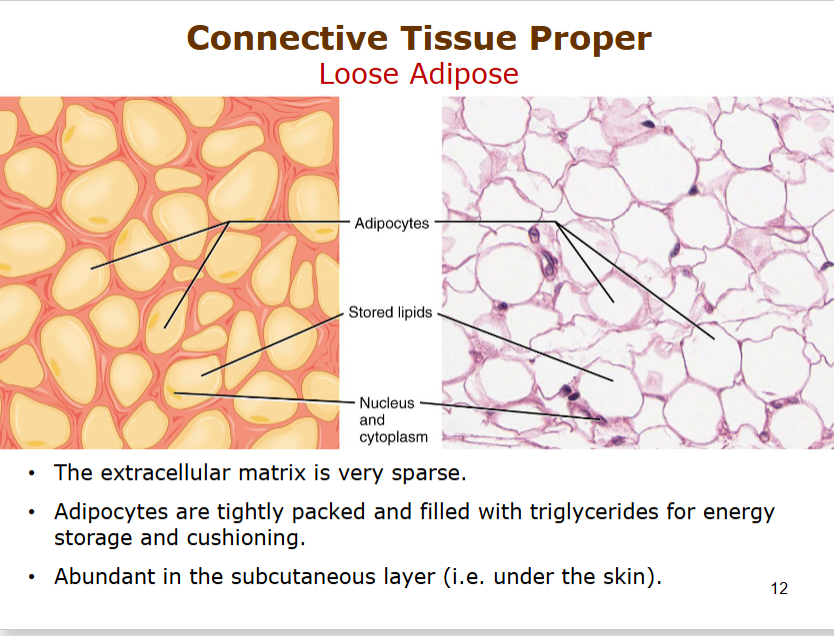
describe loose adipose (proper)
ECF is sparse (loose)
adipocytes are TIGHTLY packed, store triglyceries for energy storage and cushioning
abundant in subcutaneous(under skin) layer
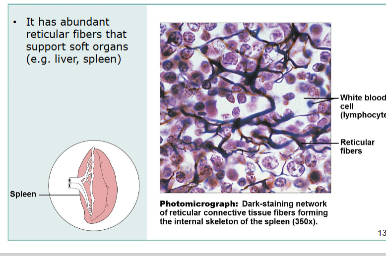
describe loose reticular (proper)
support soft organs with abundant reticular fibers
internal skeleton of spleen
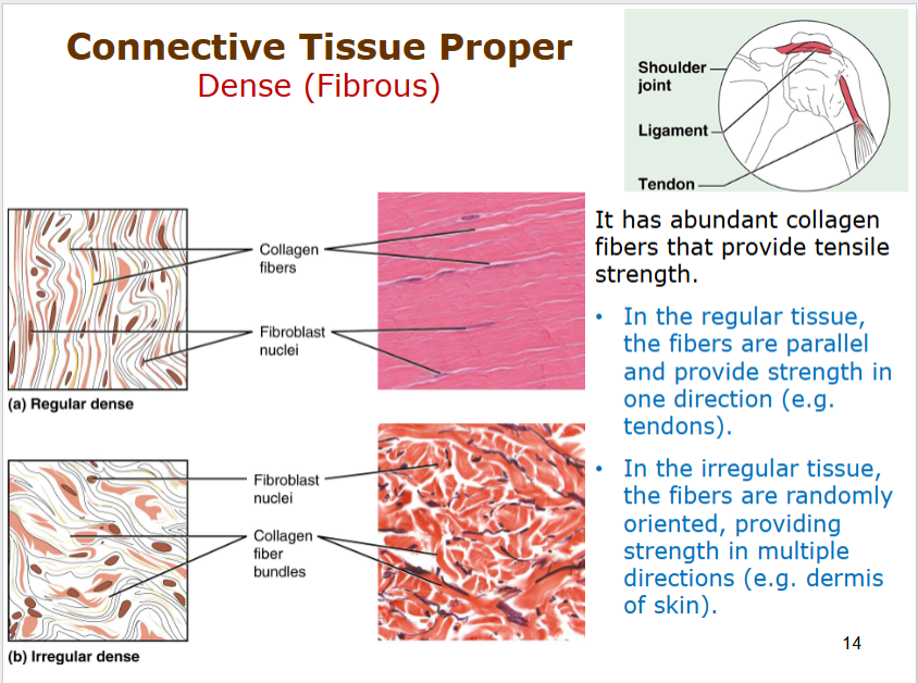
describe dense(fibrous) (proper) tissue
can be regular - parallel/strong in one direction (tendons)
irregular - random - strength in multipe directions (dermis)
provide tensile strength with abundant collagen fibers!
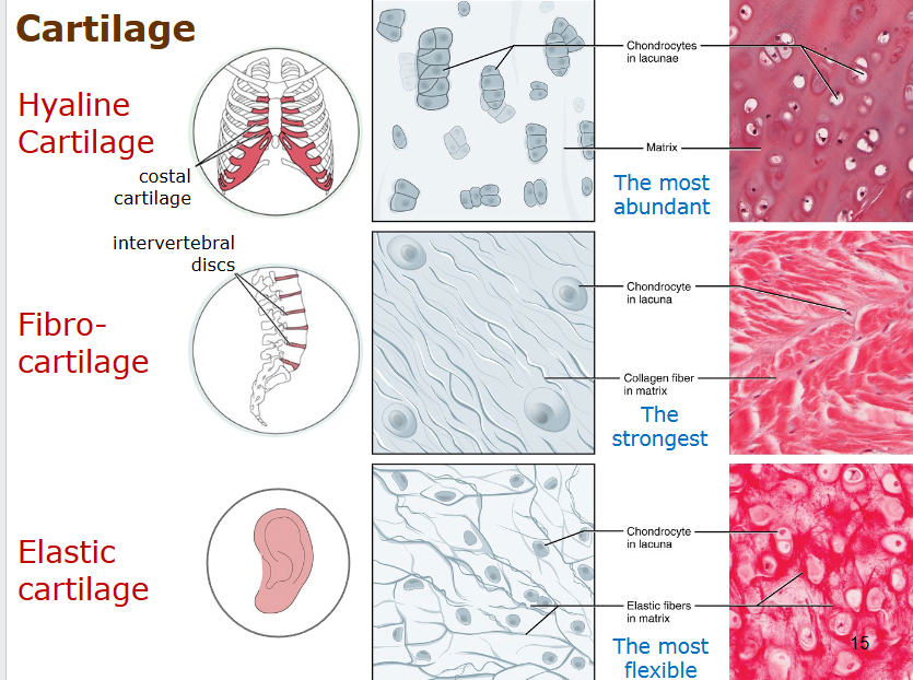
describe the types of cartilage
hyaline cartilage - most abundant, costal or trachea
fibro cartilage - strongest, intervertebral discs
elastic cartilage - most flexible, in ear
CHONDROCYTES - cell
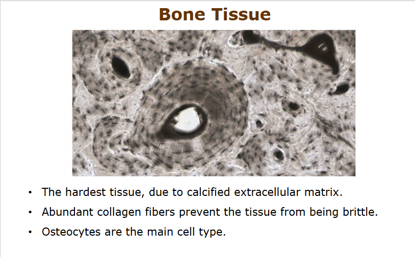
describe bone tissue
calcified extracellular matrix makes it hardest
collagen fibers present to prevent brittleness
osteocytes
describe blood
plasma is liquid ECF
RBC most abundant (erythrocytes)
transport gas, nutrients, hormones, waste
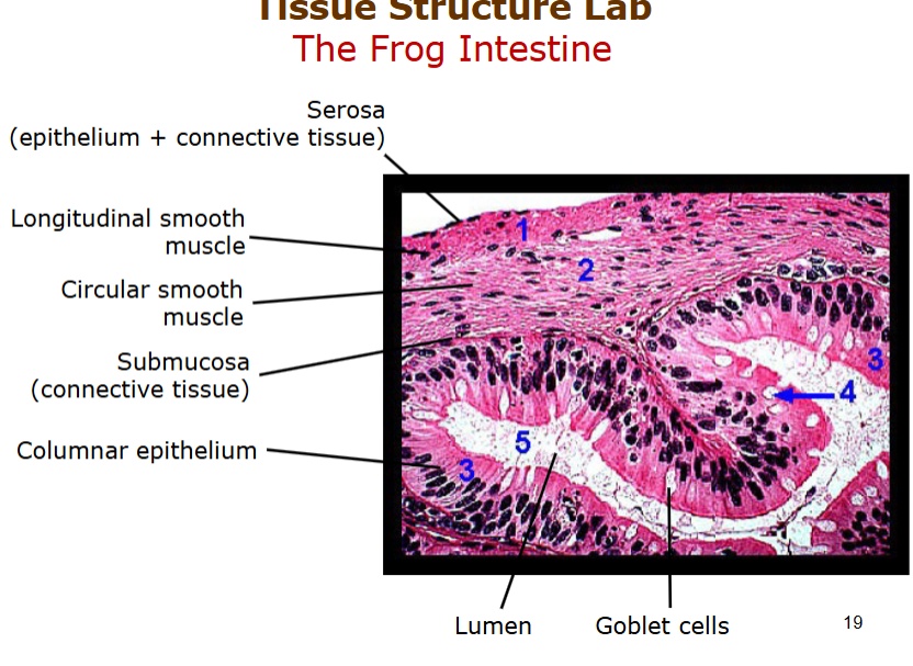
describe frog intestine
serosa - epithelium connective tissue
longitiudinal and circular smooth muscle (peristalsis)
submucosa - connective tissue
columnar epithelium - nutrient absorption
villi - creates more SA interaction
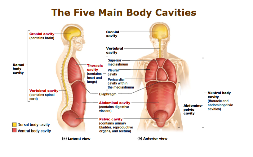
describe the 5 main body cavities
dorsal body cavity
cranial cavity - brain
vertebral cavity - spinal cord
thoracic cavity - lungs, heart
ventral body cavity
vertebral cavity
abdominal cavity - digestive viscera
pelvic cavity - urinary bladder, reproductive organs
abdominopelvic cavity
describe thoracic cavity in detail and membranes
seperated from abdominal cavity by diaphragm
superior mediastinum
pleural cavity
pericardial cavity
pleura - serous membrane
visceral pleura - cover organs
parietal - lines body cavity
pleural cavity - thin fluild filled spaces between
pleural fluid
what are the regions of the abdominopelvic cavity?
upper - hypochondriac regions
middle (horizontal) - lumbar region
middle (vertical - epigastric, umbilical, hypogastric
lower - illiac (inguinal region)
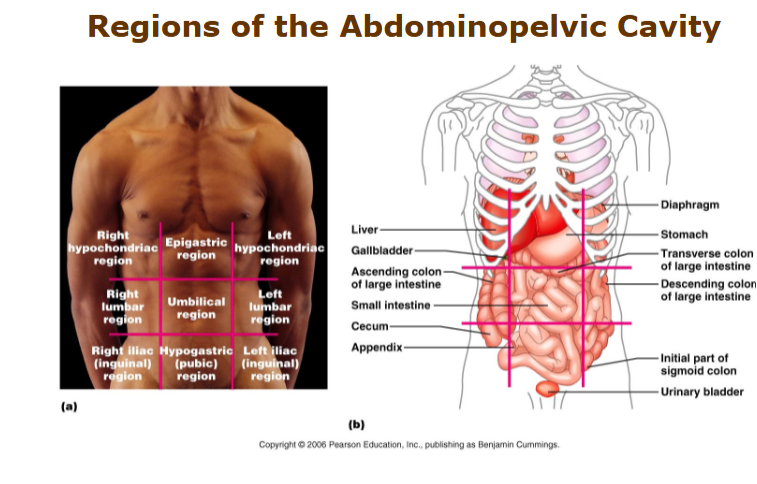
describe peritoneum and pericardium serous membranes
pericardium - heart
visercal pericardium
parietal pericaridium
pericardial cavity
peritoneum - stomach, livers, ovaries, intestines
visceral peritoneum
pariertal peritoneum
paritoneal cavity.
what are the body planes
sagittal - left and right
mid sagittal/median
parasagittal
unequal
frontal - front and back
transverse - top and bottom
what are the organ systems
integumentary - hair skin nails
skeletal - bones, joints, cartilage
muscular - muscles, tendons
nervous - brain, nerves, spinal cord
endocrine
cardiovascular - heart, vessels
respiratory
lymphatic - spleen, lymphatic vessels and nodes, thymus
digestive
urinary
reproductive (differs)
what is homeostasis
maintanence of bodys internal envrionment
temperature, pressure, concentration of substances
set point and normal range at which variable is maintained
what is the internal environment made out of?
extracellular fluid
intersitital fluid
plasma
lymph
cerebrospinal fluid
synovial fluid
aqueous humour
explain the components of negative feedback systems, what is an example?
7 steps
stimulus - change
receptor - detect change
input - information sent along afferent pathway (this is what happened)
control center afferent → efferent
determine set point
compare input within normal range
activates effector
output - info sent along efferent pathway (do this)
effector - carries out action - reduce effect of stimulus
response - return variable to set point
thermoregulation
stimulus - heat
receptor - thermoreceptors in skin/brain
input - its hot!
control center - hypothalamus (thermoregulatory center)
output - glands need to sweat, vessels must dilate
effector - glands sweat, vessels dilate
response - sweating, body temp falls!
what are 2 examples of positive feedback systems
birth
head of baby pushes against cervix
nerve impulses sent to brain
pitutiary gland seretes oxytocin
stimulates uterine contractions
breastfeeding
suckling infant sends affect impulses to hypothalamus
pitutiary gland
oxytocin released
alveolar glands response by releasing milk through ducts of nipples
ceased when baby stops suckling
what are the divisions of the nervous system?
somatic nervous system - skeletal muscle
autonomic nervous system - control of smooth muscle, cardiac muscle and glands
sympathetic - fight or flight response
parasympathetic - rest and digest
CNS - Brain, Spinal cord
PNS - Ganglion, Nerve
what is the structure of a nervous tissue and what are the functions of each
cell body - house nucleus
cell membrane - selective membrane
dendrites - receive signal from other neurons
axon - transfer/send signal to neurons
nodes of ranvier - spaces where signal jumps, speeds up
oligodendrocytes - produce myelin sheath
myelin sheath - insulate signal
synapse - junction point?
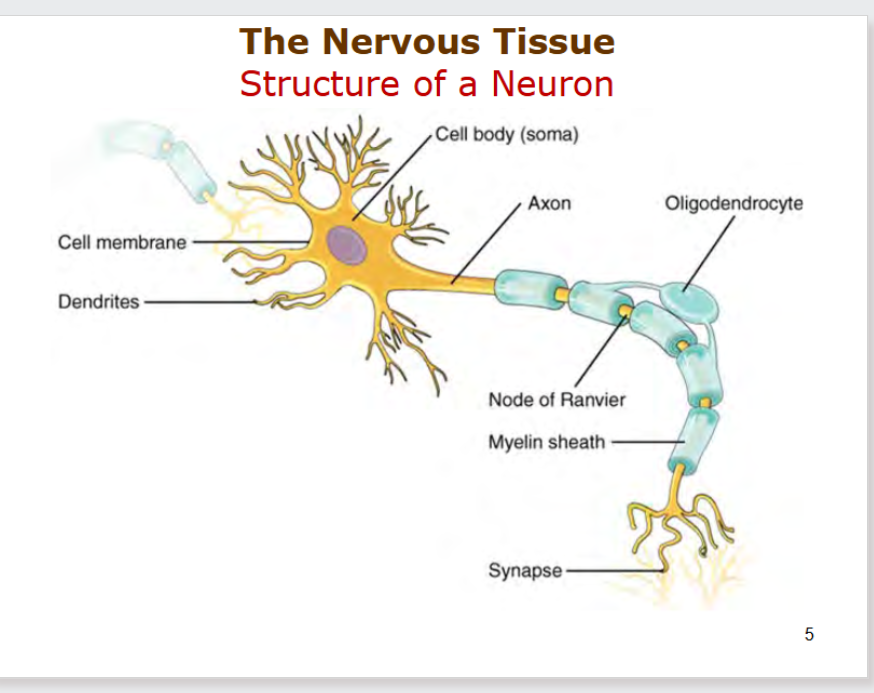
what are the types of neurons
sensory input → sensory neuron
association neuron (interneuron)
motor neuron → motor output
what are the supporting cells of the nervous tissue?
astrocyte - main supporting cell
deliever energy, maintain protective walls, clear our neurotransmitters, and control chemical baalnce
microglial cell - immunological protection
ependymal cells - production of CSF
oligodendrocytes - myelin production
whats the difference between tracts and nerves
tracts - central nervous system
bundles of axons
nerves
bundles of axons within PNS
cranial or spinal
all spinal nerves are mixed while others can be sensory, motor, or mixed.
label the nerves of the brain
olfactory bulb 1
optic nerve 2
oculomotor nerve
trochlear nerve 4
trigeminal nerve 5
abducens nerve 6
facial nerve 7
vestibulocochlear nerve 8
glossopharyngeal nerve 9
vargus nerve 10
hypoglossla nerve 12
accessory nerve 11
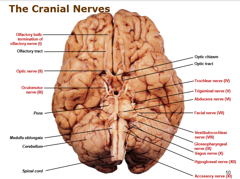
whats the difference between grey matter and white matter
gray matter
neuronal CELLBODIES within CNS
in cerebral cortex, brain nuclei, spinal “horns”
white matter
tracts within CNS!!!
exterior spinal cord
interior of brain
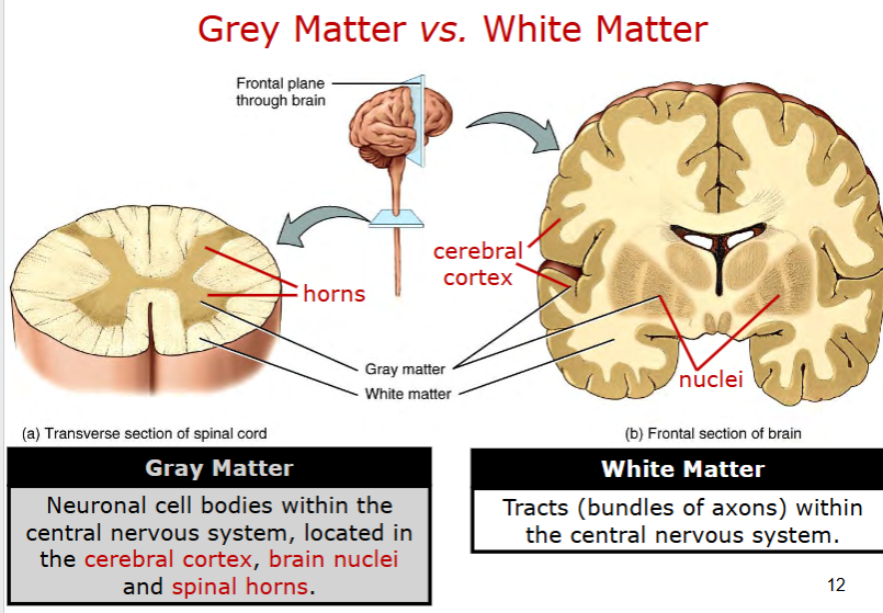
what are ganglia?
groups of neurons in PNS
sensory
autonomic
sympathetic
parasympathetic
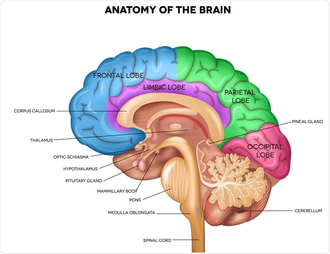
describe the brain anatomy
spinal cord connects to medulla oblongata - vital involuntary functions (hearrate)
pons - bulge - regulate sleep wake cycles, breathing rhythms
cerebellum - little brain - balance, precision
pitutary gland
thalamus - middle filters sensory data
hypothalamus - homeostasis
optic schiasma
corpus callosum - coordinate bilaterally, share data between sides
cerebrum
frontal - logical thinkning, problem solving, speech production
parietal - somatosensory
temporal - auditory processing, memory, language
occipital - visual info
what are the meninges of the brain.
duramater
periosteal
meingneal
archnoid mater
pia mater
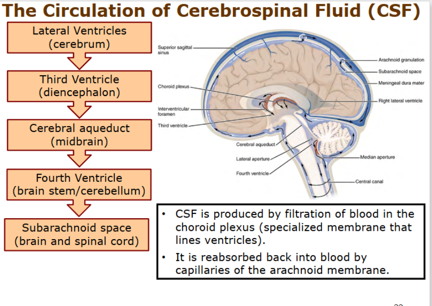
what are the ventricles of hte brain and how does CSF circulate?
lateral ventricles → third ventricles near diencephalon
→ cerebral aquaduct
→ fourth ventricle (cerebellum)
→ subarachnoid space (around brain and spinal cord)
what is spinal cord anatomy
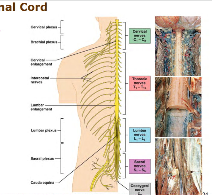
Cervical 8
Thoaracic - 12
Lumbar - 5
Sacral - 5
Coccygeal - 0
what is the function of the spinal cord and components of a reflex arc?
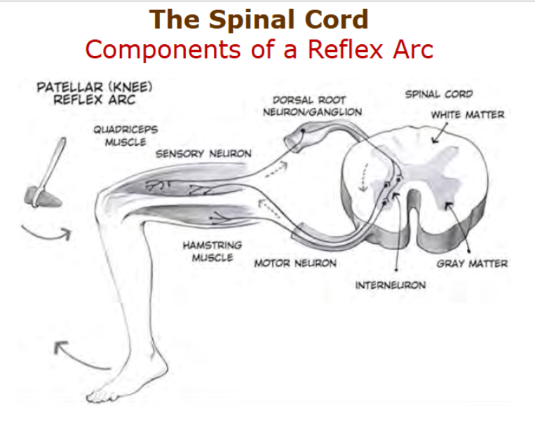
functions - communcation between brain and periphery
carry sensory impulses from bbody to brain or
motor impulses from brain to body
spinal reflexes - no ned for a brain! how does that work?
stimulus → sensory neuron → dorsal root neuron/ganglion → spinal cord → white matter (tracts) → grey mattter → interneuron → motor neuron → hamstring muscle
what is the function of an interneuron?
middlemen
between sensory and motor neurons
take in many signals at once and decides how body should respond
describe the main differences between sympathetic and parasympathetic, where are their pre and post ganglionic neurons?
sympathic - prepare organs for physical activity
norepinephrine, epinephrine
adrenal gland
prioritizes physical activity over digestive system
preganglionic neurons in spinal cord, post in sympathetic ganglia alongside vertebral column
parasympathetic - rest and digest
prioritizes digestive system over physical activity
erection
slows physical activity, heart, waste excretion
preganglionic neurons in brainstem and spinal cord, post ganglioninc neurons in parasympathetic ganglia(near or in target organs)
describe the steps of neuron signaling
resting state
depolarization
repolarization
hyperpolarization
resting state at -70mV maintained by
sodium-potassium pump 3 NA out 2 K in
protein and phosphate anions inside - negative
selective permeability, more K than Na+ leak channels, more K+ leaves, than Na+ enter
gated channels closed
depolarization - stimulus causes Na+ channels open 30/40mV
if reaches -55mV, action potential triggered (threshold)
Na+ channels open, Na+ rush in along axon, (electrochemical gradient)
peaks to +30/40mV
repolarization - Na+ channels close negative mV
at the peak of action potential, Na stops flowing n
K+ channels (open slowly) open fully, K rushes in, Na out, MEMBRANE POTENTIAL NEGATIEV
hyperpolarization - voltage k+ channels close slowly(again), drops to -90mV(below resting state)
continued outflow of K+ causes membrane potential to drop below normal resting level
refractory period, neuron is far less likely to fire action potential (signal ensured to travel in one direction)
resting state
K+ channels close, Na K pump restores resting state voltage.
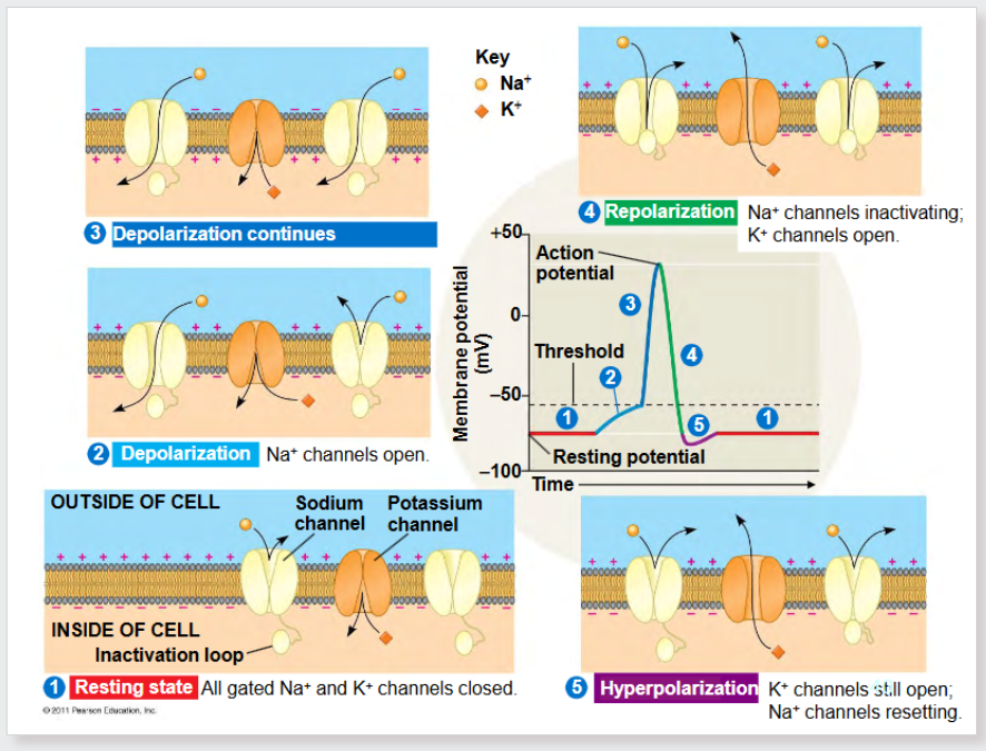
draw the graded potentials of hyperpolarization, depolarization, action potentials
explain the propagation of an action potential at a myelinated axon - saltatory conduction
formed at the nodes of ranvier, gaps in myelin sheath
voltage gated Na+ channels found here
one node’s action potential causes electrical ion currents to spread to next node
“jump” allow faster conduction
what occurs when the action potential reaches the end of axon terminal (synapse) - isimilar to muscle
release neurotransmitters into synapses at axosomatic synapses
synaptic vesicles move towards synaptic clect
voltage gated Ca2+ channels open, ca2+ enters AXON TERMINAL
calcium causes synaptic vesicles to release contents via exocytosis
neurotransmitter diffuses across cleft, binds to ligand gated ion channels on posy sypnatic membrane → neuron GRADED POTENTIALS
how does the signal between neurons get terminated/neurotransmitter
reuptake by presynapic neuron
enzymatic degradation
diffusion reduce neurotransmitter levels, kills signal
what are some neurotransmitters
acetylcholine - muscle - memory
glutamate - excitatory neurotransmitter
GABA - inhibitory neurotransmitter
glycine - inhibitory neurotransmitter
dopamine - motivation, motor control
norepinephrine - wakefulness
serotonin - arousal
what are classification and examples of sensoiry receptors
mechanoreceptors
distortation of cell membrane
photoreceptors
light
chemoreceptors
chemicals, taste, smell, blood vessels
thermoreceptors
change in termperatuire
skin and body tissues
nociceptors
pain, harmful stiuli, extreme temp, pressure chemicals
location:
exteroceptos
interoceptoirs
baroceptors, thermoreceptors
proprioceptors
golgi tendo organ - tendon stretch
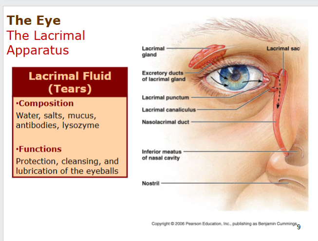
what is the lacrimal apparatus
system cointaing oribital structure
lacrimal sac
lacrimal gland → excretory ducts
lacrimal fluid
water, salts, mucus, antibodies, lysozyme
protect, cleans, lubricates eyeball
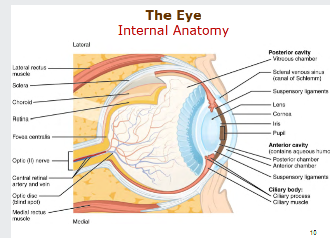
what is the internal anatomy of the eye like?
Seperated into
Posterior Cavity
Vitreous CHamber
Lens
Cornea
Iris
Pupil
Anterior Cavity
Posterior and Anterior Chambers
Aqueous humor
Cilliary body - Vascular Tunic
Cilliary process
Cilliary muscle -alter shape of lens
Back
Sclera - protection
Cornea - Refractive structure - fibrous tunic
Retina
Fovea Centralis
Optic II nerve
Optic disc
how does image form in the reitna?
Accomodation of hte lens
light rays hit the lens, diverge into the focal point on the fovea!
different in close or distant
close
parasympathetic activation
cilliary muscle contracts, leigaments loosen, lens buldge
far,
muscle relax, suspensory ligaments tense, lens flatten
constriction of the pupil
pupillary radial muscles
contract, less light intesnsity
circular muscles contract
more light intensity
convergence of the eyes
six skeletal around the eye, allow it to move in all directions
describe retina
rods
dim light, peripheral vision
highest in peripheral retina, decreaes towards fovea
rhodopsin
cones
colour vision, highest in fovea, decreaes towards peripheral retina
cone opsins
what is the visuial pathway
in retina
photoreceptors → bipolar neurons → ganglion neurons → optic nerve →optic chiasma → optic tract → thalamus → optic radiation → visual cortex
LEFT TURNS TO RIGHT AT THE OPTIC CHIASMA
left optic nerve → right optic tract, → right occipital cortex.
what are the cutaneous receptors
free nerve endings - temperatiure/pain - surface
root hair plexus - movement of hair
merkel discs - vibration
meissners corpuscle - light touch - surface - circular
pacinian corpuscle - pressure - midway
ruffini corpuscle - stretch - horizontal
what are proprioceptors
provides information about position of body parts
muscle spindles - stimulated by stretching of muscle, inform muscle length
tendon organs - stimualted by tension in tendon
helpd nervous syste mcoordinate all motor activites
how does smell work?
olfactory bulb
air goes through nasal conchae, diffuse olfactory epithelium
→ olfactory bulb, hits 400 types of olfactor receptors
dendrite
sends a signal through olfactory tract
how does taste work?
lingual papilla - full of tastebuds
gustattory cell connecting to sensory nerve fiber
sour, bitter, salty ,sweet umamis
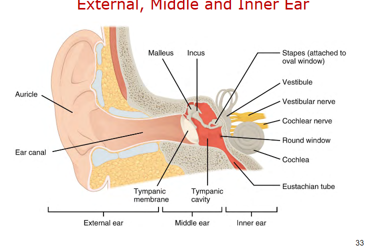
what is in the external, middle and inner eat
external - auricle, ear canal
middle - tympanic membrane, malleus, ikncus, stapes connect to tympanic cavity
inner ear - transmitted via stapes at the oval window
vestibule
vestibular nerve
cochlear nerve
round window - pressure
cochlea
oval window - scala vestibuli
vibrations of ossicles into inner ear
riound window - scala tympani
pressure valve for shock absorption for the fluid waves in cochlea
eustachain tube - pressure
what are the layers of the inner ear?
bony labyrinth
perilymph (fluid)
round window allows stapes ti displace perilymph, allow vibration
membranous labyrinth
endolymph
how does the hearing occur
sounds waves, alternate, high and low pressure, frequency measured in Hz
interacts with the tympanic membrane, vibrates
vibrations are amplified as it passes ossicles
against oval window, vibrations are transmitted to standing wave in the fluid of hte vestibuli
pressure bends the membrane of cochlear duct to a point of maximum vibration for a frequency, hair cells in basilar membrane vibrate.
whats in the organ of corti
hair cells(vibrate) attached with a tether, stereocillia, tectorial membrane
bend against the tectorial membrane
attach to dendrites of sensory organs, send signal to
axons of sensory neurons (vestibulocochlear nerve)
between the scala vestibuli an the scala tympani, surrounded by endolymph in the cochlear duct
what about different frequencies of sound? tonotopic organization
thats where the basilar membrane comes in, different areas vibrate according to different frequences
lower frequenmcies vibrate the membrane closer to the apex 20Hz
higher frequencies closer to the base 20,00-Hz
tonotopic organizatrion!
tectorial membrane stays in place/rigid to create force to move the hairs inbetween the two membranes
stereocillia bend to send a signal
what is the auditory pathway
spiral ganglion → vestibulocochlear nerve
→ brain stem
→ midbrain
thalamus
→ auditory cortex - temporal love
what is conductive deafness and sensorineural deafness
conducttiveness deafness
impaired by external or middle ear
otosclerosis - abnormal growth
injury
impacted cereumen
sensorineural deafness - organ of corti does not function or nerve is damanged
drugs
diseases
atheroschelerosis
repeated exposuire to loud soudn
how does balance work?
with the vestibular apparatus of the inner ear
patches of hair cells found in macula or utricle, sacule and cristia ampullaris of the 3 ampullae.
endolymph does not move in sync with labyrinth (inertia)
flexion and extension - anterior duct
semicircular ducts
ampulla in each canal
hair cells in with cillia, cupula
push or pull send impulse to brain
uricle and saccule
hair cells weighed down with crystals
how does your head detect rotation?
in each semicircular duct, there is a ampulla at the base
in this ampulla is a cupula surrounding the cillia cells. the cupuila bends in opposite direction of rotation due to the inertia of the endolymph. sends a signal thorugh the ampullary nerve
dtect changes in acceleration
how does your head detect head position
saccule - vertical
utricle - horizontal
in each, the macula contains hair cells that are weighed down with otoliths (hair crystals)
bend cillia, generat nerve impulse
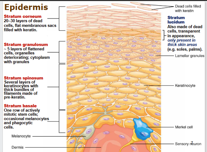
what does come lets get sun burned refer to
layers of epidermis - stratified squamous epithelium
stratum corneum - 20-30 dead cells filled with keratin
stratum luteum - dead cells, in thick skin areas
stratum granulosum flattened, dying cells
stratum spinosum - keratinocytes, make keratin
stratum basale - 1 row actively mitotic stem cells, melanocytes, phagocytic cells,
describe hair and structures
composed of shaft and root
hair follice and hair bulb at the deep end
sebaceous gland - oil
arrector pilli
apocrine sweat gland - thick secretion armpits and pubic region - mammary/earwax glands - DEEP in hypodermis
what is the dermis made up of
papillary layer - looser areolar connectivce tissue
blood vessels, sensory receptors
reticular layer - dense irregular connective tissue
fibroblasts, coillagen fibres, elastic fibres
what are some functions of the skin
mechanical
dhyradtinve
yub light porea
anbtimnicoriobva
syntehsis of vitamin D
UV light from 7-dehydrochloesteraol, pre viamitn D
Calcitriol, nedede for absorption of calcium
describe the process of intramembranous ossification
meseenchymal cell groups into clusters, osteoblasts, form ossificatrion centers.
secrete osteoid, surrounds, become osteocytes iwth osteoblats on the outside.
secrete osteoid, as trabecullar matrix forms.
compact bone starts develop at the surface of the bone, superficial to traveculae, and crowded blood vesels condense into marrow.
describe the process of endochondral ossification
hyaline cartilagem with enlarging chondrocytes - calcify matrix - leave open space
osteoblasts at the peripheral ends start laying bone
osteobats produce spongy bone at the primary ossification center (results in medullary cavity)
chondrocytes die, blood vessels bring in bone cells.
secondary ossification center at epiphyses, retains layer of articular cartilage
epiphyseal cartilage/plate remains aswell, lengthwise reguyated here
chondrocytes on one side growth, other side die, osteoblasts lay down bone
describe bone girth grow (appositional growth)
inside is emptied, outside grows
peritosteum (outside) cells difefernat into osteoblasts, laying down new bone around blood vessels, eventrualyl turn into ostecytes
in endosteum, osteovlasts erode inside, endosteum, releasing calckium into blood nad caibity enlarging.
what factors rate of ossification?
heredity
hormones - growth hormone, insulikn like ghrowth factor, thyroid hormones - stimulare osteoblasts (dwarfism, gigantism)
nutrients - vitamins A - stimulate osteoblasts C - synthesis of collagen D - absorption of calciuim, calcium is a component of bone matrix
physical actiity
whats the difference between axial and appendicular skeleton
axial - support, protection
skull, hyoid
vertrbal column thorax
Cervical 7
Thoracic 12
Lumbar 5
Sacral 5
Coccygeal 4
appendicular - movmenet, locomotion
shoulder girdle, limbs, hip girdle
what is the structure of a typical vertebra whats at the end at the top
spinous process in the middle, spinal cord in the middle
vertebral foreamen - house spinal cord
transverse process
vertebral body
forms a joint with eachother wit hthe superior and inferior articular proicesses.
at the top, atlas axis complex. axis has dens, goes into atlas at the anetrior arch, posterior arch.
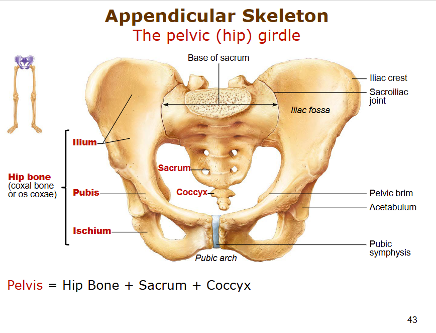
describe the anatomy of the pelvic hip girdle, what are the differences between sexes
female - pubic aerch greater than 90
wider pelvis, heart shaped, larger saccrococcygeal curvature. pelvic brim is larger, circular, wide greater sciatic notch
what are the 4 different types of bone fractures
closed - simple = skin intact
comminuted fracture - chip and splinter bone
open fracture - broken ends pierece skin
greenstin fracture - one sideof bbone bends, ulna n radius