lec 5- skeletal physiology and pathology
 Call Kai
 Learn
Learn Practice Test
 Spaced Repetition
 Match
Match Flashcards
 Knowt Play
1/58

There's no tags or description
Looks like no tags are added yet.
Name | Mastery | Learn | Test | Matching | Spaced | Call with Kai | Chat |
|---|
No analytics yet
Send a link to your students to track their progress
59 Terms
osteoblasts
produce new bone matrix, mainly collagen; help mineralize it w calcium and phosphate
osteoclasts
dissolve mineral, degrade matrix, release calcium and phosphate into bloodstream
central canals
lymphatic drainage, blood vessels, nerves
what is hydroxyapatite
bones are made of several organic/inorganic tissue- primarily made of collagen and hydroxyapatite
hydroxyapatite is made up of
two salts, calcium phosphate, calcium hydroxide
where is hydroxyapatite located
extracullular matrix (ECM); HA crystals are deposited in/around collagen fibers of the bone matrix; form along collagen fibrils, filling gaps between them
HA makes the bone ___ while collagen gives it ___
HA- rigid
collagen- tensile strengths
HA can also be found in the ___
macroscale; throughout both cortical and trabecular bone
two major phases of bone remodeling cycle
resorption, formation
resorption
old bone broken down by osteoclasts; secrete digestive enzymes and H ions to destroy bone matrix
formation
new bone built by osteoblasts; synthesize/secrete collagen fibers, initiate calcification
bone response to injury
hematoma formation, capillaries form, macrophages clean debris, fibroblasts prod. collagen fibers, chondroblasts secrete cartilage matrix, osteoblasts start forming spongy bone
parathyroid hormone (PTH)
raises serum calcium levels by stimulating osteoclast-mediated bone breakdown; causes calcium release into bloodstream
subperiosteal bone resorption
loss of cortical bone along outer surface
brown tumors
benign focal lytic lesions filled w fibrous tissue and blood products
PTH negative feedback loop
decreased blood (plasma) Ca2+, release of PTH, increased osteoclast activity, increased blood Ca2+
PTH negative feedback loop- bone resorption increasing causes
increased serum calcium
Calcitonin negative feedback loop
increased blood Ca2+, release of calcitonin, decreased osteoclast activity, increased osteoblast activity, decreased blood Ca2+
calcitonin is released by
C cells of the thyroid in response to high calcium plasma levels
primary role of calcitonin
lower serum calcium levels (PTH antagonist)
calcitonin inhibiting osteoclast activity does what?
decreases plasma calcium, promotes calcium deposition in bone
NM and PET bone scans are ___, not ___; they detect what?
sensitive, not specific; detect activity, not the cause
Tc-99m labeled phosphate drugs (MDP, HDP) : mechanism
binds to calcium in HA at sites of osteoblastic activity
Tc-99m labeled phosphate drugs (MDP, HDP) : result
sensitive for detecting abnormal bone turnover; sites of new crystal deposition show up bc it’s where HA is laid down (arthritis, lesions)
Tc-99m labeled phosphate drugs (MDP, HDP) : limitation
not specific- uptake occurs in trauma, infections, mets, etc
F-18 NaF : mechanism
fluoride ion exchanges w hydroxyl groups in HA- forms fluorapatite
F-18 NaF : result
sensitive for detecting abnormal bone turnover; better localization than phosphate
F-18 NaF : limitation
not specific
what are common physiological/pathological bone conditions
normal uptake, growth plates, injury, infection, paget’s disease, hyperparathyroidism, mets disease
normal bone scan features
symmetric upatke, high uptake in joints/growth plates, kidney/bladder visible, mild background soft tissue uptake
growth plates in children
active bone formation sites- very high osteoblastic activity; symmetry is key
most obvious growth plate sites
knees, ankles, wrists, shoulders
fracture eval
uptake seen within 24 hrs, uptake usually resolves in 24 months
shin splints
stress induced posteromedial tibia pain; increased uptake in middle to distal tibia
for injury/fracture, what is the imaging protocol
3-phase protocol to assess blood flow into area
vascularity with injury/fracture
increased or decreased perfusion at site of interest
osteomyelitis
infection in bone, increased bone turnover; increased blood flow/soft tissue activity (hyperemia)
cellulitis
soft tissue infection; increased flow/pool, no focal delayed bone uptake
infection of prosthetic joints
uptake at prosthesis interface and diffuse, extending into adjacent bone; increase activity in flow, pool, delays

aseptic loosening of prosthetic joints
linear/rim-like uptake where bone remodeling occurring around prosthesis; no marked flow/pool activity, only on delays

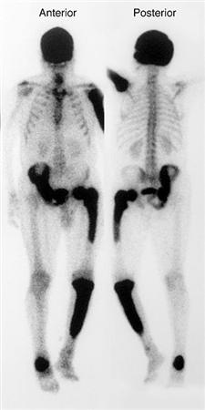
paget’s disease (metabolic bone disease)
chronic condition of disorganized bone remodeling leading to enlarged, weak, deformed bones
pathophysiology of paget’s disease
osteoclastic overactivity (excessive resorption), compensatory osteoblastic activity (chaotic new bone formation)
paget’s disease shows what on a bone scan
intense uptake involving 1+ bones; usually diffuse through entire affected bone. common sites: pelvis, spine, skull, long bones
hyperparathyroidism
chronically elevated PTH; leads to increased bone turnover (both resorption and reactive osteoblastic repair)
hyperparathyroidism on bone scan
whole skeleton looks uniformly “hot”; relative decreased renal visualization; “super scan” pattern (skeleton dominates, kidneys faint/absent)

paranasal sinus uptake suggests
sinusitis (sinus infection)

maxilla/right mandible uptake is typical for
odontogenic disease vs recent procedures/hardware
metastatic disease location: benign
common in long bones, joints, areas of known trauma
metastatic disease uptake: malignancy
frequently spine, pelvis, ribs, skull
mets disease correlative imaging
xray, ct, mri help clarify if uptake is benign/malignant lesion
history of trauma, surgery, growth supports…
benign bone-building
history of known malignancy, unexplained pain, systemic symptoms supports…
suspicion for cancer
mets to bone primarily occurs from what primary cancers?
prostate, breast, lung cancers
cold lesions
osteolytic, i.e. multiple myeloma, renal cell cancers, sarcoma
flare phenomenon
lesion may appear to worsen initially after chemotherapy
most cancers that mets to the bone stimulate…
osteoblastic repair activity around lesion
which cancers do not stimulate osteoblastic processes?
renal cell carinoma, multiple myeloma, sarcoma; these are purely lytic lesions
multiple focal lesions are common in patients with
history of prostate cancer
mult focal lesions on bone scan look like
asymmetrical uptake pattern; focal spots include right scapula, sternum, left 5th rib, several vertebral bodies, femurs