Unit 10: Somatic Sensory System | Touch & Pain (copy)
 Call Kai
 Learn
Learn Practice Test
 Spaced Repetition
 Match
Match Flashcards
 Knowt Play
1/22

There's no tags or description
Looks like no tags are added yet.
Name | Mastery | Learn | Test | Matching | Spaced | Call with Kai | Chat |
|---|
No analytics yet
Send a link to your students to track their progress
23 Terms
LO: Identify and describe different types of sensory receptors in the skin
Somatic Sensation
Enables the body to feel, ache, sense temperature and pressure (position of joints, muscles, distension of the bladder, temperature of limbs, itch, pain, etc.)
Responsible for touch and pain
Somatic sensory system: different from other systems
Receptors: broadly distributed
Responds to many kinds of stimuli - at least four senses rather than one
Five systems
Touch
Temperature
Pain
Body Position
Itch
Skin Types & Layers:
Hairy (with follicles) and glabrous (hairless, e.g., palms).
Two layers: Epidermis (outer) and Dermis (inner).
Functions of Skin:
Protects body and prevents fluid loss.
Provides direct contact with the external world.
Mechanoreceptors:
Detect mechanical stimuli (pressure, vibration, stretch, touch).
Differ by stimulus type, frequency sensitivity, and receptive field size.
Types of Sensory Receptors
Free Nerve Endings – detect pain and temperature (to be discussed later).
Merkel’s Disks – respond to gentle, sustained pressure (touch).
Found in epidermis.
Contain Merkel cells with Piezo2 mechanosensitive channels.
Both the Merkel cell and attached nerve ending are mechanically sensitive.
Meissner’s Corpuscles – respond to light touch and low-frequency vibration (1–50 Hz).
Located in ridges of glabrous skin (fingerprints).
Detect texture and fluttering sensations.
Hair Follicle Receptors – detect hair movement (touch).
Nerve endings wrap around or run along the follicle.
Hair bending deforms the follicle → alters nerve firing.
Pacinian Corpuscles – detect deep pressure and high-frequency vibration (200–300 Hz).
Found deep in the dermis.
Responsible for feeling vibrations (e.g., from a speaker).
Ruffini Corpuscles – detect skin stretch.
Have large receptive fields, slowly adapting responses.
Important for detecting sustained pressure and hand shape.
LO: Describe how a sensory event is encoded in action potentials in sensory fibers
Generator Potentials:
Act like EPSPs—graded depolarizations that vary in size with stimulus intensity.
If depolarization reaches threshold, voltage-gated Na⁺ channels open → action potential fired.
Stronger stimuli = larger generator potentials = higher AP frequency.
Mechanosensitive Ion Channels:
Convert mechanical force → ionic current in unmyelinated axon terminals.
Forces (membrane stretch, extracellular/cytoskeletal tethers, or second messengers) alter channel gating.
Many remain unidentified, but Piezo channels are key examples.
Piezo Channels (Piezo1 & Piezo2):
Mechanically gated, non-selective cation channels.
Closed: membrane curved → Open: membrane flattened/stretch → ion flow → depolarization.
Found in many tissues; involved in touch, proprioception, BP sensing, visceral reflexes, bone formation, and immunity.
Piezo2 is major in mechanoreceptors (Merkel, Meissner, Pacinian, hair follicles).
Merkel’s Disks:
Merkel cells contain Piezo2; both cell and axon terminal are mechanically sensitive.
Pressure opens Piezo2 → depolarization → transmitter release → excites sensory axon.
Unified Model for Dynamic Touch (Handler & Ginty, Neuron, 2023):
Low-threshold mechanoreceptors (LTMRs) (e.g., hair follicle, Meissner, Pacinian) share a common mechanism.
Axon protrusions (tethered via adherens junctions to non-neuronal cells) stretch during touch.
Stretching opens Piezo2 channels along the axon → neuron excitation.
Anchoring cells include terminal Schwann cells (hairy skin) and lamellar cells (Meissner/Pacinian).
Suggests a unified, structural basis for dynamic, light touch across skin types.
LO: Plan an experiment to map the receptive field of a somatic sensory receptor
Apply a small mechanical stimulus (e.g., fine brush or probe) to the skin.
Record electrical activity from a single sensory neuron.
Identify the region where stimulation evokes action potentials → this area is the receptive field.
Åke Vallbo’s Method:
Insert microelectrode into the median nerve to record action potentials from a single sensory axon.
Use a fine stimulus probe to touch and move around the skin’s surface.
Map the areas where the neuron responds to define the receptive field.
Purpose: Measure mechanoreceptor sensitivity and perception produced by mechanical stimuli.
LO: Distinguish somatic sensory receptors based on their receptive field size
Findings:
Meissner’s corpuscles and Merkel’s disks → small receptive fields (few mm wide).
Pacinian corpuscles and Ruffini’s endings → large receptive fields (can cover a finger or half the palm).
Conclusion: Small vs. large receptive fields help classify mechanoreceptors by spatial resolution.
LO: Compare and contrast rapidly-adapting and slowly-adapting receptors
Feature | Rapidly-Adapting (RA) Receptors | Slowly-Adapting (SA) Receptors |
|---|---|---|
Response to stimulus | Fire briefly at onset and sometimes offset of a stimulus; stop firing if stimulus is constant | Fire continuously as long as stimulus is present |
Sensitivity | Detect changes in stimulus (e.g., vibration, movement, texture) | Detect steady pressure, shape, and stretch |
Examples | Meissner corpuscles, Pacinian corpuscles | Merkel disks, Ruffini endings |
Receptive field size | Meissner: small; Pacinian: large | Merkel: small; Ruffini: large |
Function | Sense dynamic touch, vibration, motion | Sense static touch, pressure, skin stretch |
Adaptation speed | Fast | Slow |
Key idea: RA = detect changes, SA = monitor sustained stimuli.
In Pacinian corpuscles, the receptor potential peaks during dynamic indentation and declines during static indentation. Removing the corpuscle’s outer core prolongs the receptor potential, confirming RA behavior depends on structural properties.
LO: Relate the structure of the pacinian corpuscle to adaptation in the receptor
Capsule structure: 20–70 concentric layers of connective tissue with viscous fluid (“onion-like”), axon terminal in center.
Mechanism of RA response:
Initial compression → energy deforms axon terminal → opens mechanosensitive channels → receptor potential → action potential.
Sustained pressure → layers slip past each other → terminal no longer deformed → receptor potential dissipates → no firing.
Release of pressure → layers reverse → terminal depolarizes → firing resumes.
Evidence from Loewenstein: Removing capsule → axon becomes less sensitive to vibration, more sensitive to steady pressure → shows that RA property depends on capsule mechanics, not just the nerve ending.
Additional notes: Pacinian corpuscles have large, fast-conducting axons to transmit vibration signals efficiently to CNS.
LO: Discuss why our ability to discriminate a somatic sensory stimulus across different regions of the body varies
Two-point discrimination: Ability to detect two closely spaced stimuli; varies >20-fold across body.
Fingertips: Highest resolution. Reasons:
High receptor density → more mechanoreceptors per area.
Small receptive fields → enriched with Merkel’s disks & Meissner corpuscles.
Cortical representation → more brain tissue (“computational power”) devoted to each fingertip area.
Practice/learning → Braille readers can read ~600 letters/min; scanning motion improves sensitivity.
Potential specialized mechanisms for fine discrimination in fingertips.
LO: Describe the somatosensory pathways to the brain
Dorsal Column-Medial Lemniscal Pathway: Carries touch and vibration (not pain).
Primary sensory axons (Aβ) enter ipsilateral dorsal column of spinal cord.
Ascend to dorsal column nuclei at the junction of spinal cord and medulla.
Axons from dorsal column nuclei decussate (cross) in medulla → contralateral medial lemniscus.
Medial lemniscus ascends through medulla, pons, midbrain → ventral posterior (VP) nucleus of thalamus → S1 (primary somatosensory cortex).
Touch from right side of body is processed by left S1 cortex.
Longest axons can travel from toes to dorsal column nuclei at the base of the head.
Synaptic transformations: Information is altered at each synapse; inhibitory interactions enhance tactile responses.
Cortical feedback can modulate thalamic and dorsal column nuclei activity.
LO: Predict how a lesion in the pathway would impact sensation
Lesion of dorsal root: partial loss of sensation in dermatome due to overlapping innervation.
Lesion after decussation in medulla/medial lemniscus: contralateral loss of touch/vibration.
Lesion in VP thalamus or S1 cortex: loss of sensation for the contralateral side; detailed deficits depend on specific somatotopic region affected
Dorsal Column Touch Pathway: carries touch and vibration information.
Axons in spinal cord: travel ipsilaterally (same side as sensory input).
Damage to the spinal cord causes loss of sensation on the same side as the lesion.
Decussation (crossing) occurs in the medulla at the dorsal column nuclei.
After crossing, axons ascend to the thalamus → S1 cortex.
Result: touch from the right side of the body is processed in the left S1 cortex.
LO: Define dermatome
Dermatome = area of skin innervated by the right and left dorsal roots of a single spinal segment.
Each spinal segment has paired dorsal and ventral roots (≈30 segments total).
Dorsal roots pass through notches between vertebrae.
Cutting one dorsal root does not completely eliminate sensation due to overlap from neighboring roots; typically 3 adjacent roots must be cut.
Clinical example: Shingles infects all neurons of a single dorsal root.
Mapping shows dermatomes as bands on the body surface.
LO: Describe the organization of the primary somatosensory cortex
Somatotopy:
Adjacent body regions often mapped to adjacent cortical regions (e.g., fingers, hand, forearm).
Map is not always continuous; e.g., hand separates face and head.
Representation not scaled to body size; size of cortical area reflects density and importance of sensory input.
Example: fingers, lips, tongue are large; trunk and legs are small.
Importance/size varies by species (rats → whiskers have large representation).
Layered cortex:
S1 has layers like other neocortex areas; thalamic inputs terminate in layer IV.
Layer IV neurons project to other cortical layers.
Columnar organization:
Neurons with similar inputs/responses are stacked vertically into columns.
Example: adjacent cortical areas represent different fingers (D1, D2, D3), with alternating columns for rapidly- and slowly-adapting receptors.
S1 areas:
Areas 3a, 3b, 1, 2 in postcentral gyrus.
Further processing occurs in posterior parietal cortex (areas 5, 7).
Additional notes:
Penfield experiments mapped S1; painless because brain itself lacks somatic sensation.
Somatosensory maps are functional caricatures of the body, emphasizing areas of high sensory importance.
LO: Discuss an example of how the organization of the somatosensory cortex is plastic
Cortical plasticity: Somatosensory cortex can reorganize after changes in sensory input.
Example – digit removal:
Removing a finger does not leave its cortical area unused or cause atrophy.
Instead, the cortical area is taken over by inputs from adjacent digits.
Experimental evidence:
Michael Merzenich (1980s, UCSF) mapped S1 in adult owl monkeys before and after removing digit 3.
Months later, the area previously devoted to the removed finger responded to stimulation of neighboring fingers, showing major cortical reorganization.
Implication: Plasticity allows recovery of function and adaptation after peripheral injury.
Related observation: Training (e.g., Braille reading) can expand cortical representation of heavily used digits.
LO: Compare and contrast: retinotopy and somatotopy
Map distortion:
Both are not proportional to physical size of sensory surface.
Retinotopy → fovea overrepresented; somatotopy → lips and index finger overrepresented.
Plasticity:
Both maps can change with experience.
Ex: Losing a finger → adjacent fingers take over cortical space; losing input from one eye → visual cortex reorganizes.
Neighborhood organization:
Adjacent sensory regions are usually represented in adjacent cortical areas.
True for retinotopy (with central vision split) and somatotopy (e.g., lips near nose).
LO: Define pain. Explain why it should it be viewed as a positive adaptation
Definition of pain:
Pain is the subjective perception of sore, stinging, aching, throbbing, or unpleasant sensations.
Nociception is the sensory process that triggers pain through activation of specialized receptors (nociceptors).
Pain can occur even without nociceptor activity (e.g., phantom limb pain).
What activates pain:
Extreme temperatures, mechanical pressure, chemical irritation, inflammation, oxygen deprivation, or tissue damage.
Receptors: free nerve endings, TRP channels, specialized nociceptors.
Pathways to the brain:
Body: spinothalamic pathway.
Face: trigeminal pain pathway.
Information crosses at the spinal cord/brainstem depending on the pathway.
Processing and modulation:
Pain perception is influenced by afferent regulation, descending pain pathways, and endogenous opioids.
Cognitive and memory components affect how pain is felt.
Why pain is a positive adaptation:
Alerts the body to avoid harmful stimuli.
Promotes withdrawal and rest of injured parts to allow healing.
Prevents injury during sleep (e.g., turning to avoid bedsores).
Life without pain (congenital insensitivity to pain, CIP) leads to degeneration of joints, skeletal damage, infections, and early death.
Examples: SCN9A mutations affecting sodium channels in nociceptors prevent action potentials, causing CIP.
Evolutionary and practical significance:
Pain teaches risk assessment and prevents self-destructive behavior.
Helps in learning how to modulate activity safely.
Provides critical information for survival and health.
Clinical relevance:
Studying CIP has revealed targets like Nav1.7 and PRDM12 for potential pain treatments with minimal side effects.
LO: Explain how pain receptors work and the concept of hyperalgesia
Activation of nociceptors:
Stimuli that can cause tissue damage: strong mechanical pressure, extremes of temperature, oxygen deprivation, chemicals.
Mechanotransduction: stretching or bending of the nociceptor membrane opens mechanically gated ion channels → depolarization → action potentials.
Chemically mediated activation:
Proteases → bradykinin → opens ion channels
ATP → ATP-gated channels → depolarization
High extracellular K+ → depolarizes membrane
Histamine
TRP channels (Transient Receptor Potential) mediate responses to temperature and chemical stimuli.
Example: TRPM8 (CMR1) responds to cold and menthol.
TRPV1 responds to heat, protons, physical abrasion, and capsaicin.
Substances released from damaged cells open nociceptor channels:
Proteases → bradykinin
ATP
K+
Histamine (from mast cells) → depolarizes nociceptors, increases blood vessel permeability → swelling/redness
Exercise example: lactic acid → high extracellular H+ → activates nociceptors → dull ache.
Hyperalgesia:
Increased sensitivity to pain in damaged tissue.
Primary hyperalgesia: occurs directly at the site of tissue damage.
Secondary hyperalgesia: increased sensitivity in surrounding tissue.
"Inflammatory soup": neurotransmitters (glutamate, serotonin, ATP), peptides (substance P, bradykinin), lipids (prostaglandins, endocannabinoids), cytokines, chemokines, ions (K+, H+) → enhance nociceptor sensitivity.
Results in the cardinal signs of inflammation: pain, heat, redness, swelling.
LO: Compare the structure of various nociceptors and how this influences the perception of pain
Aδ fibers: myelinated, large, fast → sharp, immediate pain
C fibers: unmyelinated, slow → dull, aching pain, responds to cool temperatures
TRP channels (transient receptor potential channels) detect temperature and chemical stimuli
CMR1/TRPM8 → cold and menthol receptor
Structure influences:
Fiber diameter and myelination → conduction speed and pain quality
Ion channel expression → modality specificity (mechanical, thermal, chemical)
Combination of fiber type and receptor expression shapes perception of pain (sharp vs dull, fast vs slow, localized vs diffuse)
LO: Analyze sex differences in mechanical allodynia
Definition: Mechanical allodynia = perception of pain in response to normally non-painful mechanical stimuli.
Experimental evidence:
Von Frey hairs used to measure mechanical sensitivity in mice.
IL-23/IL-17A/TRPV1 axis induces mechanical allodynia in female mice only, not in males.
Effect depends on:
Sex hormones (estrogen receptor in nociceptors)
Macrophages
IL-17A release
TRPV1 sensory receptor
Outcome: female mice show decreased paw withdrawal threshold → increased sensitivity to mechanical stimuli.
Implications:
Highlights sex-specific pain mechanisms.
Suggests immune-neuronal crosstalk is differentially regulated in females vs. males.
LO: Discuss sex bias and omission in neuroscience research
Sex bias: historically, experiments conducted only in male animals.
Sex omission: failure to report the sex of animals included in studies.
Recent changes:
Funding agencies (NSF, NIH) require inclusion of female animals for grant eligibility.
Some journals now require reporting of sex and inclusion of females.
Sex differences in neurobiological diseases:
Chronic pain, migraine, anxiety, depression → more common in women.
Autism, ADHD → more common in men.
Importance: Understanding sex differences is crucial for accurate modeling of disease and drug responses.
LO: Sketch the pain pathway from periphery to cortex
1. Peripheral Nociceptors
Located in skin, muscles, joints, and viscera.
Receptor type: Free nerve endings.
Fiber types:
Aδ fibers: thinly myelinated, fast → sharp, localized pain.
C fibers: unmyelinated, slow → dull, burning, diffuse pain.
Stimulus: Mechanical, thermal, or chemical injury triggers depolarization.
2. First-Order Neurons
Cell bodies:
Dorsal root ganglia (body).
Trigeminal ganglion (face).
Axon path:
Peripheral axon detects stimulus.
Central axon enters dorsal horn of spinal cord (or trigeminal nuclei for face).
3. Dorsal Horn Synapse
Synapse site: Lamina I (marginal layer) & Lamina II (substantia gelatinosa).
Neurotransmitters: Glutamate, Substance P (enhances excitability).
Modulation: Local inhibitory interneurons and descending pathways can suppress or enhance signal.
4. Decussation (Crossing Over)
Second-order neurons cross to the contralateral side of spinal cord.
Pathway: Anterolateral system / spinothalamic tract.
5. Ascending Tracts
Spinothalamic tract: Carries discriminative pain and temperature.
Other tracts:
Spinoreticular → arousal and alertness.
Spinomesencephalic → orienting responses, emotional aspects.
6. Thalamic Relay
Second-order neurons terminate in:
Ventral posterolateral (VPL) nucleus → body.
Ventral posteromedial (VPM) nucleus → face.
Thalamus integrates signals and relays to cortex.
7. Third-Order Neurons (Cortex)
Project from thalamus to primary somatosensory cortex (S1) → conscious perception of pain (location, intensity).
Other cortical targets:
Insula → sensory-emotional integration.
Anterior cingulate cortex (ACC) → motivational/affective aspects of pain.
8. Descending Modulation
Pathway: Periaqueductal gray (PAG) → rostral ventromedial medulla → dorsal horn.
Mechanism: Releases serotonin, norepinephrine, endogenous opioids to inhibit nociceptive transmission.
Function: Adjusts intensity of pain perception, mediates placebo effects.
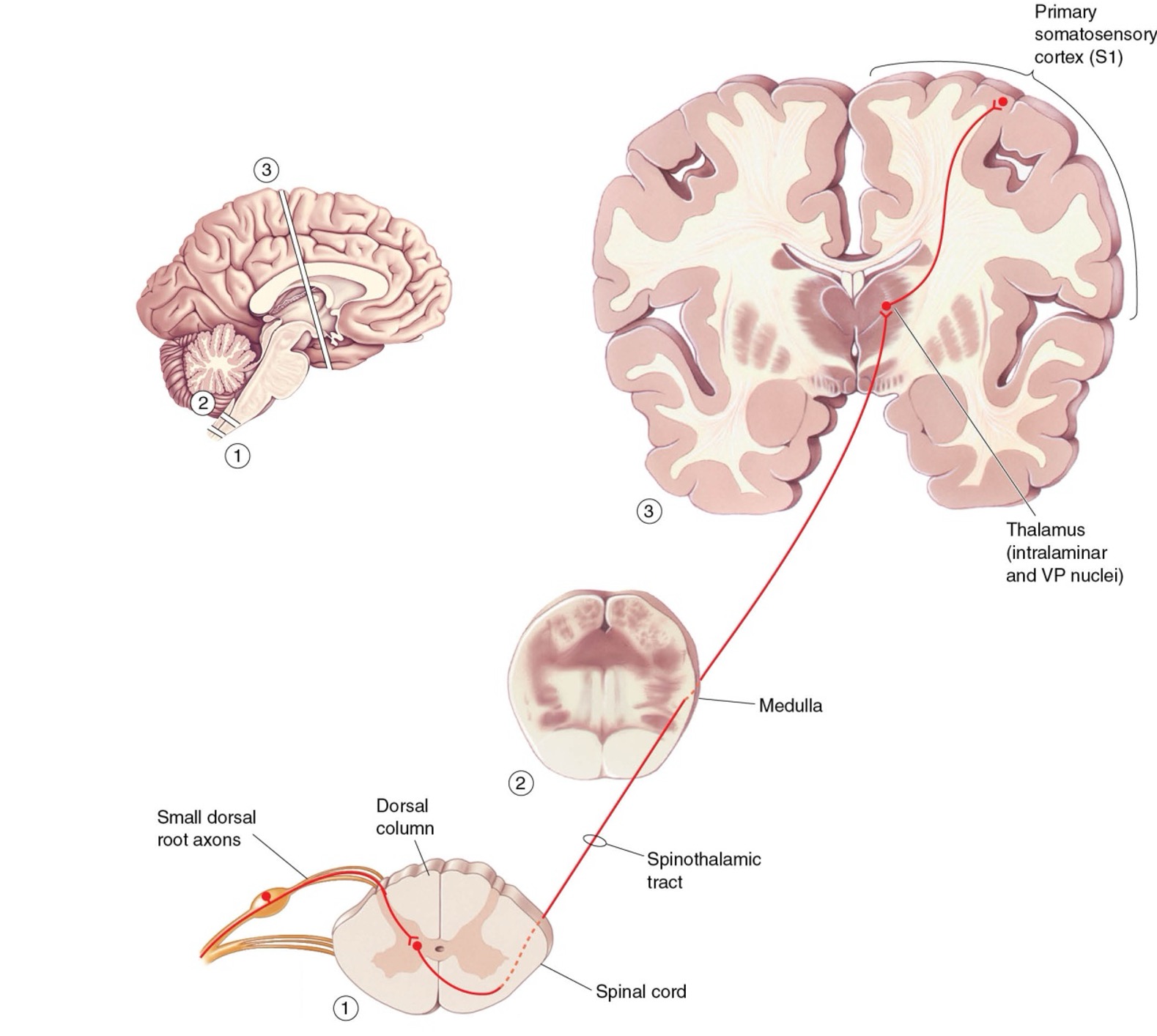
LO: Compare and contrast: spinothalamic and trigeminal pain pathways
Spinothalamic pathway:
Carries pain and temperature information from the body.
Small-diameter fibers synapse in the dorsal horn of the spinal cord.
Axons cross to the contralateral side in the spinal cord and ascend to the thalamus.
Trigeminal (trigeminothalamic) pathway:
Carries pain and temperature information from the face and head.
Small-diameter fibers in the trigeminal nerve synapse in the spinal trigeminal nucleus of the brainstem.
Second-order neurons cross and ascend to the thalamus via the trigeminal lemniscus.
Similarities:
Both pathways transmit pain and temperature information to the thalamus.
Axons of second-order neurons cross to the contralateral side.
Additional features:
Other related pathways modulate slow, burning pain and behavioral arousal.
Trigeminal pathway implicated in migraine pathophysiology.
LO: Describe three ways that pain is regulated
Afferent Regulation (Gate Control):
Pain signals from nociceptors can be reduced by simultaneous activity in Aβ mechanoreceptors (light touch).
Mechanism: activation of inhibitory interneurons in the dorsal horn suppresses nociceptive projection neurons.
Explains why rubbing a bruise or using electrical stimulators can relieve pain.
Descending Pain Pathways:
Emotional state, stress, or strong determination can suppress pain.
Periaqueductal gray (PAG) in midbrain sends descending axons to raphe nuclei in medulla (serotonin release).
Raphe neurons project to dorsal horn to inhibit nociceptive neurons.
Provides top-down modulation of pain based on context and emotion.
Endogenous Opioids:
Small peptides (endorphins, enkephalins, dynorphins) bind opioid receptors in PAG, raphe nuclei, and dorsal horn.
Mechanism: inhibit neurons by hyperpolarization and suppress glutamate release from presynaptic terminals.
Analgesic effects blocked by naloxone.
Opioid drugs (morphine, codeine, heroin) mimic this effect.
LO: Describe the placebo effect and phantom limb pain
Placebo effect:
Pain relief occurs even when treatment has no active pharmacological agent (e.g., saline injection).
Belief in the effectiveness of a treatment can activate endogenous pain-relief systems in the brain.
Examples: acupuncture, “mom’s kiss,” post-op saline injections.
Phantom limb pain:
A type of neuropathic pain experienced in a limb that is no longer present.
Likely due to inappropriate signaling or memory of pain by neurons in the brain or spinal cord.
Pain perception can occur even without nociceptor activity in the missing limb.
LO: Compare and contrast: pain and touch receptors and pathways
1. Receptors / Nerve Endings
Touch: Specialized mechanoreceptors in the skin (e.g., Merkel cells, Meissner corpuscles).
Pain: Free nerve endings (nociceptors) sensitive to mechanical, thermal, or chemical damage.
2. Axon Diameter / Conduction
Touch: Large-diameter, myelinated axons → fast conduction.
Pain: Small-diameter fibers:
Aδ fibers → thin myelinated, fast/sharp pain.
C fibers → unmyelinated, slow/dull or aching pain.
3. Spinal Cord Connections / Pathways
Touch:
Axons ascend ipsilaterally in the dorsal column.
Deep connections in the dorsal horn and medulla before crossing.
Pain:
Axons branch immediately in spinal cord → Lissauer’s tract.
Terminate in the substantia gelatinosa.
Ascend contralaterally in the spinothalamic tract.
4. Types of Pain
Nociceptive / Inflammatory (acute): Dull, shock-like, prickling, burning, aching. Activated by Aδ and C fibers.
Neuropathic (chronic): Electric, shooting, or continuous burning pain due to injury or dysfunction in nervous system.
Mechanisms: Inflammatory mediators (cytokines, bradykinin, prostaglandins, histamine) can sensitize nociceptors → hyperalgesia / allodynia.
5. Clinical Example:
Brown–Séquard syndrome: Lesion on one side of spinal cord → ipsilateral touch loss (dorsal column) & contralateral pain/temperature loss (spinothalamic).