Gene and chromosomal mutations
 Call Kai
 Learn
Learn Practice Test
 Spaced Repetition
 Match
Match Flashcards
 Knowt Play
1/31

There's no tags or description
Looks like no tags are added yet.
Name | Mastery | Learn | Test | Matching | Spaced | Call with Kai | Chat |
|---|
No analytics yet
Send a link to your students to track their progress
32 Terms
Mutations
although occasional genetic changes enhance the long-term survival of a species, the survival of an individual demands a high degree of genetic stability
when the cell’s DNA maintenance processes fail —> mutation
Definition of mutation: permanent change in DNA, which is an alteration to an organism’s characteristics that is inherited due to a change in the genetic material of a cell
it can destroy an organism if it occurs in a vital position in the DNA sequence
mutations can involve
just a single or a few nucleotide pairs
large regions of a chromosome
types of mutations in multicellular organisms
Germline mutation
mutation occurs in germline cells (consisting of germ cells and gametes) - it may be transmitted to the offspring and to successive generations (can be inherited)
if the mutation has an adverse effect on the phenotype of an organism, the mutant condition is referred to as a genetic disorder, or hereditary disease
Somatic mutation
mutation occurs in somatic cells
these mutations are not inherited by the progeny and hence not passed on to the next generation
Gene/point mutations
Gene/point mutations involve chemical changes that affect the DNA sequence of just one gene
they involve changes at specific sites in a gene - result in change in one or a few bases in the DNA sequence
in gene/point mutation, 2 basic types of changes to a gene can occur
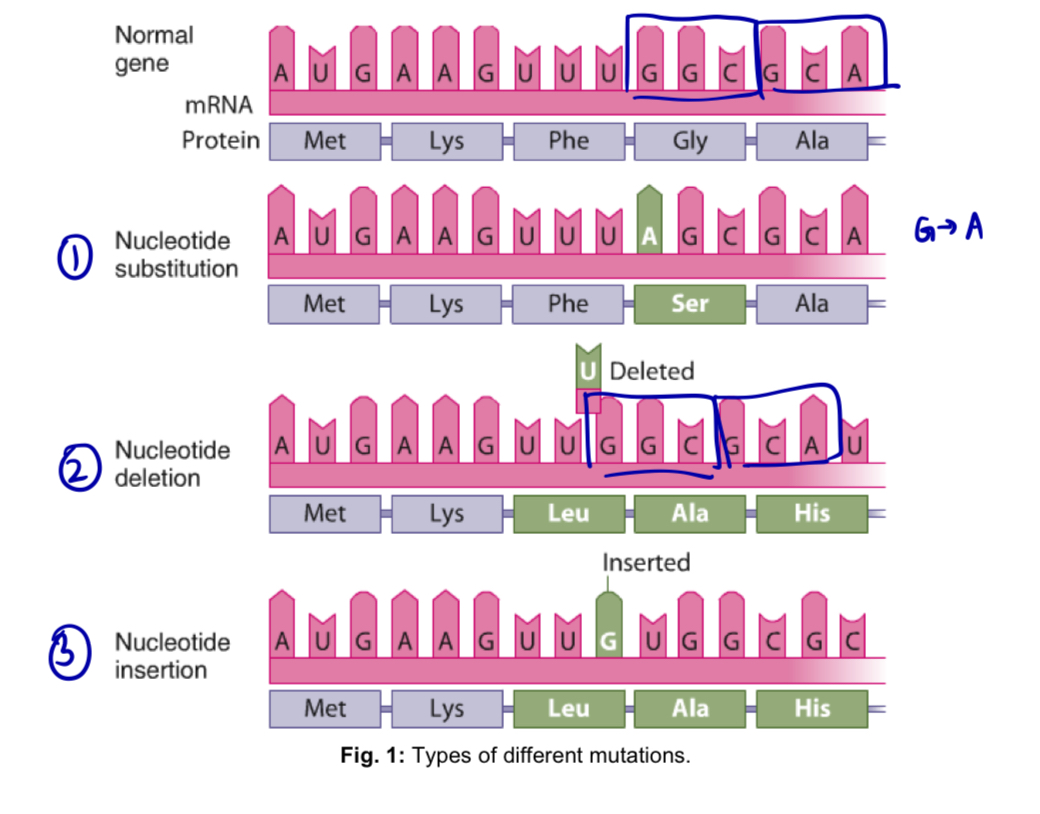
Nucleotide substitutions
replacement of one nucleotide pair with another, resulting in one of the following
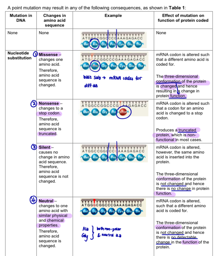
a. missense mutation: nucleotide substitution in a DNA sequence results in the translation of a different amino acid
b. nonsense mutation: nucleotide substitution in a DNA sequence results in a codon for an amino acid being changed into a stop codon, leading to premature termination of translation
c. silent mutation: nucleotide substitution in a DNA sequence changes the mRNA codon. however, the same amino acid is inserted into the protein because of the degeneracy of the genetic code
d. neutral mutation: nucleotide substitution in a DNA sequence changes the mRNA codon and amino acid translated. however, the resulting amino acid substitution produces no detectable change in the function of the protein translated.
note: these are 4 examples of mutation, but they are NOT TYPES of mutations
Nucleotide insertions or deletions
addition or deletion of one or more nucleotide pair
depending on the location in the DNA sequence and the number of nucleotide pairs added or deleted, one of the following will ensure
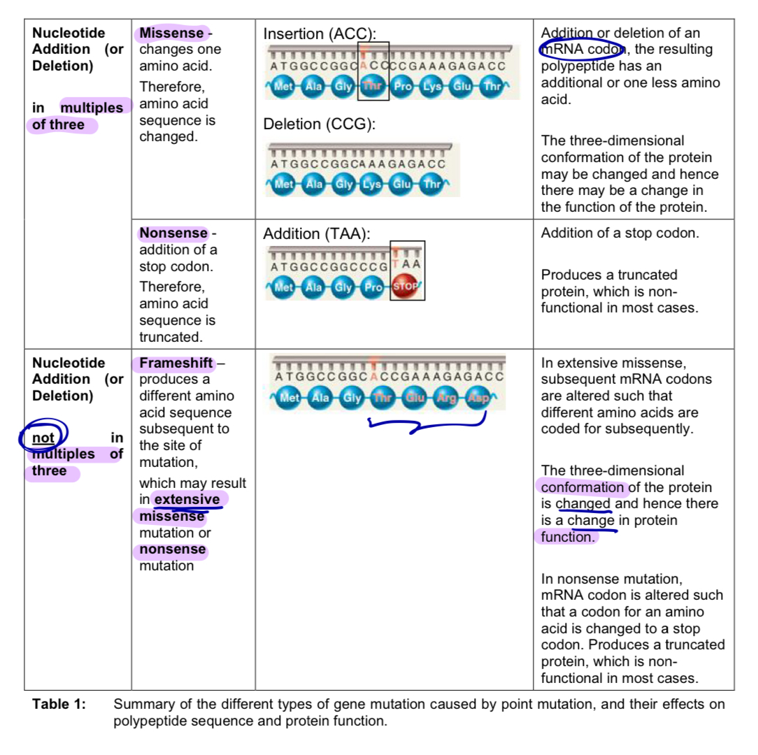
a. Addition or deletion of deoxynucleotides in multiples of 3 results in:
missense mutation - mRNA codon was added or deleted and the resulting polypeptide has an amino acid added or deleted respectively
OR
nonsense mutation - stop codon was added, leading to premature termination of translation
b. Addition or deletion of deoxynucleotides not in multiples of 3 results in:
frameshift mutation - mRNA codons subsequent to the insertion or deletion are changed, resulting in
extensive missense mutation - subsequent amino acid sequence of the polypeptide is changed OR
nonsense mutation - codon for an amino acid is changed to a stop codon, resulting in a truncated protein
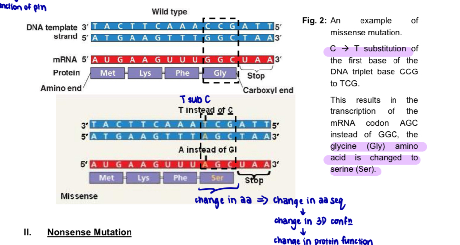
Effects of nucleotide substitutions - Missense mutation
nucleotide substitutions may or may not affect the amino acid sequence of a protein depending on the location of the substitution
Missense mutation
nucleotide substitution in DNA sequence changes the mRNA codon, resulting in the translation of a different amino acid
amino acid sequence of the polypeptide is changed = resulting in a change in the specific 3-dimensional conformation of the protein = function of the protein is altered
*change in a.a —> change in a.a sequence —> change in 3D conformation —> change in protein function
e.g. sickle cell anaemia
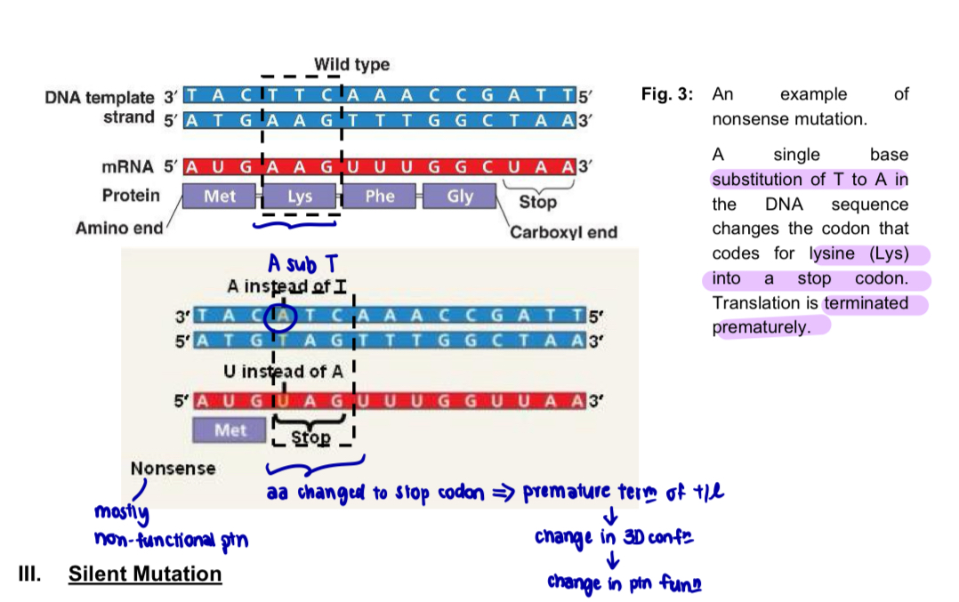
Effects of nucleotide substitutions - nonsense mutation
nucleotide substitution in a DNA sequence changes a codon for an amino acid into a stop codon
= premature termination of translation
= resulting polypeptide will be truncated (shorter) than normal polypeptide encoded
amino acid sequence of the polypeptide is shortened
= change in the specific three-dimensional conformation the protein
= function of the protein is altered
nearly all nonsense mutations result in non-functional proteins (due to truncated polypeptides)
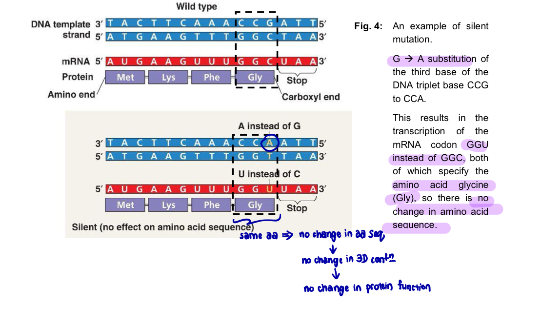
Effects of nucleotide substitution - Silent mutation
nucleotide substitution in a DNA sequence changes the mRNA codon
however, the same amino acid is inserted into the polypeptide because of the degeneracy of the genetic code
the amino acid sequence of the polypeptide is unchanged
= resulting in no change in the specific three-dimensional conformation of the protein
= no change in the function of the protein
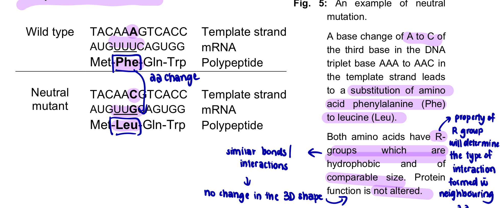
Effects of nucleotide substitution - Neutral mutation
nucleotide substitution in a DNA sequence changes the mRNA codon
however, the resulting amino acid produces no detectable change in the function of the protein translated
this could arise from
the substitution of the original amino acid with an amino acid of similar physical and chemical properties OR
the substitution of an amino acid residue that is non-essential to that protein’s structure and function
the amino acid sequence of the polypeptide is changed, BUT there is no change in the overall three-dimensional conformation of the protein and hence the function of the protein is not altered
Effects of nucleotide insertions or deletions
nucleotide insertions or deletions are additions or losses, respectively, of one or more nucleotide pairs in a gene
often has deleterious effects
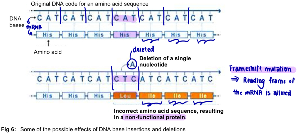
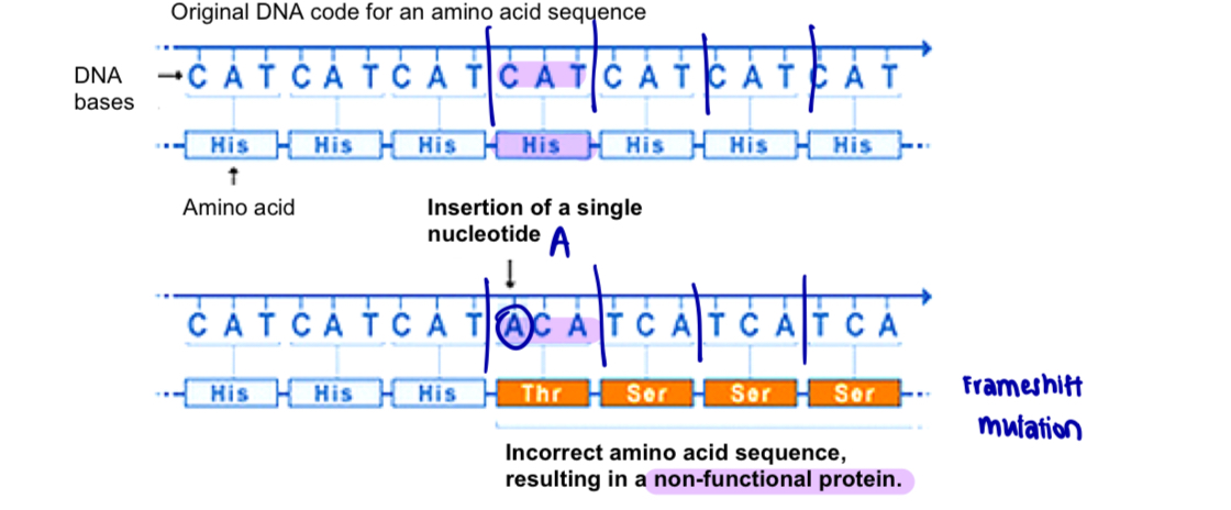
resulting mRNA is read as a series of non-overlapping codons, an insertion or deletion of nucleotides NOT in multiples of 3 will result in frameshift mutation (reading frame of the mRNA is changed)
all the nucleotides downstream of the insertion/deletion site will be improperly grouped into codons, resulting in extensive missense
frameshift may also cause a new, premature stop codon to be generated (nonsense mutation) in the reading frame, or result in a read-through of the normal stop codon, resulting polypeptides of altered lengths
a frameshift usually results in a non-functional protein
Effects of nucleotide insertion/deletion - Deletion mutation
single base deletion mutation leads to frameshift
e.g. in figure: deletion of A in the DNA sequence results in the alteration of subsequent codons in the corresponding mRNA, hence different amino acids are coded for subsequently
= incorrect amino acid sequence of the polypeptide chain, and consequently incorrect three-dimensional protein conformation and function
Effects of nucleotide insertion/deletion - Insertion mutation
single base insertion mutation leads to frameshift
e.g. in figure: insertion of A in the DNA sequence results in the alteration of subsequent codons in corresponding mRNA
= different amino acids are coded for subsequently
= incorrect amino acid sequence of the polypeptide chain, and consequently incorrect three-dimensional protein conformation and function
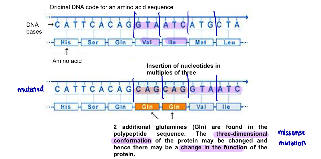
Effects of nucleotide insertion/deletion - Insertion or deletion mutation (in multiples of three)
e.g. insertion of 2 sets of nucleotides in multiples of three does not lead to frameshift
amino acid sequence contains 2 additional glutamines —> 3D conformation of the protein may be changed hence leading to a change in the functions of the protein
Summary - point mutation leading to different consequences
Summary table (2)
Case study: Gene mutations resulting in phenotypic change
—> Sickle-cell anaemia (how gene mutations can result in disease)
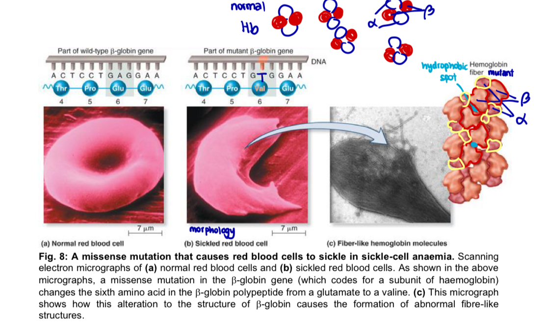
involves a mutation in the β-globin gene, which encodes one of the polypeptide subunits that make up haemoglobin (Hb) (Hb - 2α and 2β subunits)
Genetic and molecular basis
substitution of a thymine for an adenine at one position in the Hb gene (template strand), which results in a missense mutation
T —> A
6th amino acid residue in polypeptide is changed from a glutamate (hydrophilic) to a valine (hydrophobic)
change in property of amino acid —> change in the interactions/bonds formed with neighbouring a.a
= specific three-dimensional conformation and function of the Hb protein is altered
this substitution creates a hydrophobic spot on the outside of the Hb protein that sticks to the hydrophobic region of an adjacent Hb protein’s beta chain
mutant Hb subunits tend to stick to one another when the oxygen concentration is low, particularly when the red blood cells are in capillaries and veins
the aggregated proteins form fibre-like structures within red blood cells
at high oxygen concentration, haemoglobin resumes globular haemoglobin structure
Physiological effects
the fibre-like structures cause the RBC to lose their normal morphology and become sickle-shaped
sickles cells are less able to move through capillaries and can block blood flow, resulting in severe pain and cell death of the surrounding tissue due to shortage in oxygen
the sickles RBC are also fragile and easily destroyed, further decreasing the oxygen carrying capacity of the blood
*point mutation (nucleotide substitution) —> change in a.a sequence —> change in protein structure (3D conformation) —> change in protein function (decreased capacity to carry blood cos of the change in morphology of RBC) —> disease
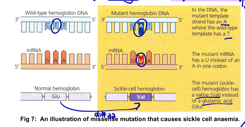
Figure showing missense mutation causing sickle-cell anaemia
Causes of gene mutations
mutagenesis: production of mutations
2 groups of causes
Spontaneous mutations
mutations that occur naturally - without the use of chemical or physical mutagenic agents
result of errors that occur during DNA replication, recombination or repair (can lead to both gene and chromosomal mutations)
DNA replication and repair
mistakes: although DNA replicates with high fidelity, DNA polymerase sometimes inserts the wrong nucleotide/too many/too few nucleotides into DNA sequence
DNA polymerase make mistakes at a rate of about 1 in every 100 000 nucleotides (~120 000 mistakes every time a cell divides with 6 billion bp in each human diploid cell)
attempts at correction: proofreading, where some of the mistakes are corrected immediately; some are corrected after replication during mismatch repair
during proofreading, DNA polymerase enzymes recognise mistakes and replace the incorrectly inserted nucleotide so that replication can continue
after replication, mismatch repair reduces the final error rate even further
—> incorrectly paired nucleotides cause deformities in the secondary structure of the final DNA molecule - during mismatch repair, enzymes recognise and fix these deformities by removing the incorrectly paired nucleotide and replacing it with the correct nucleotide
some replication errors fail to be recognised by the repair enzymes = these altered nucleotide sequences can be passed down from one cellular generation to the next
if they occur in cells that give rise to gametes, can be transmitted to subsequent generations of the organism
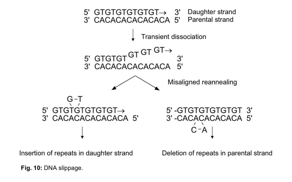
DNA slippage
daughter or parental DNA strand slips during DNA replication followed by folding back of the strand
mispairing between the daughter DNA strand and the parental template strand
causes parts of the DNA which are folded back to be copied more than once
if this duplicated DNA segment corresponds to a gene, it will result in gene duplication
Induced mutations
result of deliberate application of mutagens - chemical or physical agents - that result in increased mutation rates
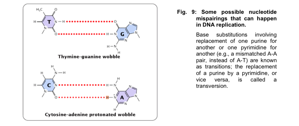
Figure of DNA slippage
Mutagens and effect on DNA
Physical agents (radiation)
X-rays
results in the production of free radicals of water (the OH radical) which are chemically very reactive
free radicals interact with DNA to produce double-stranded breaks leading to chromosomal rearrangements and deletions (KIV: Pg 18 on Chromosomal Mutations)
Ultraviolet (UV) rays
are absorbed by bases of DNA. This may result in
the production of a covalent attachment between adjacent pyrimidines in one strand, usually thymine dimers, or
base pair substitutions, insertions and deletions
DNA replication and transcription are blocked – this is lethal if unrepaired
Chemical agents
Base analogues
• are molecular structures that are similar to the bases normally found in DNA
• may be incorporated into DNA in place of the normal bases during DNA
replication, hence producing base substitutions
E.g. Bromouracil (5-BU), which
o can exist in alternate states
o each of which can pair with a different base in DNA, hence producing base substitutions
Base-modifying agents
• modify the chemical structure and properties of bases
• leads to mispairing during DNA replication and hence base substitution
E.g. Alkylating agents such as ethylmethylsulfonate (EMS) and mustard gas.
o transfer alkyl groups to bases, modifying their chemical structure
Intercalating agents
• are flat molecules with multiple ring structures
• insert themselves (intercalate) between adjacent bases in one or both strands of
the DNA helix
• leads to insertions or deletions during DNA synthesis and hence frameshift
mutations
E.g. Proflavin, acridine, ethidium bromide.
Chromosomal mutation/aberration
Definition:
a change in the structure of a chromosome (involving several gene loci) OR
a change in the number of chromosomes
Change in chromosome structure
structural changes that delete, add or rearrange substantial portions of one or more chromosomes
deletion and duplication of genes or part of a chromosome and rearrangements of genetic material in which a chromosome segment is inverted, exchanged with a segment of non-homologous chromosome, or merely transferred to another chromosome - translocation (location of gene is altered within the genome
these structural change of a chromosome result in reshuffling of alleles on chromosome
Change in chromosome structure - Deletion
Deletion
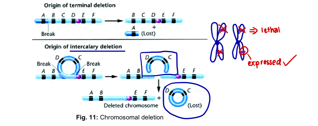
when a chromosome breaks in one or more places, and a portion of it is lost, the missing piece is referred to as deletion
deletion can occur near one end or from the interior of the chromosome —> terminal or intercalary deletions
effect of a deletion is profound, in which the genotype is altered due to the absence of certain gene loci
if a deletion affects the same gene loci on both homologous chromosomes, the effect is usually lethal
if only one of a homologous pair of chromosomes is affected, the effect on the phenotype is that the alleles on the non-deficient homologous will be expressed, even if recessive
Change in chromosome structure - Duplication
Duplication
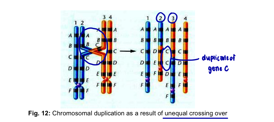
when any part of the genetic material is present more than once in the genome —> duplication
can arise as a result of unequal crossing over between synapses chromosomes during meiosis or through a replication error prior to meiosis
in the former case, both duplication and deletion are produced
effects of duplications - gene redundancy, phenotypic variations
in addition, duplications have also been deemed as an important source of genetic variation during evolution
Change in chromosome structure - Inversion
Inversion
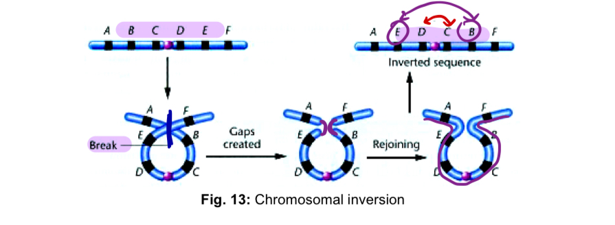
a segment of a chromosome is turned around 180º within a chromosome
an inversion does not involve a loss of genetic information, but simply rearranges the linear sequence
requires 2 breaks along the length of the chromosome and subsequent reinsertion of the inverted segment
by forming a chromosomal loop prior to breakage, the newly created “sticky” ends are brought close together and rejoined
organisms heterozygous for inversions may produce aberrant gametes that have a major impact on their offspring
in addition, inversion may also result in position effects which lead to altered gene expression due to new positioning of a gene within the genome and thus play an important role in the evolutionary process
Change in chromosome structure - Translocation
Translocation
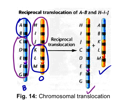
movement of a chromosomal segment to a new location in the genome
reciprocal translocation involves the exchange of segments between 2 non-homologous chromosomes
genetic consequences of a reciprocal translocation are similar to inversions
genetic information is not lost or gained —> rearrangement of genetic material
may produce position effect (may realign certain genes in relation to other genes)
Change in chromosome number
addition or loss of one or more chromosomes, addition of one or more haploid sets of chromosomes
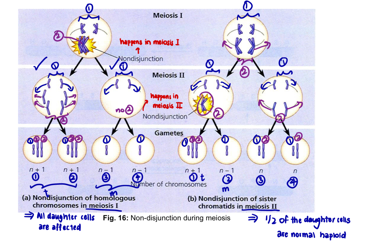
Aneuploidy
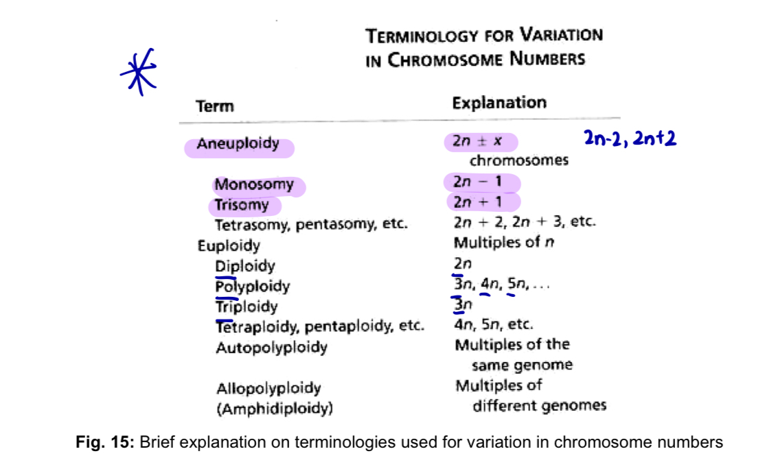
Aneuploidy: general condition in which an organism loses or gains one or more chromosomes, but not a complete set
these chromosomal variations originate as a random error during the production of gametes
non-disjunction is the failure of chromosomes or chromatids to disjoin and move to opposite poles during division
when this occurs in meiosis, the normal distribution of chromosomes into gametes is disrupted
for the affected chromosome, abnormal gametes can be formed containing either two members or none at all
the loss of a single chromosome from an otherwise diploid genome —> monosomy
gain of one chromosome —> trisomy
fertilising these with a normal haploid gamete produces a zygote with either three members (trisomy) or only one member (monosomy) of this chromosome
non-disjunction leads to a variety of autosomal aneuploidy conditions in humans and other organisms
Monosomy
monosomy in humans only occur for the X chromosome
e.g. 45, X Turner syndrome - affected individuals have only 45 chromosomes, including just a single X chromosome
45, X (only 1 X copy present in the sex chromosome)
these individuals generally have female external genitalia and internal ducts, but the ovaries are rudimentary (underdeveloped)
short stature, skin flaps on the back of the neck, underdeveloped breasts, broad shieldlike chest
intelligence normal
monosomy for any of the autosomes is usually not tolerated in humans or other animals
Trisomy
e.g. 47, XXY Klinefelter syndrome - affected individuals have more than one X chromosome
characteristics
genitalia and internal ducts that are usually male
but their testes are rudimentary and fail to produce sperm
generally tall and have long arms and legs and large hands and feet
although masculine developed does occur, feminine sexual development is not entirely suppressed
slight enlargement of breasts is common, hips are often rounded
intersexuality, which may lead to abnormal social development
intelligence often below the normal range
e.g. Down syndrome - the only human autosomal trisomy in which a significant number of individuals survive longer than a year past birth
affected individuals have an extra chromosome 21
characteristics
short
skin folds over the corner of their eyes
stocky bodies
thick necks
prone to heart abnormalities
short life expectancy
low intelligence level
Polyploidy
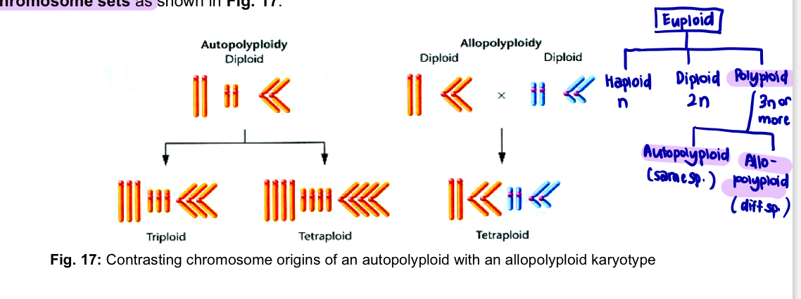
Euploidy - instances where multiples of the haploid chromosome set are found
Polyploidy - instances where more than 2 multiples of the haploid chromosome set are found
triploid - 3n chromosomes
tetraploid - 4n chromosomes
pentaploid - 5n
well known in lizards, amphibians and fish
more common in plants
Can originate in 2 ways
addition of one or more extra sets of chromosomes, identical to the normal haploid component of the same species resulting in autopolyploidy
combination of chromosome sets from different species may occur as a consequence of interspecific matings resulting in allopolyploidy
Autopolyploidy
autotriploids occur in a few ways
failure of all chromosomes to segregate during meiosis divisions (first-division or second-division non-disjunction) can produce a diploid gamete
if such a gamete survives and is fertilised by a haploid gamete —> zygote with 3 sets of chromosomes (3n)
2 sperms may fertilise an ovum —> triploid zygote
produced under experimental conditions by crossing with tetrapolyploids
diploid organisms produce gametes with n chromosomes
tetraploids produce 2n gametes
upon fertilisation, the desired triploid is produced
economically important triploid plants include several potato species of the genus Solanum, Winesap apples, commercial bananas, seedless watermelons, and the cultivated tiger lily Lilium tigrinum, which are propagated asexually
autotetrapolyploids are theoretically more likely to be found in nature than autotriploids due to their even number of chromosomes
more likely to be found in nature than autotriploids due to
their even number of chromosomes. Unlike triploids, which often produce genetically unbalanced
gametes with odd number of chromosomes, tetraploids are more likely to produce balanced
gametes when involved in sexual reproduction. Tetraploid alfalfa, coffee, peanuts and McIntosh
apples are also of economic value due to either their larger sizes or more vigorous growth than
their diploid or triploid counterparts.
Allopolyploidy
results from hybridisation of 2 closely related species
if a haploid ovum from a species with chromosome sets AA is fertilised by a haploid sperm from a species sets BB —> resulting hybrid is AB
A = a1, a2, … an
B = b1, b2, … bn
hybrid plant may be sterile because of its inability to produce viable gametes
this occurs when some or all of the a and b chromosomes are not homologous and thus are unable to synapse in meiosis
leads to unbalanced genetic conditions
however, if the new AB genetic combination undergoes a natural or induced chromosomal doubling, 2 copies of all a chromosomes and 2 copes of all b chromosomes present —> pair during meiosis —> fertile AABB tetraploid produced
since this tetraploid contains equivalent of 4 haploid gametes derived from two separate species = allotetraploid
when both species are known - amphidiploid (equivalent term)
Genetic screening
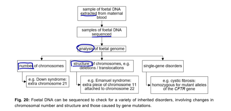
Genetic screening is the analysis of a person’s DNA to check for presence of one or more alleles that are associated with disease
samples of DNA can be obtained for testing from embryos, newborn babies, children and adults
some screenings can be carried out before a baby is born
Genetic screening and genetic testing
genetic test - link to family history
e.g. If a person from a family with Huntington’s disease, for example, begins to develop symptoms of
the disease, he or she is likely to be advised to have a genetic test (KIV: Molecular Techniques) to
confirm the diagnosis. Other people in the family who do not show any symptoms may also be
offered the opportunity to have the genetic test
is the same whether a person has symptoms or not - involves PCR to amplify the gene HTT in the case of Huntington’s diseases and electrophoresis to determine the lengths of the two alleles and to count the number of CAG repeats
genetic screening - people who are free of the symptoms of a disease
Maternal genetic screening
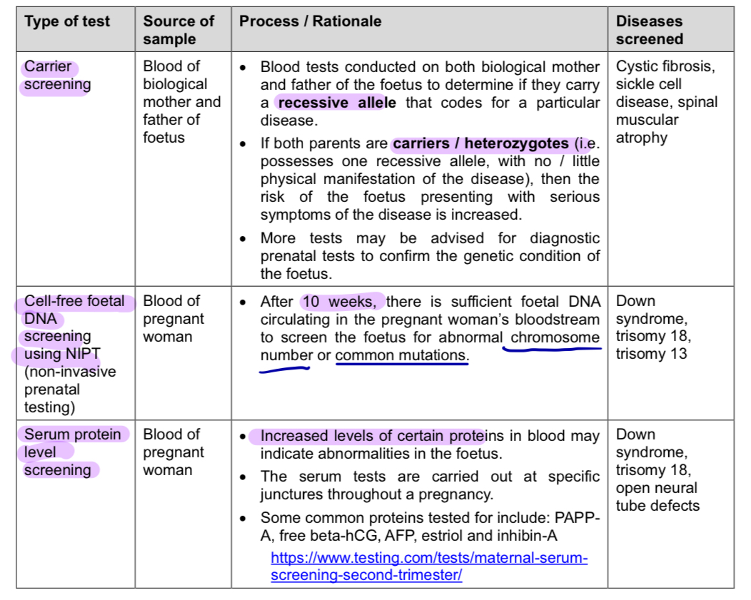
maternal genetic screening, also known as prenatal genetic screening, are tests that provide information about the health of a foetus
check for congenital conditions e.g. Down Syndrome, trisomy-13 and spinal bifida
high-risk pregnancies
over age of 35
family or personal history of genetic conditions
history of miscarriage or stillbirth
lifestyle factors e.g. exposure to cigarette smoke, radiation
results of screening tests can be used for counselling purposes to help parents make informed decisions about the outcome of pregnancy
maternal genetic screening tests do not diagnose medial conditions
these screening tests determine if the foetus is at an increased or decreased risk of a particular abnormality
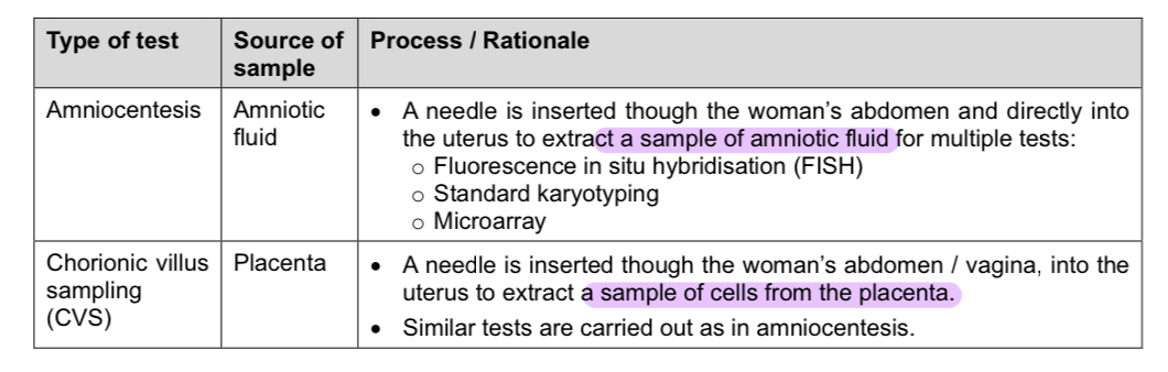
Prenatal Diagnostic tests
diagnostic test is only performed when a screening test is abnormal
prenatal diagnostic tests are usually more invasive tests that may pose a higher risk to the pregnant woman and the foetus
Maternal genetic screening for down syndrome
Down syndrome (trisomy-21)
non-disjunction is found in 95% of down syndrome, the other 5% caused by translocation, mosaicism or partial trisomy
occurs when there is additional chromosome for the 21st pair of chromosomes
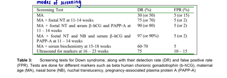
maternal genetic screening is used to look for potentially at-risk pregnancies
maternal genetic screening has significantly reduced the rate of down syndrome live briths over the years in singapore
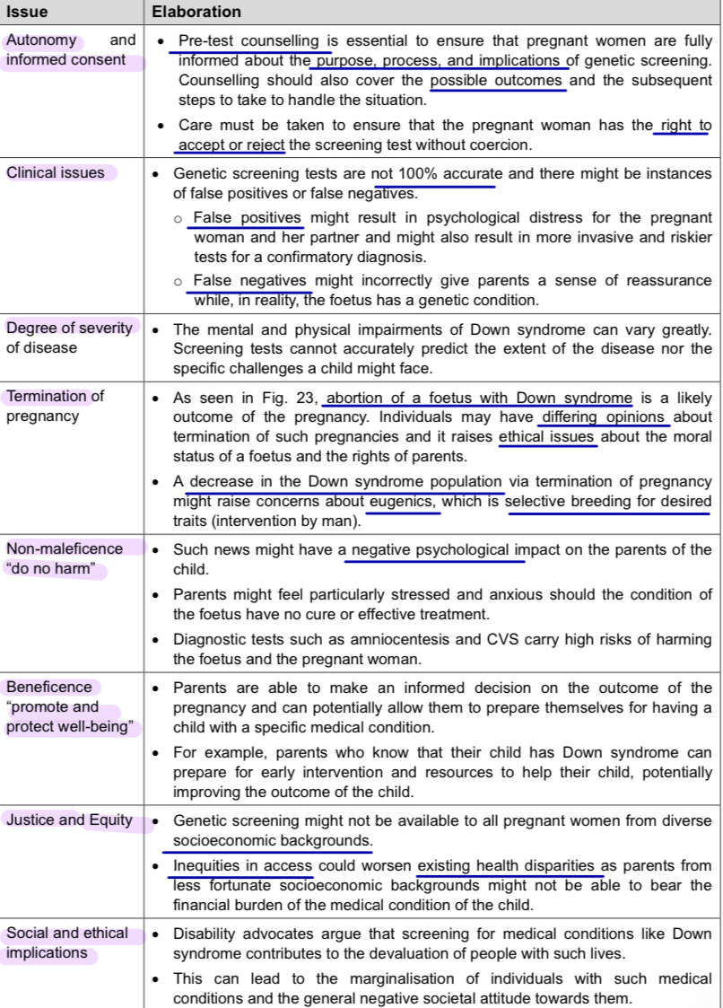
Bioethical considerations in maternal genetic screening