ECG Rhythms
1/178
There's no tags or description
Looks like no tags are added yet.
Name | Mastery | Learn | Test | Matching | Spaced | Call with Kai | Chat |
|---|
No analytics yet
Send a link to your students to track their progress
179 Terms
How do you convert bpm to ms?
Take 60,000 divided by your heart rate in bpm
How do you convert ms to bpm?
Take 60,000 and divide by the cycle length (distance between two R waves)
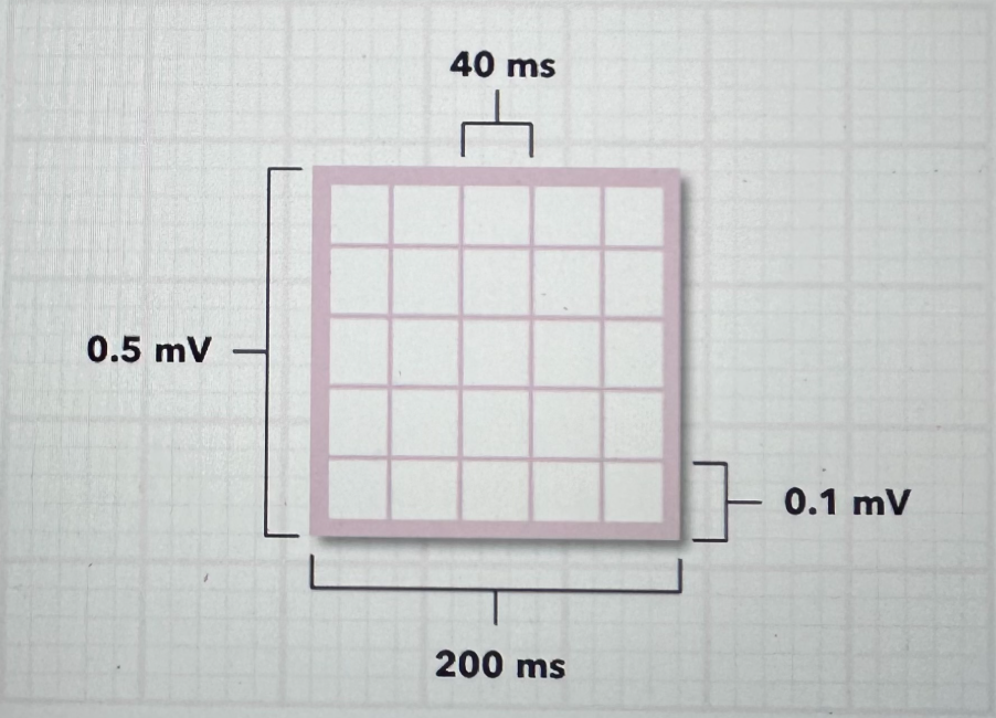
What do the dimensions of the squares on an ECG translate to?
What are the normal steps to identifying a rhythm?
Define the Rate
Normal Rate is 60-100 bpm
Define the Intervals (PR and QRS)
Normal PR: 120-200 ms
Normal QRS: 60-100 ms
Define the Rhythm
Are the P waves: absent or present? Normal (positive/upright) or abnormal?
Is the QRS normal or wide?
Is there one P wave for every QRS?
Define the Regularity
Is there consistent duration of time between QRS complexes (R waves)?
Is there consistent duration of time between the start of a P wave and the beginning of the QRS complex?
What does it mean to say a rhythm is regular?
It means that there is constant spacing between each beat, which is how you can say NSR and Sinus Brady and Sinus Tach are all regular, even if the actual rates are different
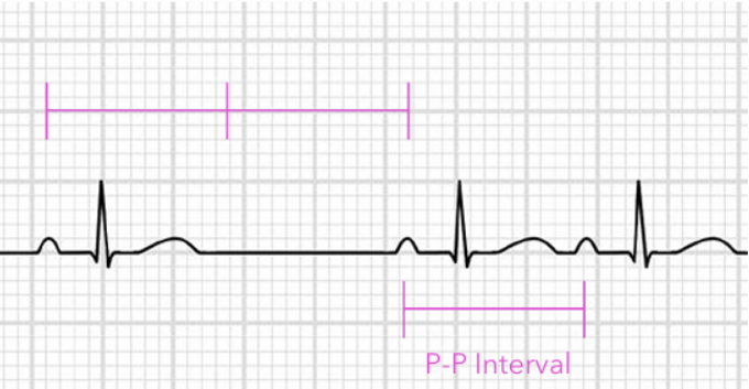
Normal Sinus Rhythm (Strip)
Regular rhythm, 60–100 bpm, normal P before every QRS
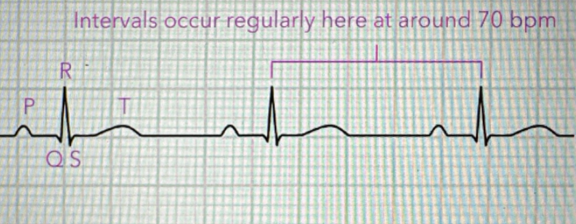
Normal Sinus Rhythm (Rate)
60-100 bpm
Normal Sinus Rhythm (P Wave)
P waves are present, upright, uniform, and occur before every QRS.
One P wave for every QRS.
Normally is less than 120 ms
Normal Sinus Rhythm (PR Interval)
PR interval is normal and constant: 120–200 ms.
Normal Sinus Rhythm (QRS Complex)
QRS is narrow and normal-looking: <120 (60-100) ms.
Each QRS follows a normal P wave.
Normal Sinus Rhythm (Regularity)
R-R interval regular. P-P interval regular. Rhythm is regular because the SA node fires at a steady rate.
Sinus Bradycardia (Strip)
Regular rhythm, normal P before every QRS, but rate <60 bpm
Occurs because the SA node fires at an abnormally slow rate due to abnormally slow automaticity (electrical impulse formation)
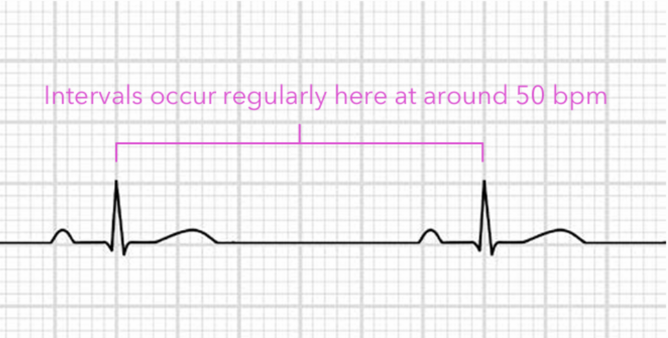
Sinus Bradycardia (Rate)
<60 bpm
Sinus Bradycardia (P Wave)
P waves are present, upright, uniform, and occur before every QRS.
Same as NSR, but rate is slow.
Sinus Bradycardia (PR Interval)
PR interval is usually normal and constant: 120–200 ms.
Same as NSR, but rate is slow.
Sinus Bradycardia (QRS Complex)
QRS is usually normal: <120 (60-100) ms.
Same as NSR, but the rate is slow.
Sinus Bradycardia (Regularity)
Regular intervals between P wave to P wave and R wave to R wave
Sinus Tachycardia (Strip)
Regular rhythm, normal P before every QRS, but rate >100 bpm
Caused by abnormally fast automaticity, so the cells are easily excited
Sinus Tachycardia (Rate)
>100 bpm
Sinus Tachycardia (P Wave)
P waves are present, upright, uniform, and occur before every QRS.
Same as NSR, but rate is fast.
At very fast rates, P waves may blend into the previous T wave.
Sinus Tachycardia (PR Interval)
PR interval is usually normal and constant: 120–200 ms.
At faster rates, P waves may blend into T waves, making PR harder to measure.
The higher the heart rate, the shorter the PR intervals tend to be
Sinus Tachycardia (QRS Complex)
QRS is usually narrow: <120 (60-100) ms.
Same as NSR, but the rate is fast.
Sinus Tachycardia (Regularity)
Regular intervals between P wave to P wave and R wave to R wave
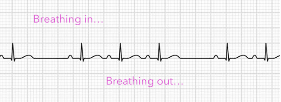
Sinus Arrhythmia (Strip)
Normal sinus P waves, but R-R intervals vary
Typically occurs in young, healthy people, in which the reflex changes in vagal tone during breathing
Sinus rate increases with inhaling, and decreases with exhaling
Sinus Arrhythmia (Rate)
Usually 60-100 bpm, but rhythm is irregular
Sinus Arrhythmia (P Wave)
P waves are normal: present, upright, uniform, and occur before every QRS.
The rhythm is irregular, but the P waves still look sinus.
Sinus Arrhythmia (PR Interval)
PR interval is normal and constant: 120–200 ms.
The rhythm is irregular, but conduction from atria to ventricles is still normal.
Sinus Arrhythmia (QRS Complex)
QRS is normal: <120 (60-100) ms.
QRS complexes occur irregularly because the sinus rate varies, but conduction through the ventricles is normal.
Sinus Arrhythmia (Regularity)
R-R & P-P are irregular.
P waves are still sinus and occur 1:1 with every QRS.
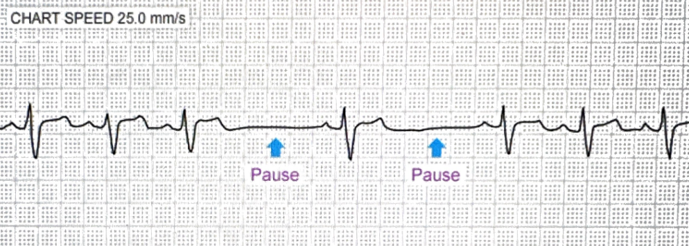
Sinus Pause (Strip)
The pause will usually terminate with a sinus or escape beat from the atrium or ventricle, where the sinus node will typically resume pacing after the escape beat occurs
Sinus Pause (Rate)
Rate varies depending on pause length, though underlying rate is typically 60-100 bpm
Sinus Pause (P Wave)
P waves are normal when present, but there is a temporary missing P-QRS-T complex during the pause.
After the pause, sinus P waves resume.
Sinus Pause (PR Interval)
PR interval is usually normal when beats are present: 120–200 ms.
During the pause, there is no P wave or QRS, so no PR interval to measure.
Sinus Pause (QRS Complex)
QRS is normal and narrow: <120 (60-100) ms when beats are present.
During the pause, the expected QRS complex is missing.
Sinus Pause (Regularity)
Underlying rhythm is usually regular, then there is a temporary pause in which the R-R and P-P intervals become irregular.
R-R and P-P are interrupted by a missing beat.
The pause is usually not an exact multiple of the normal P-P interval.
When does a Sinus Pause become a Sinus Arrest?
A Sinus Pause becomes a Sinus Arrest when the SA node fails to generate an electrical impulse for 3 seconds or longer
Sinus Exit Block / SA Block (Strip)
The underlying mechanism for this is a conduction issue
Sinus Exit Block / SA Block (Rate)
Rate varies but tends to stay around 60-100 bpm; pause causes slower overall rate
Sinus Exit Block / SA Block (P Wave)
A whole P-QRS-T complex is dropped.
P waves are normal before and after the block, but one expected P wave is missing because the impulse does not exit the SA node.
Sinus Exit Block / SA Block (PR Interval)
PR interval is usually normal on conducted beats: 120–200 ms.
Sinus Exit Block / SA Block (QRS Complex)
QRS is usually normal and narrow: <120 (60-100) ms on conducted beats.
Sinus Exit Block / SA Block (Regularity)
Underlying rhythm is usually regular, then a full P-QRS-T complex is dropped.
The pause is usually an exact multiple of the normal P-P interval.
The pauses R-R and P-P intervals to briefly become irregular
How does Sinus Exit Block differ from Sinus Pause/Arrest?
It differs in that sinus exit block the underlying P-P or R-R intervals of sinus exit block resume or “march out” on time after the pause
What is Ectopy?
Ectopy means an electrical impulse starts from an abnormal location outside the heart’s normal pacemaker, the SA node.
What causes premature beats in the heart?
Premature beats can initiate from atrial, junctional (AV node), or ventricular tissue, usually through increased automaticity
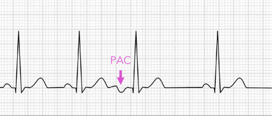
Sinus Rhythm w/ Premature Atrial Complexes (PAC) (Strip)
Occurs when atrial tissue other than the sinus node spontaneously depolarizes earlier than the sinus node and produces this premature contraction
Sinus Rhythm w/ PAC (Rate)
Underlying rate usually 60-100 BPM; PAC occurs early
Sinus Rhythm w/ PAC (P Wave)
Normal sinus P waves are present, but the PAC has an early abnormal-looking P wave.
The PAC P wave may be hidden in the previous T wave.
The shape of the P wave depends on the site of origin, and the closer the ectopic (non-SA node) site is to the SA node, the more it will look like a standard P wave
The further away the ectopic site is from the SA node, the more different it will look, i.e. inverted on the ECG
Sinus Rhythm w/ PAC (PR Interval)
Underlying sinus beats usually have a normal PR: 120–200 ms. The PAC may have a different PR interval, often slightly abnormal depending on where the ectopic atrial impulse starts.
Sinus Rhythm w/ PAC (QRS Complex)
Underlying QRS complexes are usually narrow: <120 ms. The PAC usually has a narrow QRS too because it comes from above the ventricles.
Sinus Rhythm w/ PAC (Regularity)
Underlying rhythm is regular, but the PAC occurs early, making the rhythm irregular.
The R-R interval before the PAC is shortened, often followed by a pause.
What is a Non-conducted PAC?
It is when the PAC occurs so early that conduction through the AV node is blocked due to it still being in refractory, thereby not conducting the PAC any further
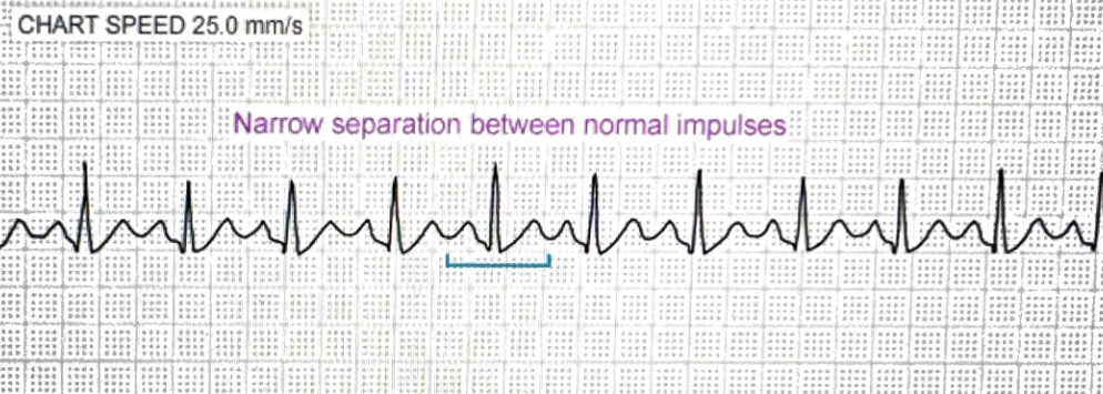
Atrial Tachycardia (Strip)
Usually paroxysmal, meaning it starts and ends abruptly
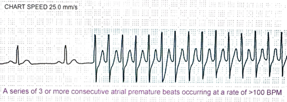
Atrial Tachycardia (Rate)
Usually 100-250 bpm
Atrial Tachycardia (P Wave)
P waves are usually present but abnormal in shape because the impulse starts from an ectopic atrial focus instead of the SA node.
There is usually one P wave before each QRS, but they may be hard to see at fast rates.
The P waves can be inverted, pointed, oddly shaped, or buried in the previous T wave
Atrial Tachycardia (PR Interval)
PR interval may be normal or slightly abnormal, but usually measurable if P waves are visible. Often around 120–200 ms, but exact value can vary.
Atrial Tachycardia (QRS Complex)
QRS is usually narrow: <120 ms because the rhythm starts above the ventricles and conducts normally through the His-Purkinje system.
Atrial Tachycardia (Regularity)
Usually regular R-R intervals. P-P intervals are usually regular if P waves are visible.
It is a fast, regular atrial rhythm.
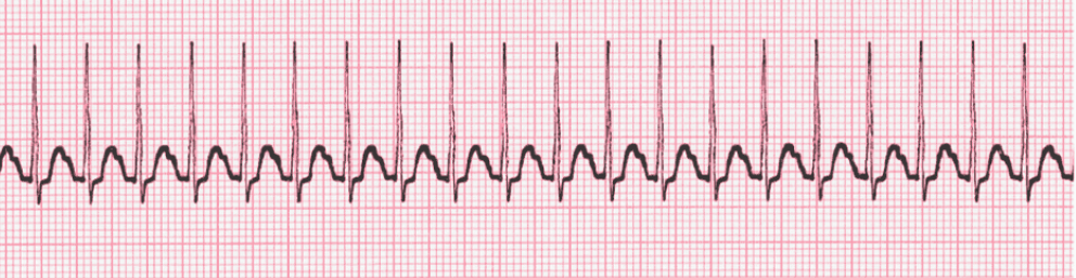
Supraventricular Tachycardia (SVT) (Strip)
Supraventricular Tachycardia (SVT) (Rate)
Usually 150-250 bpm
Supraventricular Tachycardia (SVT) (P Wave)
P waves are often absent, hidden, or buried in the T waves/QRS complexes. If visible, they may be abnormal or retrograde.
Supraventricular Tachycardia (SVT) (PR Interval)
PR interval is often not measurable because P waves are hidden in the QRS or T waves. If visible, retrograde P waves may make the PR interval abnormal.
Supraventricular Tachycardia (SVT) (QRS Complex)
QRS is usually narrow: <120 ms. Key clue: very fast, regular, narrow-complex rhythm. QRS can be wide if there is aberrant conduction, but basic SVT is narrow.
Supraventricular Tachycardia (Regularity)
Usually very regular R-R intervals. P-P intervals are often not measurable because P waves are hidden. Key clue: fast, regular, narrow-complex rhythm.
Atrial Flutter (Strip)
Sawtooth flutter waves
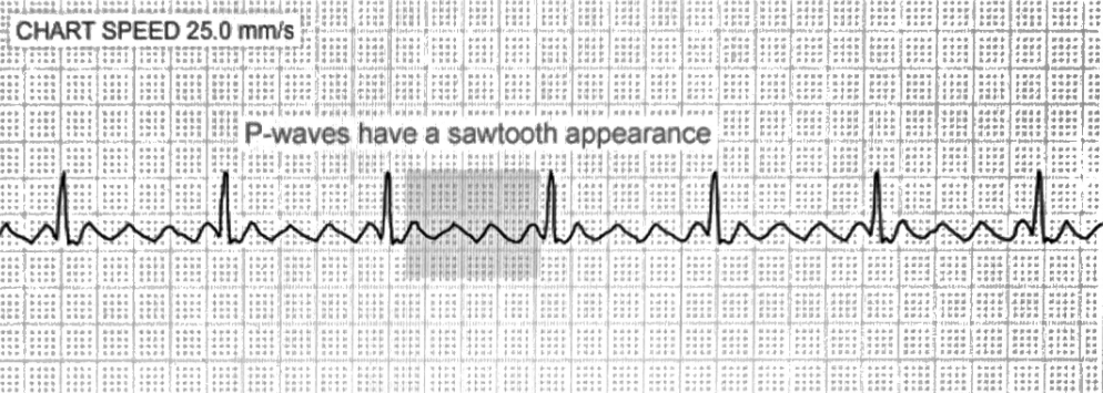
Atrial Flutter (Rate)
Atrial rate 250-350 BPM
Ventricular rate is often, but not always, around 150 BPM with 2:1 conduction
Atrial Flutter (P Wave)
No normal P waves. Instead, there are flutter waves, often described as sawtooth-shaped F waves.
Atrial Flutter (PR Interval)
PR interval is usually not measurable because there are no normal P waves; flutter waves replace P waves.
Atrial Flutter (QRS Complex)
QRS is usually narrow: <120 ms.
Multiple flutter waves may appear between QRS complexes.
These waves appear in a ratio of, usually, 2:1 or 4:1
Atrial Flutter (Regularity)
Can be regular or irregular depending on AV conduction. With fixed conduction, like 2:1 or 3:1, R-R is regular. With variable conduction, R-R is irregular.
Flutter waves usually occur at a regular atrial rate.
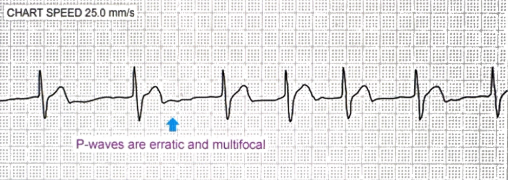
Atrial Fibrillation (Strip)
Irregularly irregular rhythm with no distinct P waves
Atrial Fibrillation (Rate)
Atrial rate chaotic, often 350-600 BPM; ventricular rate varies with AV conduction
Atrial Fibrillation (P Wave)
No distinct P waves.
Baseline is chaotic, wavy, or fibrillatory due to disorganized atrial activity.
Atrial Fibrillation (PR Interval)
PR interval is not measurable because there are no distinct P waves.
Atrial Fibrillation (QRS Complex)
QRS is usually narrow: <120 ms, but the R-R intervals are irregularly irregular. QRS can be wide if there is a bundle branch block or aberrant conduction.
Atrial Fibrillation (Regularity)
R-R intervals are irregularly irregular.
P-P intervals are not measurable because there are no true P waves.
This is the classic irregularly irregular rhythm.
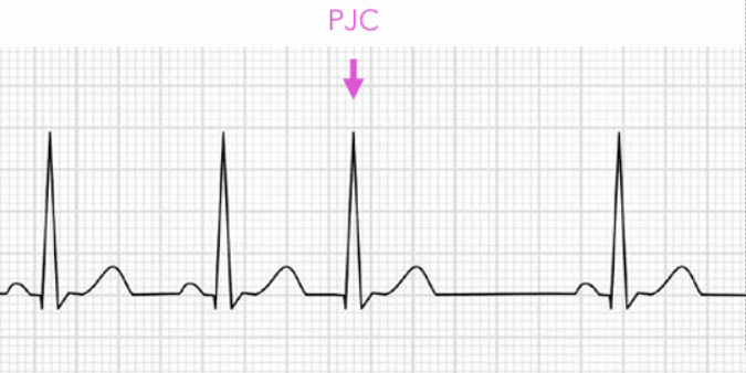
Premature Junctional Contractions (Strip)
If the AV node’s rate of automaticity is faster than the sinus rate, the AV junction will then be the site of activation, and the signals travels backward (retrograde) to the atria
Premature Junctional Contractions (Rate)
The underlying rate is considered the rate, with the PJC briefly deviating from that
Premature Junctional Contractions (P Wave)
The P wave is typically not visible because it is covered by the QRS complex, but it can also appear inverted when seen
Premature Junctional Contractions (PR Interval)
If the P wave appears before the QRS, the PR interval is usually short: <120 (60-100) ms.
If the P wave is hidden in the QRS or occurs after the QRS, the PR interval is not measurable.
Premature Junctional Contractions (QRS Complex)
The QRS complex appears as normal since the ventricles are activated by the normal conduction pathway
It will simply appear premature
Premature Junctional Contractions (Regularity)
The underlying rhythm may be regular, but the PJC makes it irregular because the junctional beat comes early.
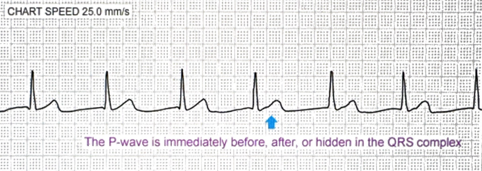
Junctional Rhythm (Strip)
Narrow regular rhythm with absent/inverted/after-QRS P waves
When the SA node activity is blocked or < the automaticity of the AV node/His bundle (where these rhythms occur), this rhythm presents
It is also known as a Junctional Escape Rhythm since it comes from the AV node when there is no signal from the atria and it depolarizes enough
Junctional Rhythm (Rate)
40-60 bpm for junctional escape rhythm
Junctional Rhythm (P Wave)
P waves may be absent, hidden in the QRS, or appear after the QRS.
If before the QRS, the PR interval is usually short.
It will ALWAYS be inverted.
Junctional Rhythm (PR Interval)
PR interval depends on where the P wave appears. If P wave is before QRS, PR is usually short: <120 ms. If P wave is hidden or after QRS, PR is not measurable.
Junctional Rhythm (QRS Complex)
QRS is usually narrow: <120 ms because the impulse still travels normally through the ventricles.
Junctional Rhythm (Regularity)
Usually regular R-R intervals. P-P intervals may be absent or not measurable because P waves may be hidden, inverted, or after the QRS.
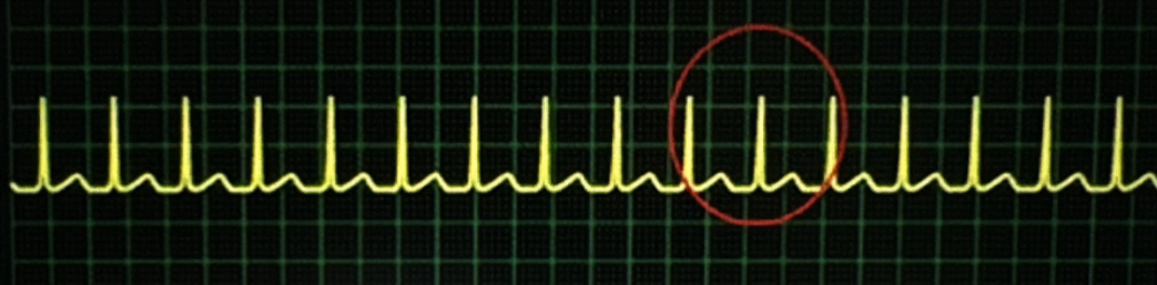
Accelerated Junctional Rhythm (Strip)
Notice the inverted P wave

Accelerated Junctional Rhythm (Rate)
60-100 bpm
Accelerated Junctional Rhythm (P Wave)
P waves may be absent, hidden, or after the QRS.
The P wave will ALWAYS be inverted
Difference is the rate is faster, 60–100 bpm.
Accelerated Junctional Rhythm (PR Interval)
Same as junctional rhythm.
If P wave is before QRS, PR is usually short: <120 ms.
If hidden or after QRS, PR is not measurable.
Accelerated Junctional Rhythm (QRS Complex)
QRS is usually narrow: <120 ms. Same QRS appearance as junctional rhythm, but rate is faster.
Accelerated Junctional Rhythm (Regularity)
Usually regular R-R intervals. Same regularity pattern as junctional rhythm, but rate is 60–100 bpm.
Junctional Tachycardia (Strip)
Junctional Tachycardia (Rate)
>100 bpm
Junctional Tachycardia (P Wave)
Same junctional P wave pattern: P waves may be absent, inverted, hidden, or after the QRS. At faster rates, they are often hard to see.
Junctional Tachycardia (PR Interval)
Same junctional pattern. PR interval is often not measurable because P waves are hidden. If P wave appears before QRS, PR is usually short: <120 ms.
Junctional Tachycardia (QRS Complex)
QRS is usually narrow: <120 ms. Fast, regular rhythm with absent/inverted/hidden P waves and narrow QRS.
Junctional Tachycardia (Regularity)
Usually regular R-R intervals. Same junctional pattern, but rate is greater than 100 bpm.