Bacteriology Comp Exam
 Call Kai
 Learn
Learn Practice Test
 Spaced Repetition
 Match
Match Flashcards
 Knowt Play
1/118

There's no tags or description
Looks like no tags are added yet.
Name | Mastery | Learn | Test | Matching | Spaced | Call with Kai | Chat |
|---|
No analytics yet
Send a link to your students to track their progress
119 Terms
How does the age of bacterial cells affect Gram Stain results?
Age
Old cells die, breaking down the peptidoglycan layer, compromising cell’s ability to retain CV. May also stain Gram-variable
Antibiotics
Antibiotics that break down the cell wall or inhibit cell wall synthesis compromise cell’s ability to retain CV
Endotoxins (Lipopolysaccharides, (LPS))
Toxic structural molecules that make up the outer membrane of Gram-negative bacteria
They are passively released into the environment or a host's bloodstream when the bacterial cell dies, ruptures, or undergoes mechanical damage
Exotoxins
Highly potent, soluble proteins secreted by living bacteria (and some fungi or algae) into their surrounding environment or injected directly into host cells.
Act as targeted virulence factors that disrupt cellular metabolism, destroy tissue, or trigger severe, systemic immune responses.
Enterotoxins
Poisonous proteins secreted by bacteria that specifically target the intestines = food poisoning
What is thioglycolate broth used for?
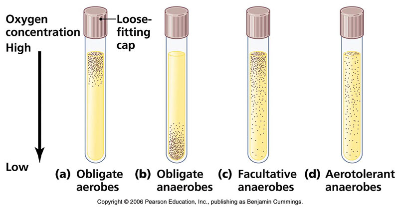
To determine the oxygen requirements of microorganisms and to cultivate fastidious, aerobic, and anaerobic bacteria. The broth naturally forms an oxygen gradient, with high concentrations at the top and zero oxygen at the bottom. This allows microbiologists to classify bacteria as obligate aerobes, obligate anaerobes, facultative anaerobes, or microaerophiles based on where they grow in the tube.
Explain the principle of the acid-fast stain procedure
Identifies bacteria with thick, waxy cell walls, containing mycolic acid (appears bright red, retaining carbol fuchsin; non acid-fast: blue)
How do Beta-lactams work? + Examples
Inhibit wall synthesis
Penicillin: GP
Cephalosporins: GP and GN
Monobactam: GN aerobes
Carbapenem: Broad spectrum
Cephamycin: GN and anaerobes
How do aminoglycosides work?
Inhibit protein synthesis
Targets aerobic GNs
How do glycopeptides work?
Inhibit cell wall synthesis
Targets GP + MRSA
What mechanism of resistance does beta-lactamase have?
Enzyme inactivation
Breaks the beta-lactam ring of the antibiotic
Can be inhibited by: Clavulanic acid, Tazobactam, Sulbactam, and Avibactam
What mechanism of resistance does carbapenemase have?
Enzyme inactivation
Enzyme destroys carbapenems, making broad spectrum β-lactams ineffective
What are the AST standards?
Use 4-5 colonies, emulsify to reach a 0.5 McFarland standard density
Mueller-Hinton agar base
pH 7.2-7.4
35C
Ambient
16-18 horus
E-test
AST test
A plastic strip contains a gradient of antibiotic concentrations. The point where the ellipse of inhibition intersects the strip is the MIC.
Microdilution
AST test
Organism is exposed to serial two-fold dilutions of antibiotics in broth to determine the MIC.
What are the possible mechanisms of resistance?
Enzyme inactivation
Altered receptors
Altered permeability
Altered metabolic pathways
Biofilm production
Draw the pathway for Staphylococcus/Micrococcus
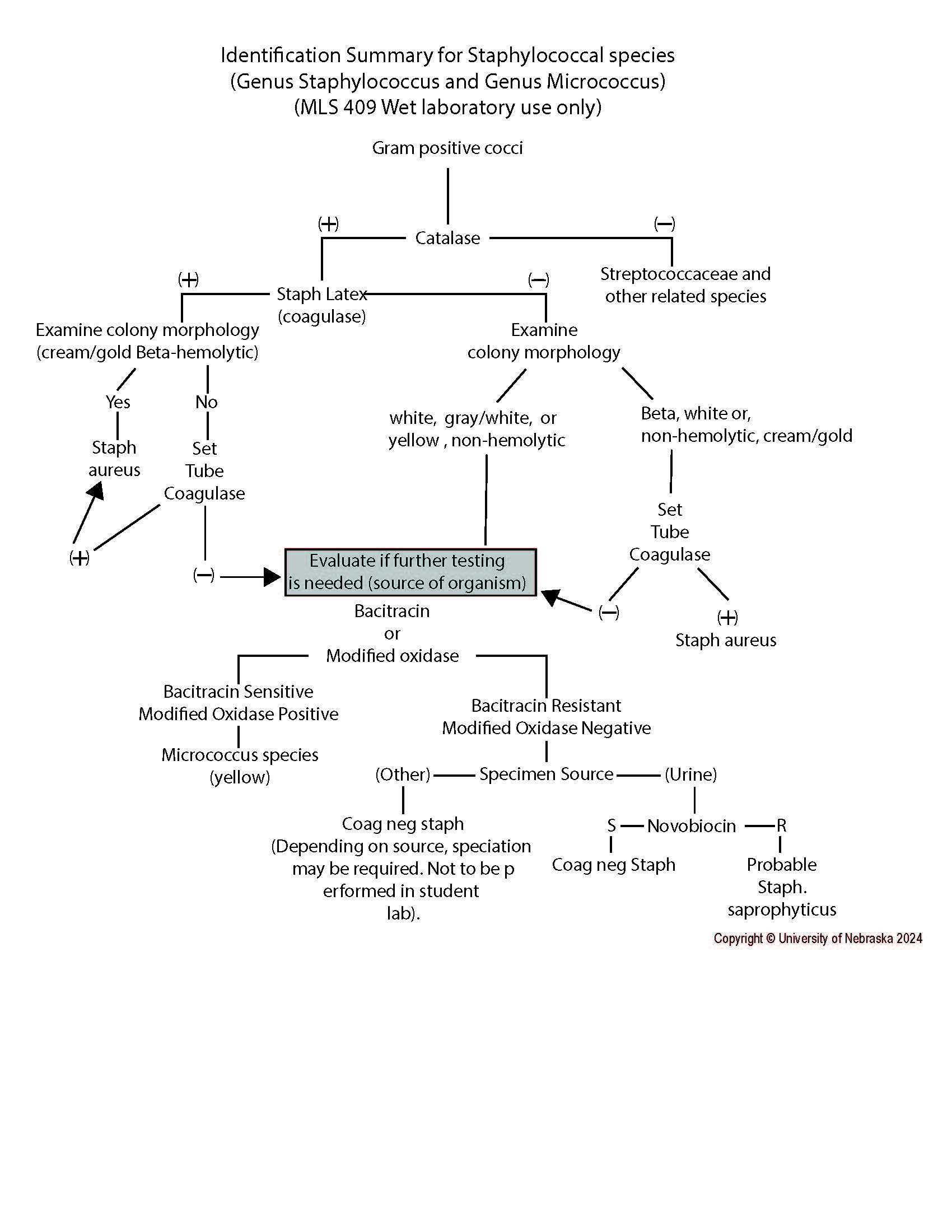
Compare: morphology, novobiocin susceptibility, coagulase production (tube/latex)
S. aureus
S. epidermidis
S. saprophyticus
S. lugdunensis
Micrococcus
| S. aureus | S. epidermidis | S. saprophyticus | S. lugdunensis | Micrococcus |
Morph | cream/gold, beta-hemolytic | gray-white, nonhemolytic | white, creamy, nonhemolytic | very white, nonhemolytic | white/yellow/pink |
Novobiocin | Susceptible | Susceptible | Resistant | Susceptible | N/A |
Coagulase | Positive | Negative | Negative | Negative | Negative |
What does S. aureus cause?
1) Skin infections
2) Pneumonia
3) Osteomyelitis
4) Wound infections
5) Toxin mediated illness
6) Opportunistic infections
What does S. epidermidis cause?
1) Subacute bacterial endocarditis (SBE)
2) Infections associated with prosthetic devices
What does S. saprophyticus cause?
UTIs (especially in young women)
What virulence factors does S. aureus have?
Clumping factor
Coagulase
Fibrinolysin
Hyaluronidase
Beta-lactamase
Exotoxins
Protein A
What is clumping factor?
a VF: surface protein that binds fibrinogen, causing bacteria to clump
Helps adhesion and avoid phagocytosis
What is coagulase?
A VF: converts fibrinogen into fibrin, forming a clot
Protects bacteria from immune cells
What is fibrinolysin/staphylokinase?
A VF: breaks down fibrin clots
Allows bacteria to spread
What is hyaluronidase?
A VF: Breaks down hyaluronic acid in connective tissue
Allows bacteria to spread
What is Protein A?
A VF: binds the Fc portion of IgG antibodies
Prevents antibodies being recognized by phagocytes
What virulence factors does S. epidermidis have?
PGA capsule
Biofilm formation
What is modified oxidase (Microdase) test used for?
To differentiate Micrococcus (pos) from Staphylococcus (neg)
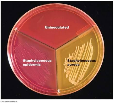
Describe the use of Mannitol Salt agar in the isolation of Staphylococcus aureus.
Staphylococcus thrive in salt, but S. aureus can ferment the mannitol (other Staph cannot)
How would you identify MRSA?
MRSA
Resistance to penicillin, oxacillin, vancomycin, and cefoxitin
Has mecA and PBP2a
Use cefoxitin (30ug) and oxacillin (1ug) disk diffusion tets to predict mecA-mediated resistance
Utilize AST standards
What does Group A (Strep pyogenes) cause?
Pharyngitis
Erysipelas
Impetigo
Scarlet fever
Necrotizing fasciitis
Toxic shock syndrome
Post-streptococcal sequelae (nonsuppurative complications)
Acute glomerulonephritis
Rheumatic fever
Wounds
Opportunistic infections
What does Group B (Strep agalactiae) cause?
Neonatal pneumonia
Neonatal meningitis
Postpartum infections
Urinary tract infections (UTI)
Osteomyelitis
What does Strep bovis group (Group D) cause?
Bacteremia associated with colon malignancy
What does Enterococcus cause?
Urinary tract infections (UTI)
Mixed infections of the abdomen and pelvis
Wounds
What does Viridans strep cause?
Subacute bacterial endocarditis
What does Strep pneumoniae cause?
Pneumonia
Meningitis
Otitis media
Sinusitis
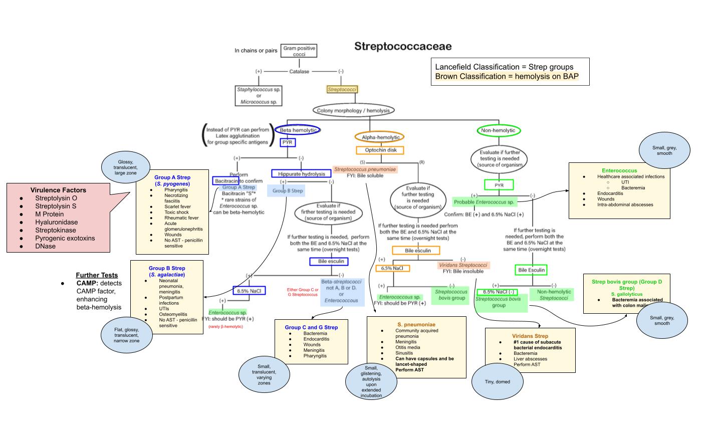
Draw the Streptococcaceae flowchart
Hippurate Hydrolysis
detects ability to hydrolyze hippurate
Bile-esculin
ability to hydrolyze esculin in presence of bile
Bile solubility
bile activates enzymes that lyse susceptible bacteria
What virulence factors does Group A Strep have?
Streptolysin O: oxygen-labile hemolysin, causes tissue damage
Streptolysin S: oxygen-stable hemolysin, responsible for beta-hemolysis
M protein: surface protein that prevents phagocytosis by inhibiting complement
What virulence factor(s) does Strep pnuemoniae have?
Capsule - prevents phagocytosis
For which organism should you perform AST?
Beta-hemolytic strep
Viridians strep
Strep pneumoniae
For virdians and strep pneumoniae
How do you set up culture for urine specimens?
For clean-catch/catheter: use 0.001 loop, streak for colony count, BAP and MAC, ambient, 35C
What are the expected test results from a BactiCard for Neisseria sp and Moraxella catarrhalis?
BactiCard Performed on isolates from enriched selective media only | ||||
| N. gonorrhoeae | N. meningitidis | N. lactamica | M. catarrhalis |
IB Pos: teal | Negative | Negative | Negative | Positive |
PRO Pos: purp | Positive | Variable | N/A | N/A |
GLUT Pos: purp | Negative | Positive | N/A | N/A |
BGAL Pos: teal | Negative | Negative | Positive | N/A |
What is Thayer-Martin media used for?
CHOC agar with added antibiotics (Vancomycin, colistin, nystatin, trimethoprim)
Selective for pathogenic Neisseria sp.
N. gonorrhoeae: small, gray to tan, translucent, raised
N. meningitidis: small, tan, sometimes mucoid, convex
N. lactamica (non-pathogenic, but still grows)
Culture requirements for Neisseriaceae
Transported at room temp, susceptible to drying
Capnophilic (2-8% CO2)
N. gonorrhoeae
Media: CHOC and modified Thayer-Martin
Inoculate with dacron or rayon swabs
N. meningitidis
Media: SBA, CHOC, modified Thayer-Martin
What does Neisseria gonorrhoeae cause?
1) Gonorrhea
2) Ophthalmia neonatorum
3) Pelvic inflammatory disease (PID)
4) Disseminated gonococcal infections (DGI)
What does Neisseria meningitidis cause?
1) Meningitis
2) Septicemia
What does Moraxella catarrhalis cause?
1) Otitis media/maxillary sinusitis in children
2) Pneumonia/bronchitis in immunocompromised
Describe the following characteristics common to the Enterobacterales
GNR / GNCB
Oxidase negative (except Plesiomonas)
Glucose fermenters
Nitrate reduction positive
Growth on MAC + HE/XLD agar
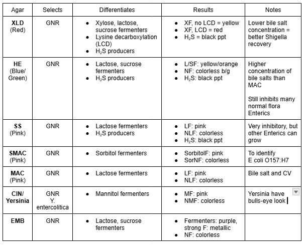
Compare all of the GNR selective agars
Agar | Selects | Differentiates | Results | Notes |
XLD (Red) | GNR |
|
| Lower bile salt concentration = better Shigella recovery |
HE (Blue/ Green) | GNR |
|
| Higher concentration of bile salts than MAC Still inhibits many normal flora Enterics |
SS (Pink) | GNR |
|
| Very inhibitory, but other Enterics can grow |
SMAC (Pink) | GNR |
|
| |
MAC (Pink) | GNR |
|
| Bile salt and CV |
CIN/ Yersinia | GNR Y. entercolitica |
|
| Yersinia have bulls-eye look
|
EMB | GNR |
|
|
What is the purpose of using Gram-negative broth?
Selectively enriched to promote Salmonella and Shigella sp
Clinically significant lactose-fermenting Enterobacterales
Escherichia coli
Klebsiella pneumoniae
Klebsiella oxytoca
Klebsiella (Enterobacter) aerogenes
Enterobacter cloacae
Citrobacter freundii
Clinically significant non-lactose fermenting, LDA/TDA positive, ONPG negative Enterobacterales
Proteus vulgaris
Proteus mirabilis
Morganella morganii
Clinically significant non-lactose fermenting, LDA/TDA negative Enterobacterales
Shigella sonnei
Shigella species
Salmonella species
Salmonella typhi
Serratia species
Citrobacter koseri
Yersinia entercolitica
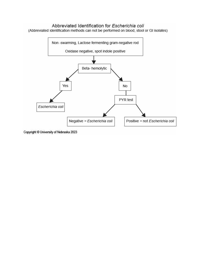
Draw out the abbreviated ID scheme for E. coli
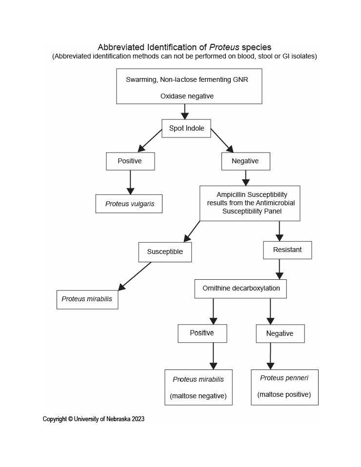
Draw out the abbreviated ID scheme for Proteus sp
What is the only lactose fermenting GNR that also produces H2S?
Citrobacter freundii
Define the following terms:
a. Capsular antigen (K)
b. Somatic antigen (O)
c. Flagellar antigen (H)
d. Vi antigen
These are specific surface structures used to classify and identify bacteria
Capsular antigen (K): surrounds bacterial cell outside the O antigen, protection from phagocytosis and complement
Somatic antigen (O): heat-stable polysaccharide portion of the lipopolysaccharide (LPS) in the outer membrane of GN bacteria
Flagellar antigen (H): heat-labile protein (flagellin) that makes up flagella
Vi antigen: surface capsular polysaccharide antigen that overlies O antigen, inhibiting phagocytosis
Which Enterics are always pathogens?
Shigella sp
Salmonella sp
Yersinia
E. coli O157:H7
What does E. coli cause?
UTI
Wound infections
Hemolytic uremic syndrome (HUS)
Neonatal meningitis
GI infections
What do Salmonella/Shigella sp cause?
GI infections
Typhoid fever (Salmonella typhi)
What do Proteus sp cause?
Wound infections
UTI
Opportunistic/nosocomial infections
What does K. pneumoniae cause?
Opportunistic/nosocomial infections
Pneumonia
Wound infections
What does Yersinia cause?
GI infections
Which Enterics are commonly responsible for opportunistic/nosocomial infections?
K. pneumoniae
Enterobacter cloacae
Serratia sp
Citrobacter sp
Proteus sp
Morganella morganni
Culture setup for Haemophilus
CHOC (fastidious organisms)
X (Hemin) and V (NAD) factor
CO2 (Capnophilic)
Moist environment
Incubation for 24-48 hours
Gram stain and colonial morphology of Haemophilus species.
GNCB
Tan, translucent, mousy odor
H. ducreyi: small, grey, yellow/tan, hockey-puck
Describe the difference in test results between H. influenzae and H. parainfluenzae
(X/Y test; Porphyrin; Horse blood; Satellitism)
Influenzae | Parainfluenzae | |
X/Y Test (Growth) | XV | V & XV |
Porphyrin | Negative | Positive |
Horse Blood | No | No |
Satellitism | Positive | Positive |
What is H. influenzae’s most significant virulence factor?
Capsule
Encapsulated strains, especially Hib cause severe invasive infections, especially in unvaccinated children younger than 5
Un-encapsulated species can still cause disease
Localized infections (elderly and immunocompromised)
What kind of diseases do H. influenzae cause?
Respiratory infections
Meningitis
Otitis media
Conjunctivitis (ssp. aegypticus
Fill in this table for Glucose NF GNRs
| Oxidase | 42C | ADH | ODC | LDC | Gelatin | Polymyxin B |
P. aeruginosa | |||||||
S. maltophilia | |||||||
B. cepacia |
| Oxidase | 42C | ADH | ODC | LDC | Gelatin | Polymyxin B |
P. aeruginosa | + | Growth | + | - | - | +/- | Susceptible |
S. maltophilia | - | No | - | - | + | + | Susceptible |
B. cepacia | +/- | +/- | - | +/- | + | +/- | Resistant |
What are the glucose non-fermenting GNRs?
P. aeruginosa
S. maltophilia
B. cepacia
Why are P. aeruginosa and S. maltophilia not considered Enterobacterales?
They are:
Oxidase positive
Glucose utilizers, not fermenters
Compare the morphologies of P. aeruginosa vs. S. maltophilia
Characteristic | Pseudomonas aeruginosa | Stenotrophomonas maltophilia |
|---|---|---|
Colony morphology | Large, flat, spreading colonies; may have irregular edges, metallic sheen, and beta-hemolysis | Smooth, glistening colonies that may appear yellow to lavender-green; usually nonhemolytic |
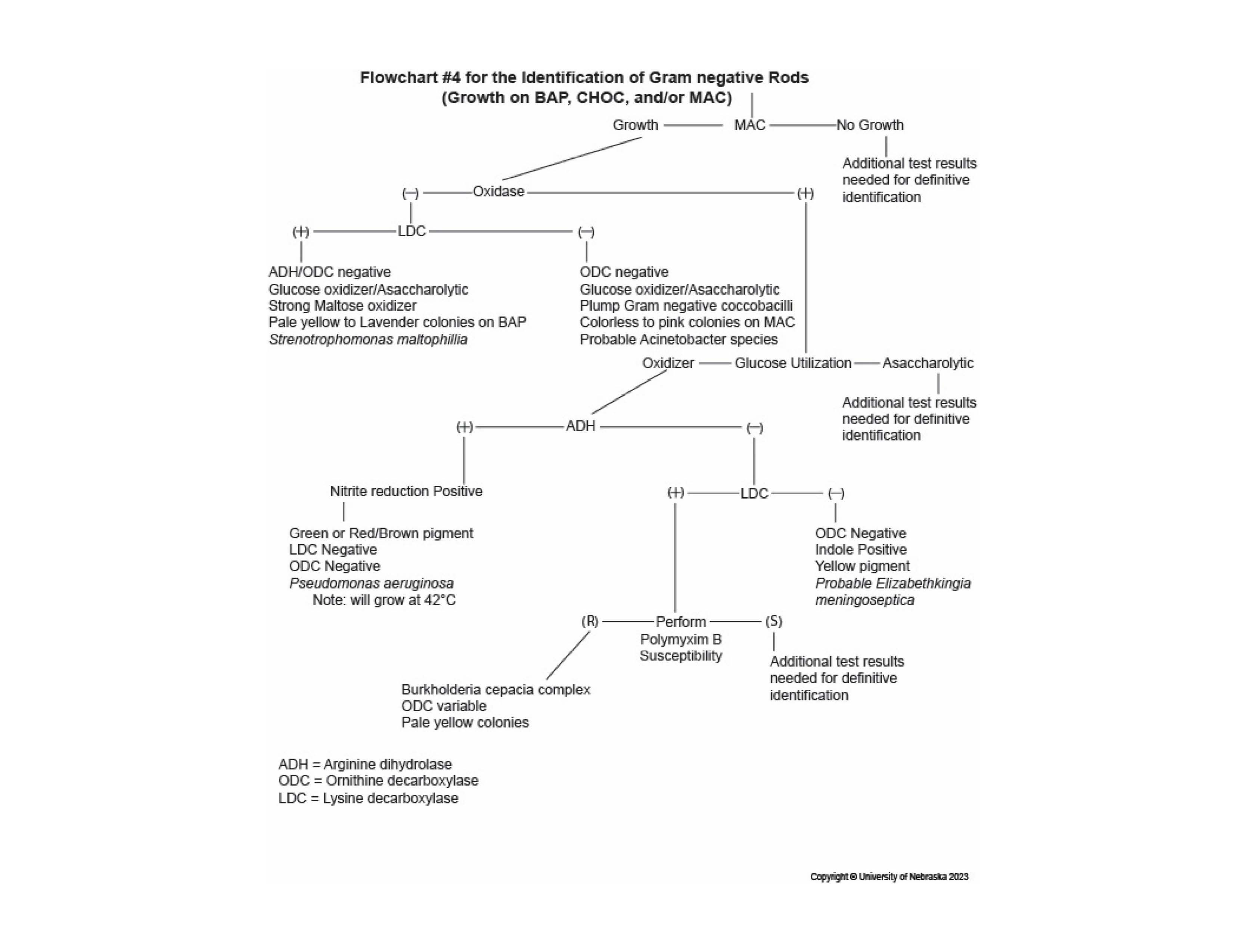
Draw the flowchart for GNF GNR
GNF GNRs are primarily ____ pathogens
opportunistic
What kind of diseases can P. aeruginosa cause?
Nosocomial infections
Wounds, burns, respiratory
Otitis externa
Corneal ulcers
UTI
Cystic fibrosis
Osteomyelitis
What kind of diseases can S. maltophilia cause?
Respiratory
Wound
Nosocomial
What is the difference between culture set up for a throat vs sputum specimen?
SPUTUM
BAP, CHOC, MAC
THROAT
Strep selective blood agar (anaerobic)
To find Group A strep only, streptolysin
Both require a direct gram stain, streak for isolation, 48 hours minimum
Pathogens vs normal flora of the respiratory tracts
Flora (upper)
Viridans strep
CoagN Staph
Nonpathogenic Neisseria
Diphtheroids
Anaerobes
Yeast
Pathogens (lower; normally sterile)
S. pneumoniae
H. influenzae
M. catarrhalis
K. pneumoniae
E. coli
Enterobacter spp
Serratia marcescens,
S. aureus
P. aeruginosa
S. maltophilia
Acinetobacter sp
S. pyogenes
B. cepacia
Should you perform a gram stain or acridine orange stain on sterile body fluids/tissues?
A gram stain should be performed ASAP, only to guide empiric treatment
Acridine orange can be used when gram stain is negative, but infection is suspected or the specimen contains few organisms (supplemental to gram stain)
Describe the proper procedure for setting up each of the following cultures, including proper specimen preparation, types of media used, and incubation requirements:
Cerebrospinal fluid
Sterile body fluids
Solid tissues
Specimen | Preparation | Media | Incubation |
|---|---|---|---|
CSF | Process ASAP; never refrigerate. >1 mL: centrifuge; <1 mL: plate directly | BAP, CHOC, MAC, Thio | 35°C; aerobic: 48 hr; Thio: 7 days |
Sterile body fluids | Process ASAP. >1 mL: centrifuge; <1 mL: plate directly. Use anaerobic transport if needed | BAP, CHOC, MAC, Thio ± anaerobic media | 35°C; aerobic: 48 hr; anaerobic: 72 hr; Thio: 7 days |
Solid tissues | Keep moist; touch prep, then grind/homogenize. If fungi suspected: mince, do not grind | BAP, CHOC, MAC, Thio ± anaerobic media | 35°C; aerobic: 48 hr; anaerobic: 72 hr; Thio: 7 days |
Where are obligate anaerobes are found in the environment and the human body?
Location | Examples |
|---|---|
Environment | Soil, deep water sediments, sewage, and decaying organic material |
Body | Mouth, colon, vagina, and deep skin |
Predisposing Conditions of Anaerobic Infections
Predisposing Conditions
Trauma of mucosal membranes
Vascular stasis
Tissue necrosis
Aspiration of oral contents into lungs
Decrease in redox potential of tissues
What types of specimens may be submitted for anaerobic cultures? What can you collect them in?
Tissues
Sterile body fluids
Blood and bone marrow
Suprapubic/Lung/Transtracheal aspirate
Wound drainage
Collect with:
Anaerobic transport tubes
Biobag
Syringe with cap
Blood culture bottles
Process immediately in a BSC or anaerobic chamber
Signs of an anaerobic infection
Purulent specimen
Gas, foul odors
Black exudate w/ possible brick red fluorescence
Sulfur granules
Necrosis or gangrene
Infections close to mucosal surfaces
Infection persists despite aminoglycoside therapy
Characteristics morphology: double zone of hemolysis, molar tooth/breadcrumb colony, ground glass/fried egg colony
What is the purpose of using ABAP?
Isolate and grow obligate anaerobes
Enriched with sheep blood, hemin, and vitamin K
What is the purpose of pre-reduced media?
Pre-reduced media have oxygen removed to grow anaerobes
Aerotolerance testing
Subculture a suspected anaerobic colony to BAP, CHOC, and AnaBAP to prove colony is true obligate anaerobe vs facultative anaerobe
CHOC | AnaBAP | Obligate Anaerobe |
Yes | Yes | No |
No | Yes | Yes |
What is the most common anaerobic organism isolated in the clinical lab?
Bacteroides fragilis
What are examples of Gram-positive Bacilli? (GPR) + GS morphology, catalase reaction, oxygen requirements
Bacillus sp - spore forming
Corynebacterium sp - irregular, palisading
Listeria monocytogenes - regular, small
All are catalase positive and facultative anaerobes
Compare GS, colony morphology, and key tests for different GPRs
Organism | Gram stain | Colony | Key tests |
|---|---|---|---|
B. anthracis | Large GP rods with spores | Nonhemolytic | Nonmotile, catalase + |
Other Bacillus | Large GP rods with spores | Usually beta-hemolytic | Usually motile, catalase + |
Diphtheroids | Irregular GP rods; Chinese letters | Small, usually nonhemolytic | Nonmotile catalase + |
L. monocytogenes | Small GP rods | Narrow beta-hemolysis | Motile hippurate + catalase + |
What diseases does Listeria monocytogenes cause?
Meningitis
Listeriosis
Should you direct gram stain a wound culture?
Yes, correlate with culture
Describe the procedure for setting up a wound culture using the following specimens:
Aerobic swab
Aerobic swab and anaerobic transport system
Aspiration
Inoculate with swab, streak for isolation
Aerobic plates
Chocolate agar (CHOC)
Sheep blood agar (BAP)
PEA or CNA
MacConkey agar (MAC)
Anaerobic plates
Supplemented anaerobic blood agar (ABAP)
Kanamycin, Vancomycin (KV) agar
Anaerobic PEA (APEA)
Thioglycollate broth (THIO)
Incubate media
Temperature: 35ºC
Atmosphere:
BAP, CHOC, CNA, PEA - CO2
MAC, THIO - ambient air
ABAP, KV agar, and Ana PEA - anaerobically
Time:
Aerobic media: total 48 hours
Anaerobic: read at 48 hours, 72 hours total or until ID complete
Thioglycollate broth: typically 5 to 7 days or longer if serious infection suspected (2 weeks)
What is Campylobacter blood agar (CVA) or Skirrow blood agar used for?
Selects for Campylobacter sp from stool
Incubated under microaerophilic, capnophilic conditions at 42C for 48-72 hours
Contains blood and antibiotics (cefoperazone, vancomycin, and amphotericin B) to inhibit other organisms
Colony: Small, gray, moist colonies that may appear flat, spreading, or “water-droplet-like”
Describe identifying characteristics of Campylobacter jejuni and what diseases it causes
Growth on CVA/Skirrow agar
Microaerophilic, capnophilic
Gram-negative thin, curved rods
Only grows at 37C or 42C
Gray, moist, flat, spreading
Catalase: positive
Oxidase: positive
Sodium Hippurate: positive
Motility: positive
Causes: gastroenteritis
What media should you use for a stool culture?
Salmonella/Shigella species
Campylobacter jejuni
Escherichia coli O157:H7
Yersinia enterocolitica
Staphylococcus aureus
Salmonella/Shigella species: MAC, HE/XLD
Campylobacter jejuni: CVA/Skirrow (42C)
Escherichia coli O157:H7: SMAC
Yersinia enterocolitica: CIN (incubate at 25C)
Staphylococcus aureus, Plesiomonas shigelloides, Aeromonas hydrophila : BAP