neck, back and thorax COMPLETE
1/245
Earn XP
Description and Tags
Name | Mastery | Learn | Test | Matching | Spaced | Call with Kai |
|---|
No analytics yet
Send a link to your students to track their progress
246 Terms
Regio scapularis is a … region.
back
The … take part in the formation of cervical plexus.
anterior rami of C1 through C4
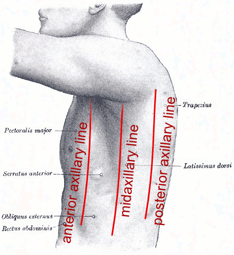
The boundaries between the thoracic region and the back are the … .
midaxillary line
mcq said anterior axillary line and this is false

The boundaries between the thoracic region and the chest are the … .
anterior axillary lines
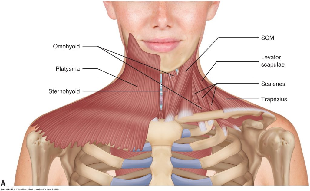
Sternocleidomastoid is a superficial muscle of the … helping in … .
neck
head rotation and flexion
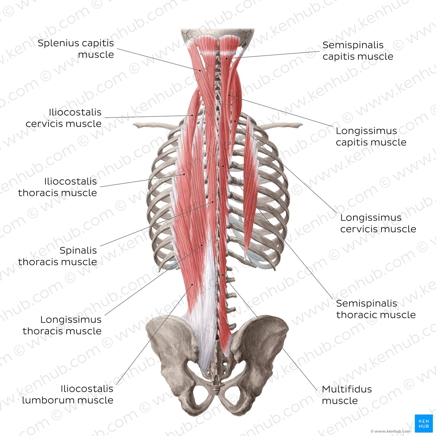
M. latissimus dorsi is a powerful … .
extensor of the arm
Superficial muscles of the back are supplied by … of spinal nerves.
posterior rami
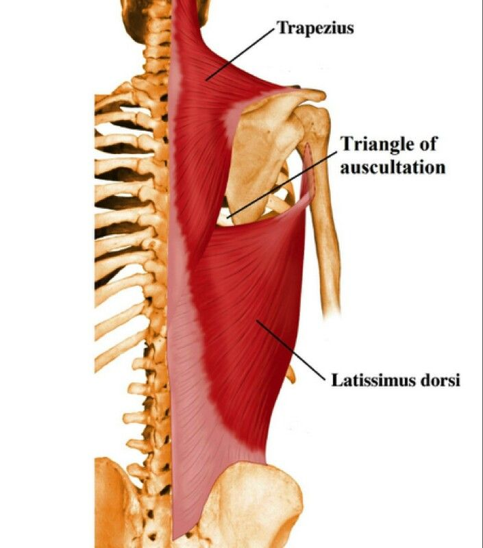
Auscultation triangle on the back is located … .
medial to the scapula
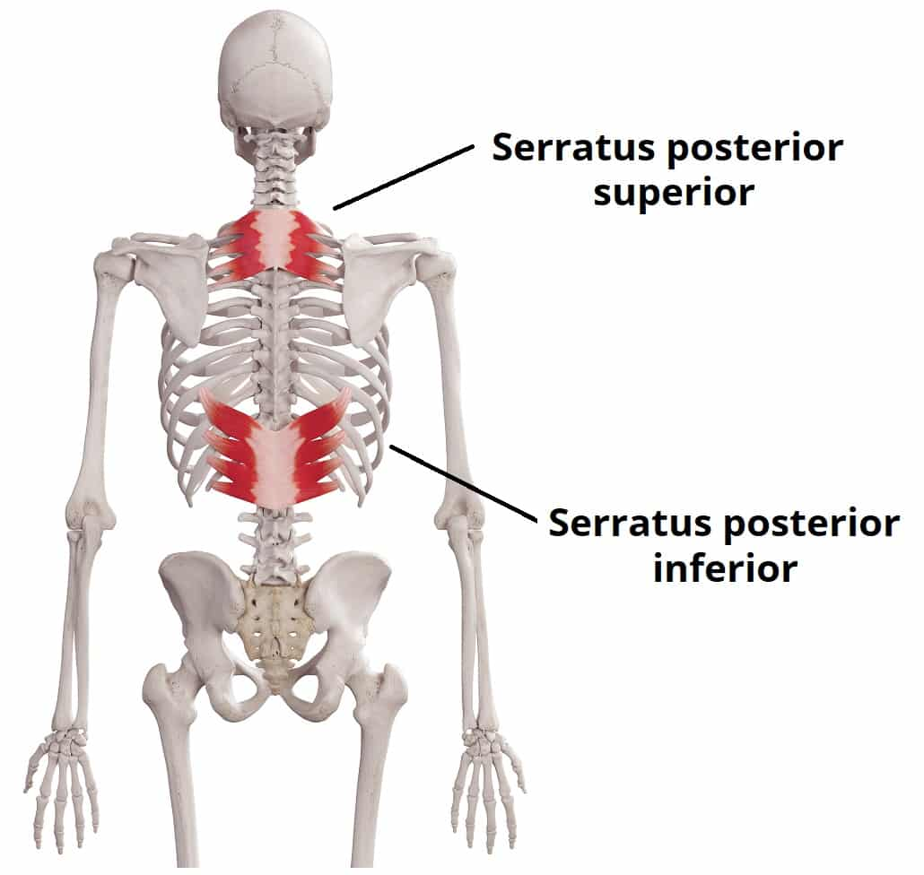
Serratus posterior superior muscle is a muscle of … .
inspiration
Muscles of the back are arranged in … groups with distinct functions
three
superficial (move the upper limbs)
intermediate (respiratory)
deep (support the spine and posture)
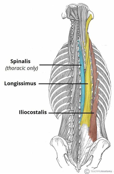
Erector spinae muscle is made of … columns.
three
iliocostalis
longissimus
spinalis
Intermediate muscles of the back are … muscles.
respiratory
Platysma is innervated by a branch of the … .
facial nerve
The platysma is a superficial muscle that overlaps the … .
sternocleidomastoid
Accessory nerve (cranial nerve ?) is a branch of … .
cranial nerve 11
It has two parts:
A spinal root (from the upper cervical spinal cord)
A cranial root ( Originates from the medulla oblongata, which briefly joins the vagus nerve)
NOT cervical plexus
IS from the spinal cord and medulla oblongata and innervates the sternocleidomastoid and trapezius muscles
Phrenic nerve (C3-C5 (primarily C4))-innervates … .
thoracic diaphragm
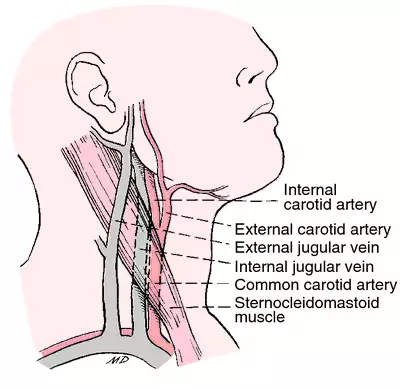
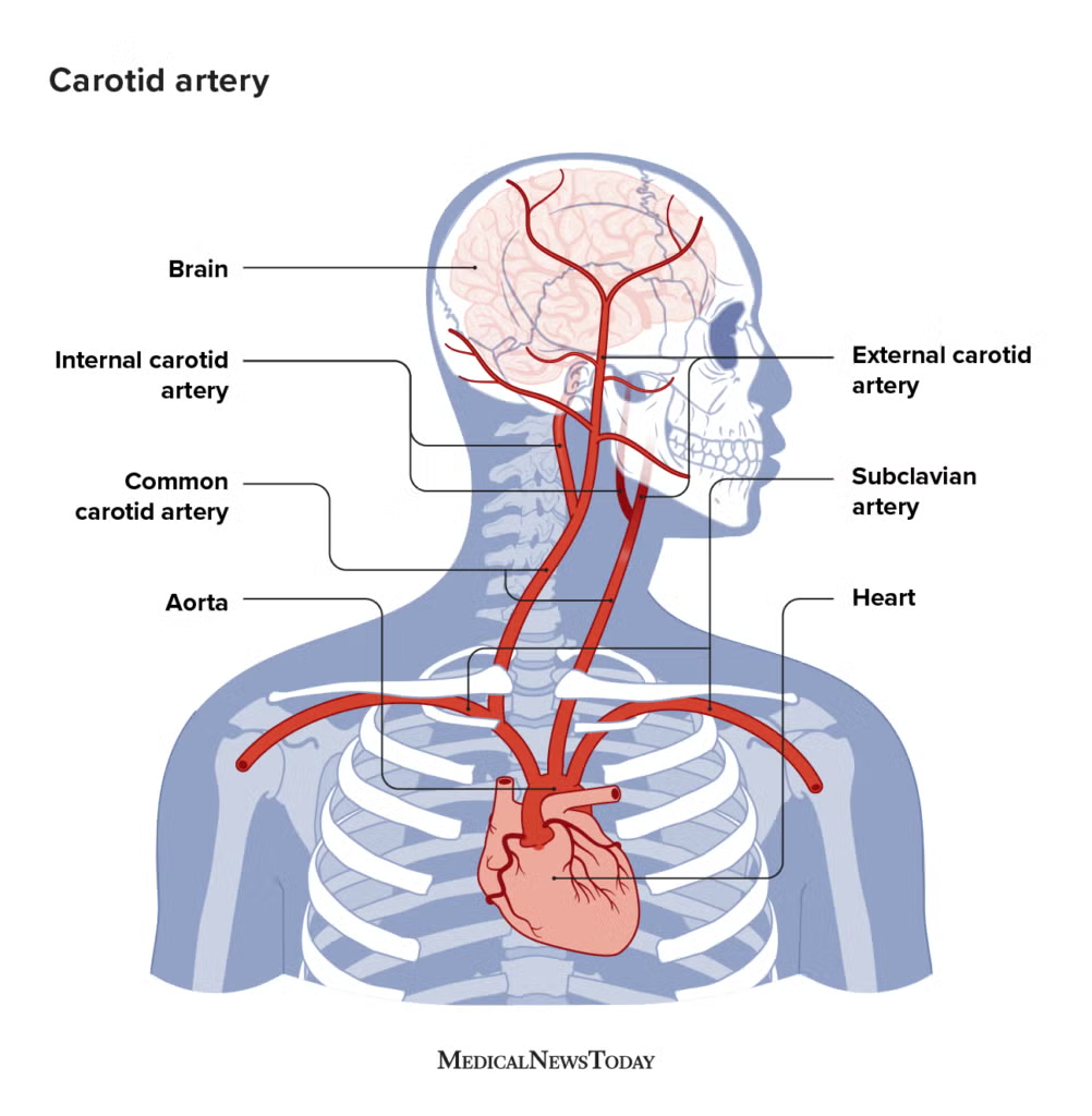
Internal carotid artery has two cervical branches.
A. Yes
B. No
and supplies what?
Internal carotid artery has two cervical branches.
B. No
internal carotid artery typically has no branches in the neck (cervical region) - enters skull thru carotid canal
mainly supplies structures inside the skull
internal carotid artery supplies the brain, eyes, orbit, and parts of the forehead and nose
via its intracranial branches (like the ophthalmic artery, anterior cerebral artery, middle cerebral artery)
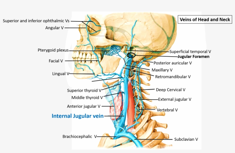
The upper end of internal jugular vein dilates into … .
internal jugular fossa
superior bulb?
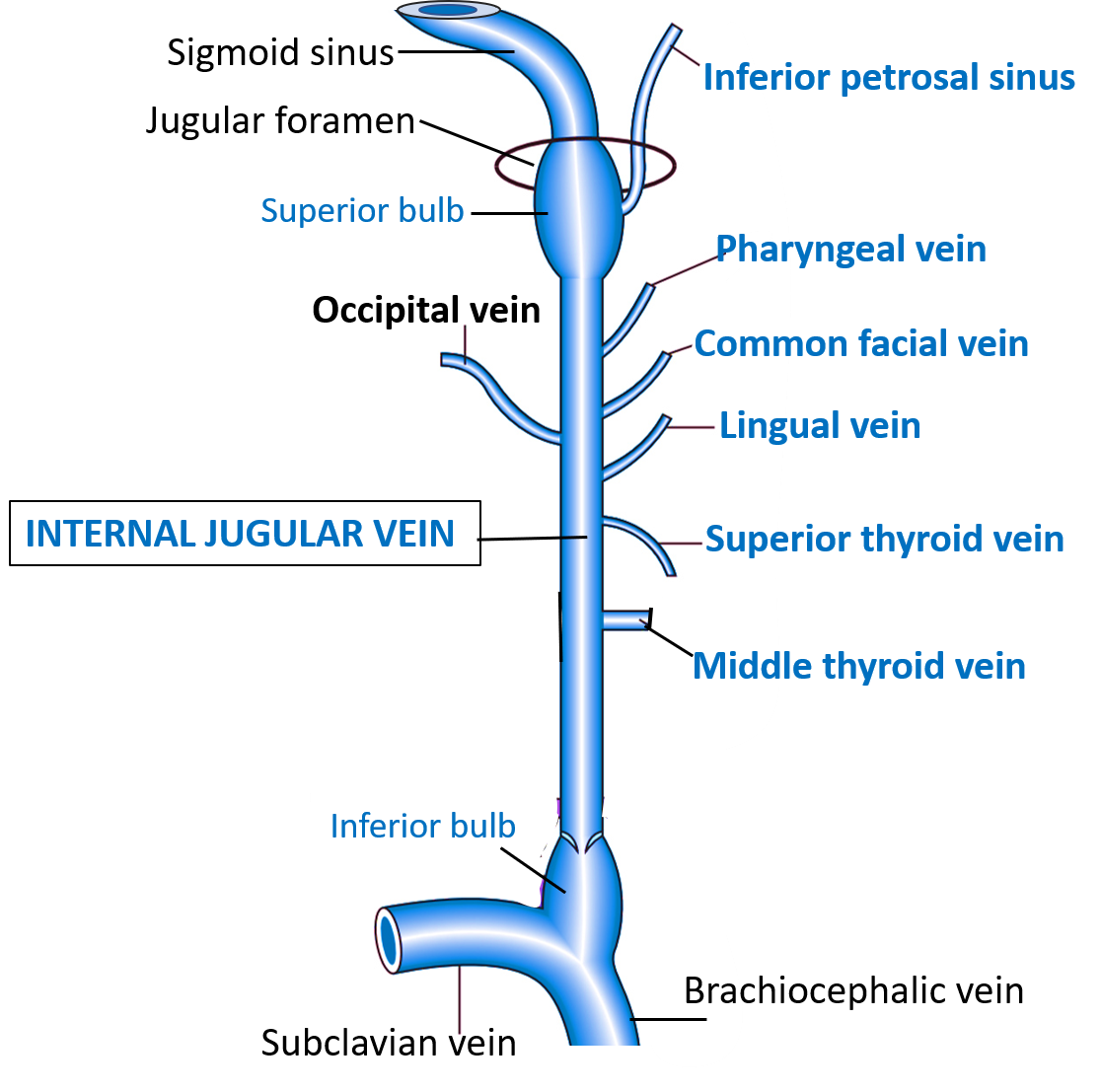
Near the termination of the internal jugular vein is… .
a smaller dilatation, the inferior bulb
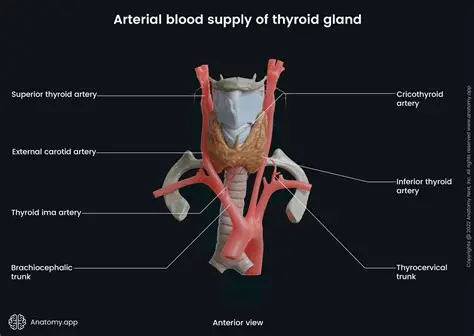
The inferior thyroid artery arises from …?
NOT a branch of the external carotid artery
arises from the thyrocervical trunk
supplies the thyroid gland
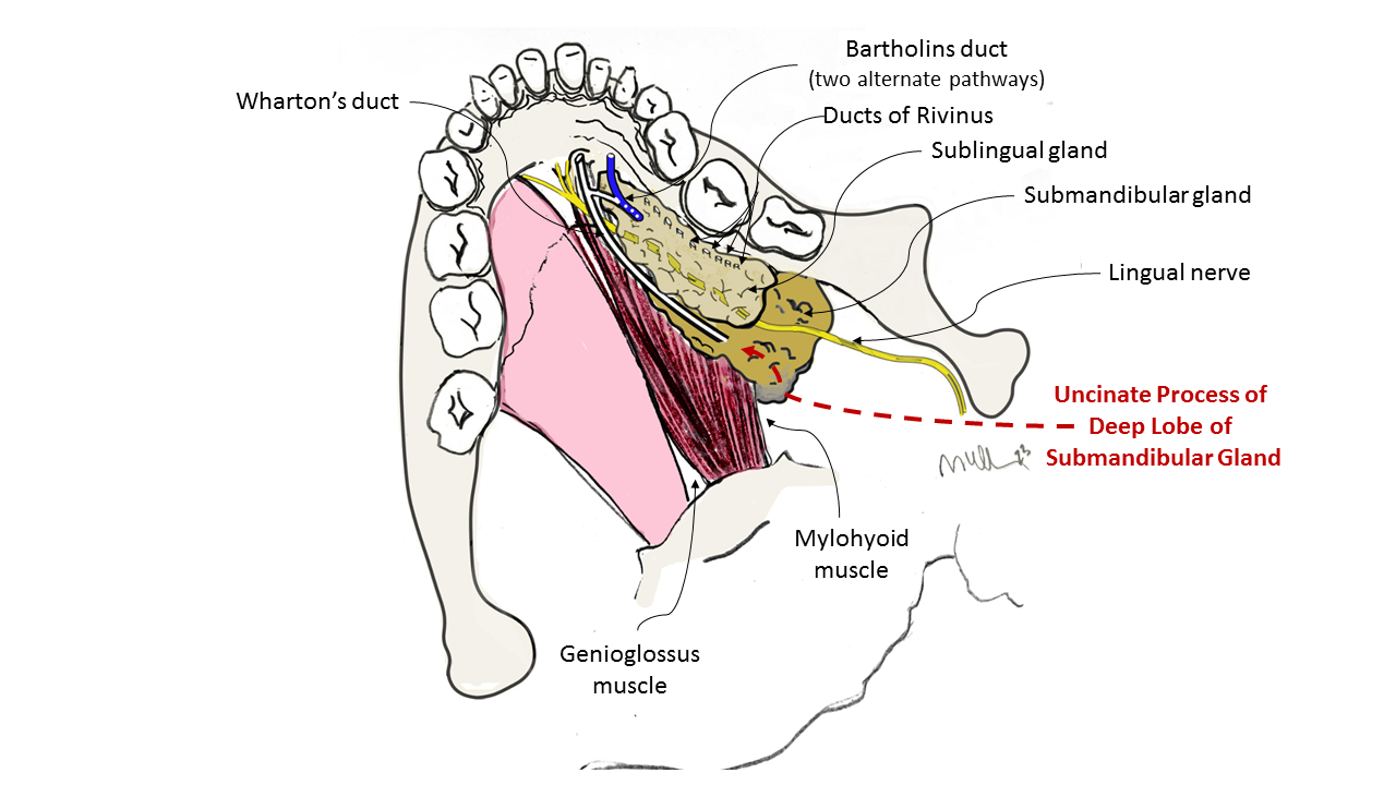
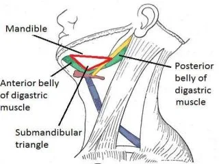
The submandibular gland is in located … .
beneath the lower jaw - SUBMANDIBULAR TRIANGLE (of neck)
NOT infrahyoid region BECAUSE THIS IS WHERE THE THYROID GLAND IS
Which one of the listed is not a muscle of the back:
A. M. serratus posterior superior
B. M. serratus anterior
C. M. iliocostalis
D. M. longissimus
E. M. spinalis
Which one of the listed is not a muscle of the back:
A. M. serratus posterior superior
B. M. serratus anterior
C. M. iliocostalis
D. M. longissimus
E. M. spinalis
Which of the muscles listed below is a deep muscle of the back
A. Levator costae
B. Latissimus dorsi
C. Levator scapulae
D. Rhomboidei
E. Splenius
Which of the muscles listed below is a deep muscle of the back
A. Levator costae - deep
B. Latissimus dorsi - superficial
C. Levator scapulae - superficial but deeper than B.
D. Rhomboidei - superficial but deeper than B.
E. Splenius
Interruption of cranial nerve XI would paralyze which muscle?
A. deltoid
B. latissimus dorsi
C. levator scapulae
D. rhomboideus major
E. trapezius
Interruption of cranial nerve XI would paralyze which muscle? ACCESSORY NERVE
A. deltoid
B. latissimus dorsi
C. levator scapulae
D. rhomboideus major
E. trapezius OR STERNOCLEIDOMASTOID
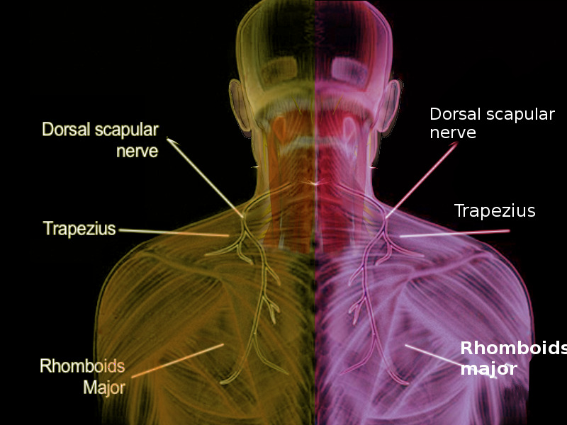
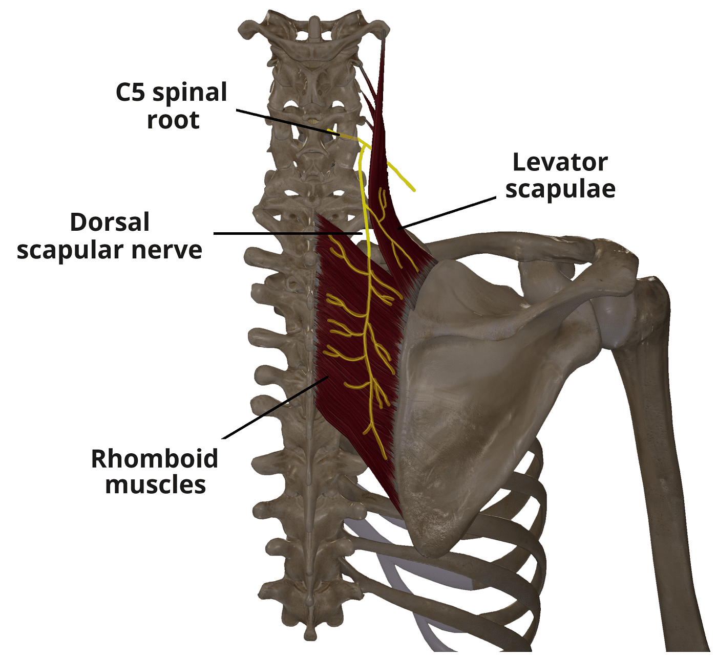
If the right dorsal scapular nerve was cut near its origin, what would result:
A. Skin of the upper back on the right side would be numb
B. The point of the right shoulder would droop
C. Scapular retraction on the right would be weakened
D. Extension of the right arm would be weakened
E. Inability to adduct the right arm
If the right dorsal scapular nerve was cut near its origin, what would result:
A. Skin of the upper back on the right side would be numb
B. The point of the right shoulder would droop
C. Scapular retraction on the right would be weakened
right dorsal scapular nerve → rhomboid major and minor muscles + levator scapulae
responsible for movements like:
scapular retraction (pulling the shoulder blades together)
elevation of the scapula
D. Extension of the right arm would be weakened
E. Inability to adduct the right arm
The cutaneous branch of the posterior primary ramus of C2 is called the:
A. Accessory nerve
B. Great auricular nerve
C. Greater occipital nerve
D. Lesser occipital nerve
E. Superior ramus of the ansa cervicalis
The cutaneous branch of the posterior primary ramus of C2 is called the:
A. Accessory nerve
B. Great auricular nerve
C. Greater occipital nerve
emerges between the first and second cervical vertebrae
travels superiorly to provide sensory innervation to the posterior scalp and upper neck
sensory innervation of the back of the head
The greater occipital nerve is the cutaneous branch of the posterior primary ramus of C2.
It supplies sensation to the back of the scalp up to the vertex.
It can be involved in conditions like occipital neuralgia, causing headaches at the back of the head
The greater occipital nerve is the skin (cutaneous) nerve that comes from the back branch (posterior ramus) of the C2 spinal nerve, and it supplies sensation to the back of the scalp
D. Lesser occipital nerve
E. Superior ramus of the ansa cervicalis
Which muscle is innervated by posterior primary rami?
A. Latissimus dorsi
B. Levator scapulae
C. Rhomboideus major
D. Erector spinae
E. Trapezius
Which muscle is innervated by posterior primary rami? - THIS RAMI IS ONLY FOR DEEP MUSCLES
A. Latissimus dorsi - THORACODORSAL NERVE
B. Levator scapulae - DORSAL SCAPULAR NERVE
C. Rhomboideus major - NO, BY THE DORSAL SCAPULAR NERVE
D. Erector spinae OR LONGISSIMUS
E. Trapezius - ACCESSORY NERVE
THE OTHERS ARE SUPERFICIAL AND INNERVATED BY VENTRAL RAMI
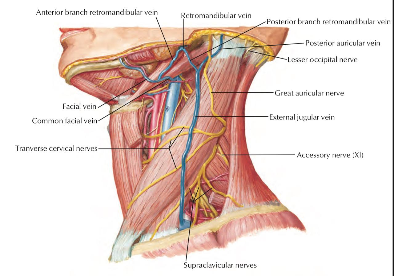
Which of the elements listed below is not in the subcutaneous layer of the neck?
A. M. platysma
B. V. jugularis anterior
C. V. jugularis externa
D. Plexus cervicalis
E. Transverse cervical nerve
Which of the elements listed below is not in the subcutaneous layer of the neck?
A. M. platysma
B. V. jugularis anterior
C. V. jugularis externa
D. Plexus cervicalis OR ANSA CERVICALIS
E. Transverse cervical nerve
F. GREATER AURICULAR NERVE
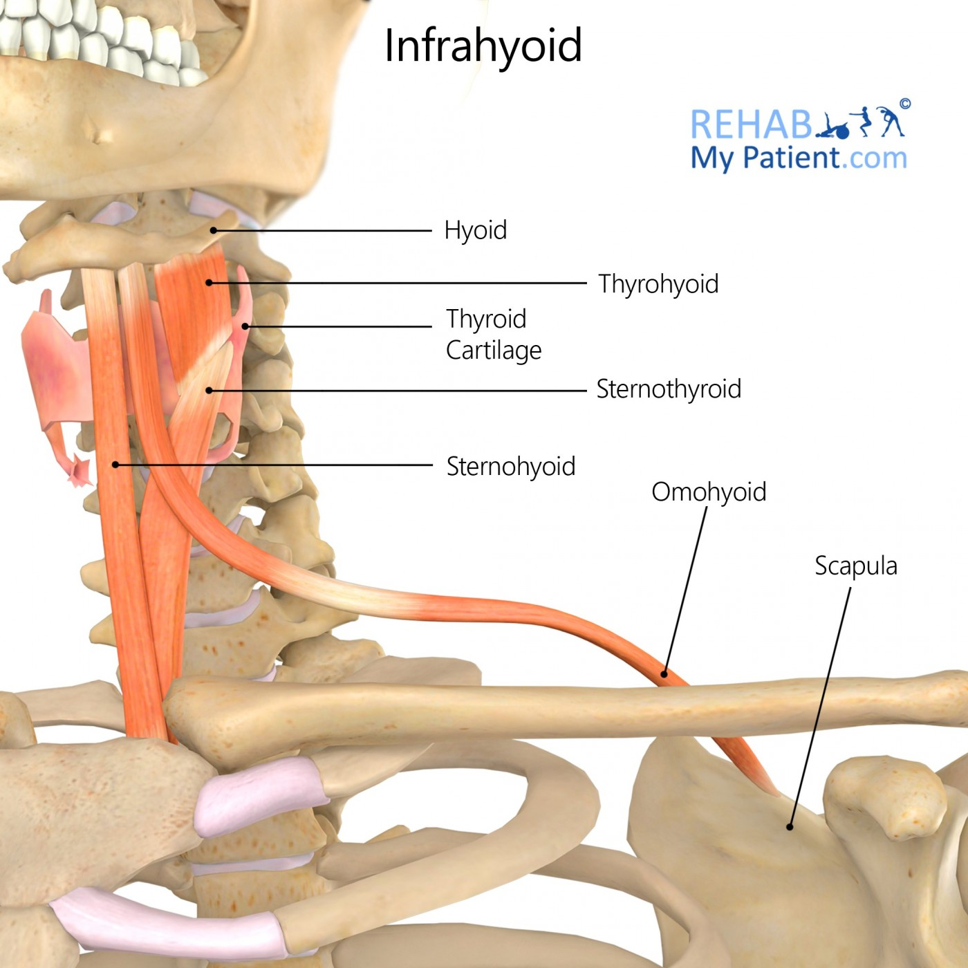
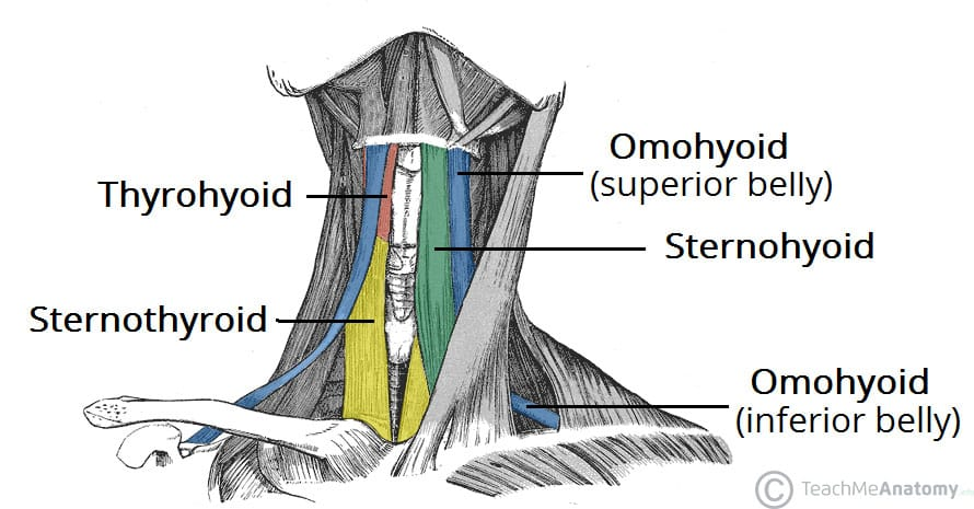
Which of the following does not belong to the infrahyoid muscles?
A. M. sternothyroideus
B. M. omohyoideus
C. M. sternocleidomastoideus
D. M. sternohyoideus
E. M. thyrohyoideus
Which of the following does not belong to the infrahyoid muscles?
A. M. sternothyroideus
B. M. omohyoideus
C. M. sternocleidomastoideus
D. M. sternohyoideus
E. M. thyrohyoideus
NOT SCALENUS ANTERIOR M. EITHER
Which one of the following structures is NOT related to infrahyoid region?
A. gl. thyroidea
B. m. thyrohyoideus
C. n. vagus
D. m. cricothyroideus
E. v. jugularis anterior
Which one of the following structures is NOT related to infrahyoid region?
A. gl. thyroidea
B. m. thyrohyoideus
C. n. vagus
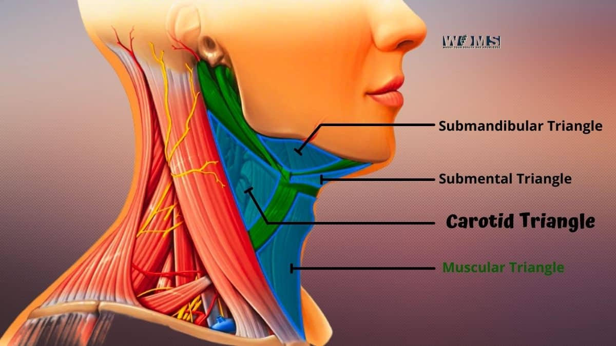
The infrahyoid region (also known as the anterior cervical region or muscular triangle) contains structures located below the hyoid bone. It includes:
Infrahyoid muscles:
m. sternohyoideus
m. omohyoideus
m. sternothyroideus
m. thyrohyoideus
Gl. thyroidea (thyroid gland)
v. jugularis anterior
m. cricothyroideus
These are all found in or near the midline of the anterior neck
runs deep in the carotid sheath along with the common carotid artery and internal jugular vein, which places it in the carotid triangle
D. m. cricothyroideus
E. v. jugularis anterior
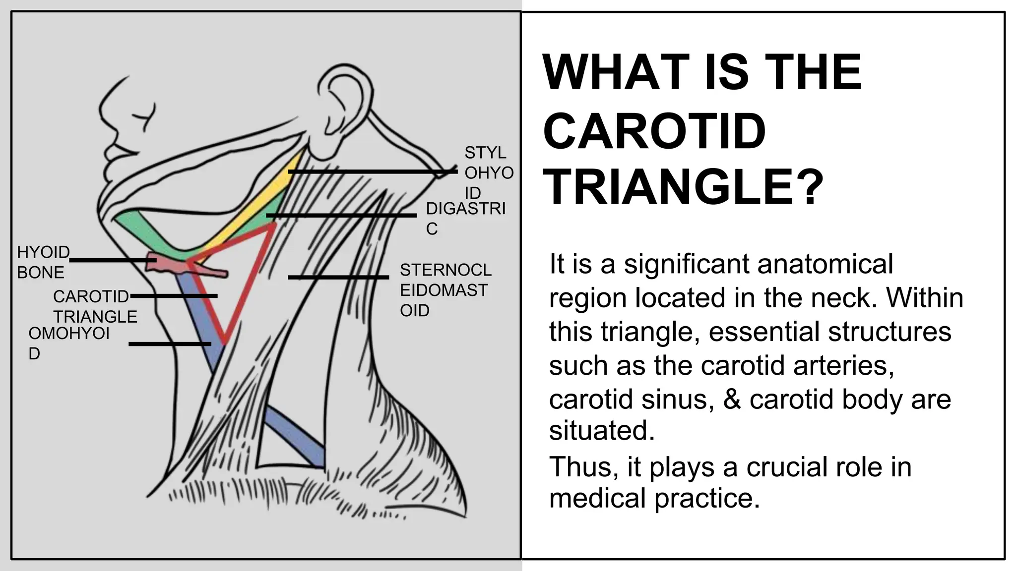
Which one of the following structures is not related to the carotid triangle?
A. hypoglossal nerve
B. superior laryngeal nerve
C. facial artery
D. thyrohyoid muscle
E. sternohyoid muscle
Which one of the following structures is not related to the carotid triangle?
A. hypoglossal nerve
B. superior laryngeal nerve
C. facial artery
and vein, as they branch from the external carotid
D. thyrohyoid muscle
superiorly, near the hyoid bone
E. sternohyoid muscle
located more medially and anteriorly, in the muscular triangle
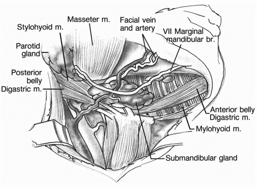
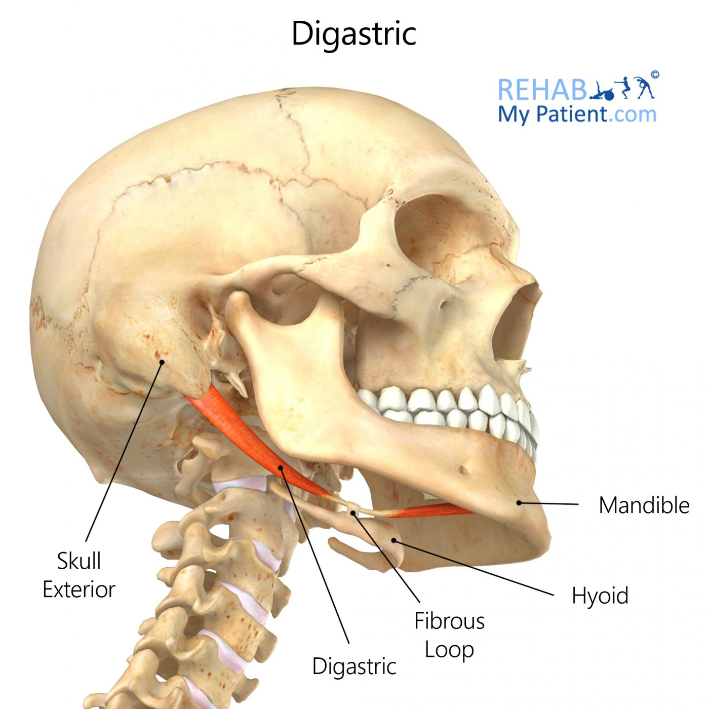
To study the compensatory response of selective suprahyoid muscles in elevating the hyoid bone, an experiment was designed in which the posterior belly of the digastric and stylohyoid muscles were paralyzed by drugs. The muscular branches of which of the following nerves must be chemically interrupted to produce paralysis in both muscles?
A. inferior alveolar
B. facial
C. hypoglossal
D. glossopharyngeal
E. lingual
To study the compensatory response of selective suprahyoid muscles in elevating the hyoid bone, an experiment was designed in which the posterior belly of the digastric and stylohyoid muscles were paralyzed by drugs. The muscular branches of which of the following nerves must be chemically interrupted to produce paralysis in both muscles?
A. inferior alveolar
branch of the mandibular division of CN V (V3); it gives motor fibers to the mylohyoid and anterior belly of digastric
B. facial
To paralyze the posterior belly of the digastric and the stylohyoid muscles, you must interrupt the motor branches of the facial nerve (cranial nerve VII), because:
Posterior belly of digastric is innervated by a branch of the facial nerve
Stylohyoid muscle is also innervated by the facial nerve
C. hypoglossal
Hypoglossal: Supplies intrinsic and extrinsic muscles of the tongue, not suprahyoid muscles like stylohyoid or posterior digastric
D. glossopharyngeal
Involved in innervating the stylopharyngeus and sensory functions
E. lingual
sensory branch of V3 - mandibular division of trigeminal nerve
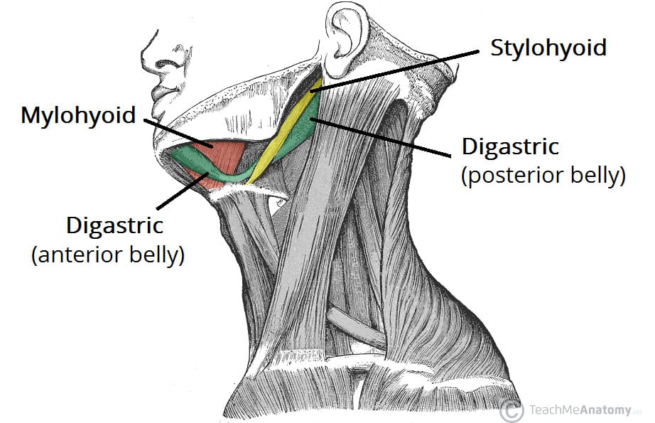
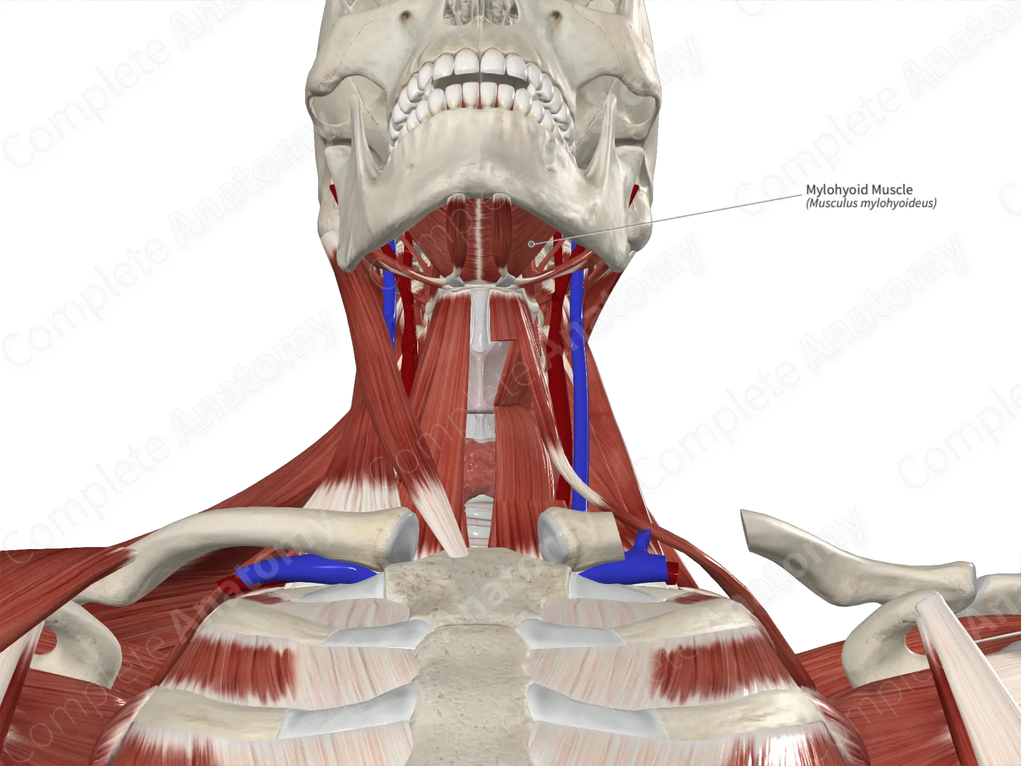
The muscle which separates the submandibular triangle from the paralingual space is the:
A. Digastric, posterior belly
B. Hyoglossus
C. Mylohyoid
D. Stylohyoid
E. Styloglossus
The muscle which separates the submandibular triangle from the paralingual space is the:
A. Digastric, posterior belly
B. Hyoglossus
C. Mylohyoid
D. Stylohyoid
E. Styloglossus
What bony feature of the mandible can be used to find and palpate the facial artery?
A. Oblique line
B. Mental trigone
C. Angle
D. Premasseteric notch
What bony feature of the mandible can be used to find and palpate the facial artery?
A. Oblique line
A ridge on the mandible, not related to the artery’s palpation point
B. Mental trigone
triangular area over the chin, not where the artery crosses.
C. Angle
Located posteriorly, not the common crossing site of the facial artery.
D. Premasseteric notch
facial artery winds around the inferior border of the mandible, typically at the premasseteric notch
small depression located just anterior to the masseter muscle, near the anterior border of the masseter.
Which of the following suprahyoid muscles would be paralyzed if the inferior alveolar nerve was severed at its origin?
A. Geniohyoid m.
B. Hyoglossus m.
C. Mylohyoid m.
D. Stylohyoid m.
Which of the following suprahyoid muscles would be paralyzed if the inferior alveolar nerve was severed at its origin?
A. Geniohyoid m.
innervated by C1 fibers via the hypoglossal nerve (CN XII)
B. Hyoglossus m.
innervated by hypoglossal nerve (CN XII)
C. Mylohyoid m.
inferior alveolar nerve, a branch of the mandibular division (V3) of the trigeminal nerve, gives off a branch called the nerve to mylohyoid before entering the mandibular foramen. This nerve to mylohyoid innervates:
Mylohyoid muscle
Anterior belly of the digastric muscle
D. Stylohyoid m.
innervated by the facial nerve (CN VII)
In accessing the submandibular gland in the submandibular triangle, what vessel coursing through the gland and triangle would need to be protected?
A. External jugular vein
B. Facial artery
C. Maxillary artery
D. Retromandibular vein
E. Superior thyroid artery
In accessing the submandibular gland in the submandibular triangle, what vessel coursing through the gland and triangle would need to be protected?
A. External jugular vein
B. Facial artery
C. Maxillary artery
D. Retromandibular vein
E. Superior thyroid artery
T/F Lamina superficialis of the deep cervical fascia
A. Covers entire neck
B. Forms fascia masseterica
C. Extends from the skull base to the bodies of T3-T4
D. Forms fascia of submandibular gland
E. Extends posteriorly to proc. transversi
T/F Lamina superficialis of the deep cervical fascia
T - A. Covers entire neck
F - B. Forms fascia masseterica
masseteric fascia is part of the temporal fascia
F - C. Extends from the skull base to the bodies of T3-T4
extends from the skull base to approximately the sternal notch (T1 level)
T - D. Forms fascia of submandibular gland
T - E. extends posteriorly to proc. transversi
to the transverse processes of the cervical vertebrae
T/F Which of the following structures are boundaries of lateral cervical region?
A. Posterior border of m. sternocleidomastoideus.
B. Venter anterior of m. digastricus
C. Anterior border of m. trapezius
D. Venter superior of m. omohyoideus
E. Middle third of clavicle.
T/F Which of the following structures are boundaries of lateral cervical region?
T - A. Posterior border of m. sternocleidomastoideus.
F - B. Venter anterior of m. digastricus
T - C. Anterior border of m. trapezius
F - D. Venter superior of m. omohyoideus
T - E. Middle third of clavicle.
T/F Which of the following structures are elements of lateral cervical region?
A. Mm. scaleni
B. A. carotis communis
C. V. jugularis interna
D. A. subclavia
E. V. subclavia
T/F Which of the following structures are elements of lateral cervical region?
T - A. Mm. scaleni
F - B. A. carotis communis
medially in the neck- anterior cervical region
F - C. V. jugularis interna
in the anterior aspect
T - D. A. subclavia
T - E. V. subclavia
T/F Which of the following are from the superficial muscles of the back?
A. M. trapezius
B. M. pectoralis major
C. M. latissimus dorsi
D. M. rectus abdominis
E. M. levator scapulae
LEARN FOR FILL IN BLANKS TOO
T/F Which of the following are from the superficial muscles of the back?
T - A. M. trapezius
F - B. M. pectoralis major - THORAX
T - C. M. latissimus dorsi
F - D. M. rectus abdominis - ABDOMEN
T - E. M. levator scapulae
T/F The deep muscles of the back
A. divide into three subgroups
B. erect the body and the neck in bilateral contraction
C. are located dorsally to the vertebral column
D. are supplied by the ventral branches of the spinal nerves
E. are autochtonous (own) muscles of the back.
T/F The deep muscles of the back
T - A. divide into three subgroups
splenius, erector spinae, and transversospinalis groups
T - B. erect the body and the neck in bilateral contraction
Bilateral contraction extends the spine and neck
T - C. are located dorsally to the vertebral column
lie posterior/dorsal to the vertebrae
F - D. are supplied by the ventral branches of the spinal nerves
Deep back muscles are innervated by dorsal rami of spinal nerves
T - E. are autochtonous (own) muscles of the back.
They are intrinsic muscles, meaning they originate and insert within the back
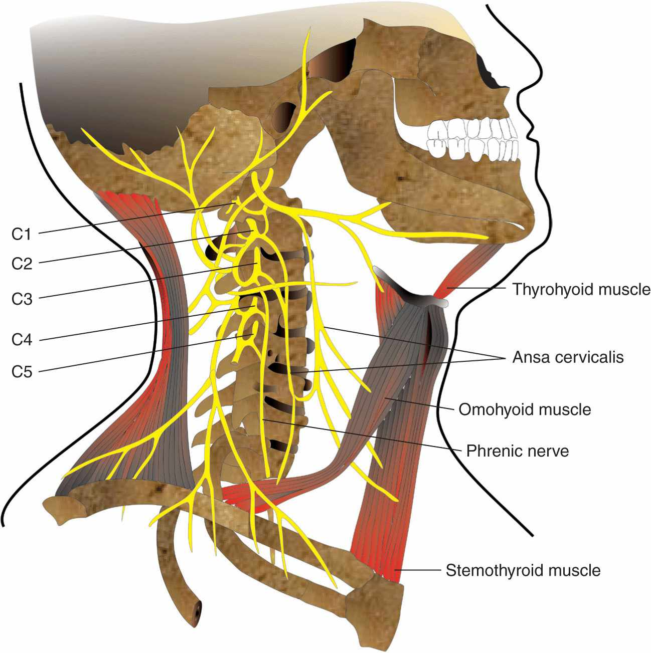
T/F The cervical plexus of nerves
A. supplies motor branches to the infrahyoid muscles
B. supplies motor branches to the muscles of the suboccipital triangle
C. supplies branches to the trapezius muscle
D. supplies sensory branches to the diaphragm
E. supplies sensory branches to the front of the scalp.
T/F The cervical plexus of nerves
T - A. supplies motor branches to the infrahyoid muscles
via ansa cervicalis (loop formed from C1, C2, C3)
F - B. supplies motor branches to the muscles of the suboccipital triangle
suboccipital triangle is mainly innervated by the suboccipital nerve (C1), which is a motor branch of the dorsal ramus of C1 (not part of the cervical plexus)
T - C. supplies branches to the trapezius muscle
via the accessory nerve
T - D. supplies sensory branches to the diaphragm
phrenic nerve (arising from C3, C4, and C5 of the cervical plexus)
F - E. supplies sensory branches to the front of the scalp.
anterior scalp is primarily innervated by branches from the trigeminal nerve (CN V)
T/F The sternocleidomastoid muscle
A. is attached to the temporal bone deep to the splenius capitis muscle
B. is active if the head is flexed against resistance
C. has a nerve supply from the cervical plexus
D. is an anterior relation of the scalenus anterior muscle
E. is crossed superficially by the external jugular vein.
T/F The sternocleidomastoid muscle
F - A. is attached to the temporal bone deep to the splenius capitis muscle
attached to the mastoid process of the temporal bone
superficial to the splenius capitis muscle
T - B. is active if the head is flexed against resistance
T - C. has a nerve supply from the cervical plexus
sensory branches from the cervical plexus (C2 and C3)
T - D. is an anterior relation of the scalenus anterior muscle
T - E. is crossed superficially by the external jugular vein.
T/F The thyroid gland
A. clasps the upper part of trachea.
B. is highly vascular.
C. doesn't move with the larynx.
D. is ductless gland.
E. consists of only one lobe.
T/F The thyroid gland
T - A. clasps the upper part of trachea.
thyroid gland wraps around the anterior and lateral aspects of the upper trachea, typically from about the 2nd to 4th tracheal rings.
T - B. is highly vascular.
receives blood from superior and inferior thyroid arteries and has rich venous drainage, making it one of the most vascular organs.
F - C. doesn't move with the larynx.
gland is attached to the larynx and trachea, so it moves up and down during swallowing.
T - D. is ductless gland.
Like all endocrine glands, the thyroid has no ducts; it secretes hormones (e.g., thyroxine) directly into the bloodstream
F - E. consists of only one lobe.
two lobes: right and left, connected by an isthmus. Sometimes a pyramidal lobe is also present
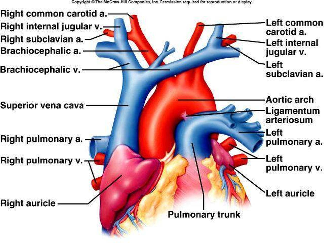
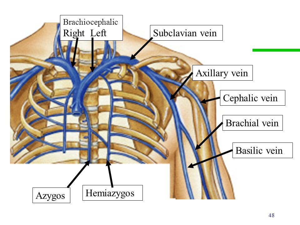
T/F The brachiocephalic vein
A. collects blood only from the head and neck.
B. ends by joining the opposite one to form the superior vena cava.
C. has no valves.
D. the right one crosses the median plain.
E. the right one is laterally to the brachiocephalic artery
T/F The brachiocephalic vein
F - A. collects blood only from the head and neck.
also collects blood from upper limb (via subclavian v.)
blood from head and neck via internal jugular vein
T - B. ends by joining the opposite one to form the superior vena cava.
T - C. has no valves.
F - D. the right one crosses the median plain.
right one runs vertically from the right side
T - E. the right one is laterally to the brachiocephalic artery
right brachiocephalic artery passes medially, while the vein lies more laterally in the thoracic cavity
T/F The vertical neurovascular bundle of the neck
A. lies on each side of the median airway and foodway.
B. extends from the base of the skull to the root of the neck.
C. contains glossopharyngeal nerve in its lower part.
D. is enclosed by the layers of the deep cervical fascia.
E. lies on the sympathetic trunk.
T/F The vertical neurovascular bundle of the neck
T - A. lies on each side of the median airway and foodway.
lies on each side of the median airway (trachea) and foodway (esophagus).
T - B. extends from the base of the skull to the root of the neck.
F - C. contains glossopharyngeal nerve in its lower part.
not part of this bundle
T - D. is enclosed by the layers of the deep cervical fascia.
bundle is enclosed within the carotid sheath, which is part of the deep cervical fascia.
T - E. lies on the sympathetic trunk.
because the sympathetic trunk is positioned posterior and medial to the carotid sheath but is considered closely related to it anatomically
sympathetic trunk (or sympathetic chain) is a paired bundle of nerve fibers that runs along each side of the vertebral column from the base of the skull down to the coccyx
It consists of a chain of interconnected sympathetic ganglia (nerve cell clusters) linked by nerve fibers.
It runs posterior and medial to the carotid sheath in the neck.
It sends branches (called gray and white rami communicantes) to spinal nerves, helping control smooth muscles and glands throughout the body.
In the neck, it gives off branches that contribute to cardiac, pulmonary, and cervical plexuses, influencing functions like heart rate and blood flow
vertical neurovascular bundle in the neck refers mainly to the contents of the carotid sheath, which run vertically on each side of the neck. It includes three key structures enclosed in the deep cervical fascia:
Common carotid artery (or internal carotid artery superiorly)
Internal jugular vein
Vagus nerve (cranial nerve X)
T/F The internal jugular vein
A. in the upper part of the neck is posterolateral to the internal carotid a.
B. is accompanied superiorly by the last four cranial nerves.
C. is posterior to vagus nerve
D. has inferiorly the sympathetic trunk lying between the vein and common carotid artery.
E. lies on the cervical plexus.
T/F The internal jugular vein
T - A. in the upper part of the neck is posterolateral to the internal carotid a.
T - B. is accompanied superiorly by the last four cranial nerves.
cranial nerves:
IX (glossopharyngeal),
X (vagus)
XI (accessory)
XII (hypoglossal) in the upper neck.
found in the carotid sheath with the IJV and ICA.
F - C. is posterior to vagus nerve
IJV is lateral, the carotid artery is medial, and the vagus nerve is in between them.
F - D. has inferiorly the sympathetic trunk lying between the vein and common carotid artery.
sympathetic trunk runs posterior to the carotid sheath
T - E. lies on the cervical plexus.
cervical plexus is deep to the sternocleidomastoid
cervical plexus formed by anterior rami of C1-C4
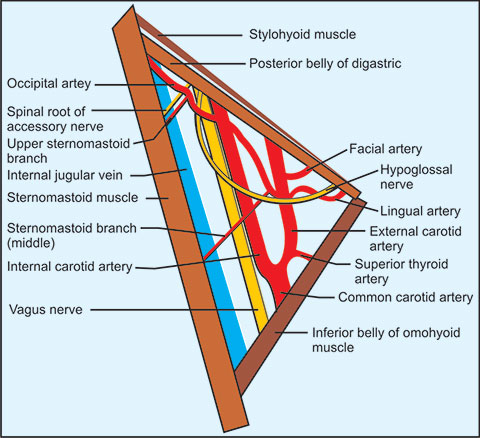
T/F A. carotis externa:
A. is in the carotid triangle
B. gives off a. thyroidea inferior
C. supplies head and neck structures
D. has baroreceptors at its origin - the bifurcation of the common carotid artery
E. occurs at the upper border of the thyroid cartilage.
T/F A. carotis externa:
T - A. is in the carotid triangle
F - B. gives off a. thyroidea inferior
arises from the subclavian artery supplies thyroid gland and surrounding structures
T - C. supplies head and neck structures
F - D. has baroreceptors at its origin - the bifurcation of the common carotid artery
baroreceptors are part of the common carotid artery
not the external carotid artery
T - E. occurs at the upper border of the thyroid cartilage.
T/F Which of the following are NOT anterior branches of external carotid artery?
A. A. pharyngea ascendens
B. A. thyroidea superior
C. A. sternocleidomastoidea
D. A. lingualis
E. A. occipitalis
T/F Which of the following are NOT anterior branches of external carotid artery?
T - A. A. pharyngea ascendens
F - B. A. thyroidea superior
T - C. A. sternocleidomastoidea
F - D. A. lingualis
T - E. A. occipitalis
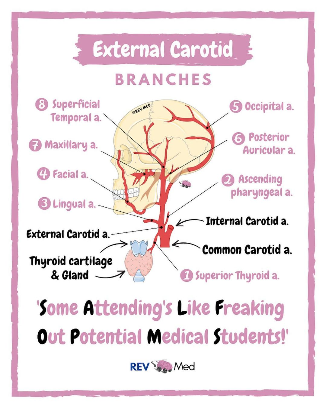
T/F Anterior branches of external carotid artery are:
A. A. thyroidea superior
B. A. occipitalis
C. A. lingualis
D. A. subscapularis
E. A. facialis
LEARN FOR FILL IN BLANKS TOO
T/F Anterior branches of external carotid artery are:
T - A. A. thyroidea superior
F - B. A. occipitalis
posterior branch
T - C. A. lingualis
F - D. A. subscapularis
branch of axillary artery
T - E. A. facialis
T/F The following elements are located in the carotid triangle:
A. N. laringeus superior
B. N. hypoglossus
C. Glandula thyroidea
D. Ansa cervicalis
E. A. thyroidea inferior
T/F The following elements are located in the carotid triangle:
T - A. N. laringeus superior
T - B. N. hypoglossus
F - C. Glandula thyroidea
inferiorly to the carotid triangle, below the cricoid cartilage
T - D. Ansa cervicalis
F - E. A. thyroidea inferior
arises from the subclavian artery and supplies the thyroid gland
T/F The internal carotid artery
A. enters the skull through the foramen lacerum
B. divides into the anterior and middle cerebral arteries
C. gives off the ophthalmic artery
D. is accompanied within the skull by preganglionic sympathetic nerve fibres
E. usually begins about the level of the cricoid cartilage.
T/F The internal carotid artery
F - A. enters the skull through the foramen lacerum
passes over the foramen lacerum
enters the skull via the carotid canal
T - B. divides into the anterior and middle cerebral arteries
T - C. gives off the ophthalmic artery
F - D. is accompanied within the skull by preganglionic sympathetic nerve fibres
accompanied by postganglionic sympathetic fibres
from the superior cervical ganglion
F - E. usually begins about the level of the cricoid cartilage.
begins at the level of the upper border of the thyroid cartilage (higher than cricoid)
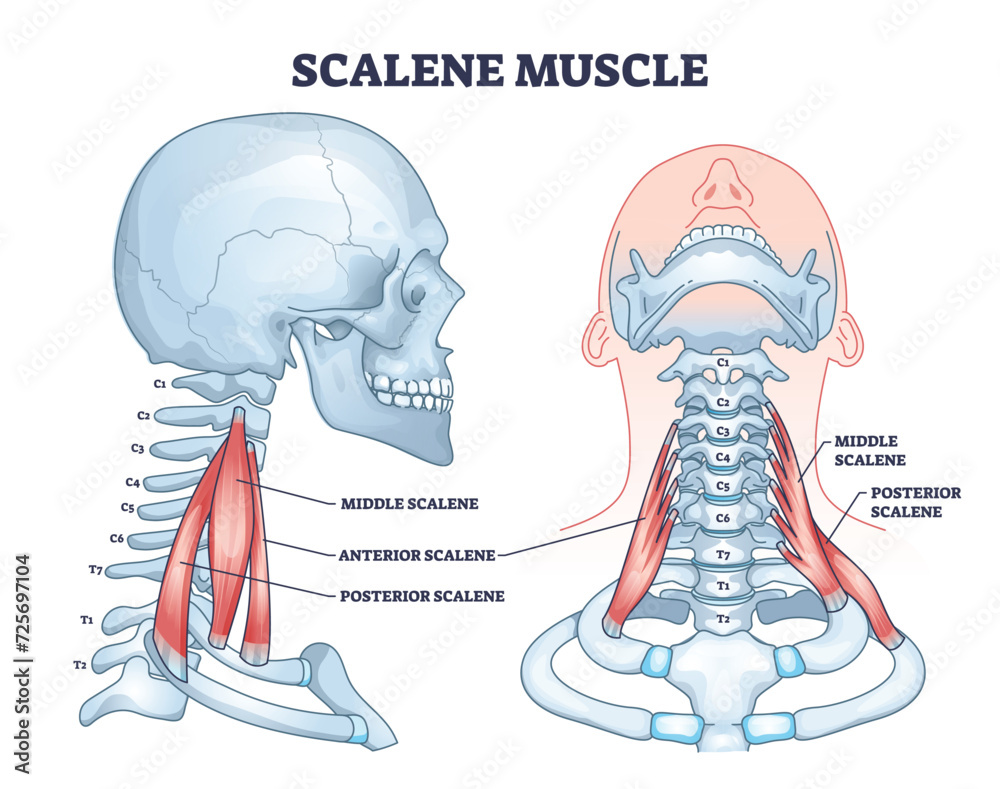
T/F The scalenus anterior muscle
A. is anterior to the nerves forming the brachial plexus
B. is attached to the posterior tubercles of the transverse processes of some of the cervical vertebrae
C. is medial to the vertebral artery
D. is anterior to the subclavian artery
E. is lateral to the inferior cervical ganglion.
T/F The scalenus anterior muscle
T - A. is anterior to the nerves forming the brachial plexus
F - B. is attached to the posterior tubercles of the transverse processes of some of the cervical vertebrae
scalenus anterior originates from the anterior tubercles of the C3-C6 transverse processes
F - C. is medial to the vertebral artery
vertebral artery runs medial to the scalenus anterior
The scalenus anterior is lateral to it
T - D. is anterior to the subclavian artery
and the subclavian vein is in front of scalenus anterior
T - E. is lateral to the inferior cervical ganglion.
T/F The external carotid artery
A. is crossed anteriorly by the hypoglossal nerve
B. usually divides into its terminal branches at the level of the angle of the jaw
C. at its origin is lateral to the internal carotid artery
D. is the only source of blood to the thyroid gland
E. is superficial to the glossopharyngeal nerve.
T/F The external carotid artery
T - A. is crossed anteriorly by the hypoglossal nerve
F - B. usually divides into its terminal branches at the level of the angle of the jaw
level of the neck of the mandible
terminal branches:
superficial temporal artery
maxillary artery
F - C. at its origin is lateral to the internal carotid artery
external carotid artery arises medial to the internal carotid artery
at the bifurcation of the common carotid artery
F - D. is the only source of blood to the thyroid gland
inferior thyroid artery, a branch of the subclavian artery, is also an important blood supply
T - E. is superficial to the glossopharyngeal nerve.
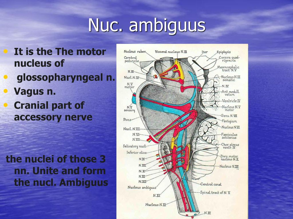
T/F The recurrent laryngeal nerve
A. has fibres whose cell bodies are in the nucleus ambiguus of the hindbrain
B. is entirely a motor nerve
C. is a close relation of the inferior thyroid artery
D. supplies all the muscles of the larynx
E. supplies some of the intrinsic muscles of the larynx.
T/F The recurrent laryngeal nerve
T - A. has fibres whose cell bodies are in the nucleus ambiguus of the hindbrain
branch of the vagus nerve
motor fibers originate from the nucleus ambiguus
medulla oblongata of the brainstem
F - B. is entirely a motor nerve
has some sensory fibers
T - C. is a close relation of the inferior thyroid artery
particularly in the neck, where it loops under the aortic arch (on the left)
F - D. supplies all the muscles of the larynx
most intrinsic muscles of the larynx, but not all
T - E. supplies some of the intrinsic muscles of the larynx.
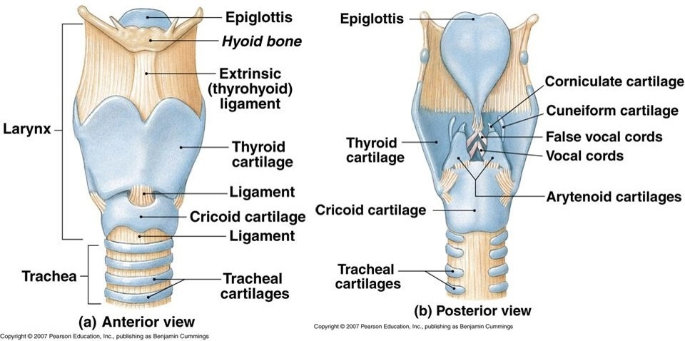
T/F The cricoid cartilage
A. has an anterior arch which moves upwards and backwards due to the contraction of the cricothyroid muscle
B. lengthens the vocal fold (true vocal cord) when its anterior part moves upwards and backwards
C. has the vocal folds attached to it
D. gives attachment to the inferior constrictor muscle of the pharynx
E. is at the level of the fourth cervical vertebra.
T/F The cricoid cartilage
T - A. has an anterior arch which moves upwards and backwards due to the contraction of the cricothyroid muscle
cricothyroid muscle tilts the thyroid cartilage forward - causes the anterior arch of the cricoid cartilage to move upwards and backwards
T - B. lengthens the vocal fold (true vocal cord) when its anterior part moves upwards and backwards
F - C. has the vocal folds attached to it
serves as a foundation for the laryngeal framework - does not directly attach to the vocal folds.
vocal folds (true vocal cords) are attached to:
Anteriorly: The thyroid cartilage
Posteriorly: The vocal processes of the arytenoid cartilages
T - D. gives attachment to the inferior constrictor muscle of the pharynx
specifically to its lateral aspect
F - E. is at the level of the fourth cervical vertebra.
located at the C6 vertebral level
T/F The scalenus medius muscle
A. is posterior to the nerves forming the brachial plexus
B. is attached to the scalene tubercle
C. is used in deep breathing
D. is posterior to the subclavian artery
E. is crossed anteriorly by the omohyoid muscle.
T/F The scalenus medius muscle
T - A. is posterior to the nerves forming the brachial plexus
plexus emerges between the scalenus anterior and medius muscles
F - B. is attached to the scalene tubercle
scalene tubercle on the 1st rib is the attachment for the scalenus anterior
Scalenus medius attaches more posteriorly on the 1st rib
T - C. is used in deep breathing
scalenus medius elevates the first rib during forced inspiration, aiding in deep breathing
T - D. is posterior to the subclavian artery
subclavian artery passes between the anterior and medius scalene muscles
T - E. is crossed anteriorly by the omohyoid muscle.
inferior belly of the omohyoid crosses the posterior triangle, passing superficial (anterior) to the scalenus medius

T/F The internal jugular vein
A. is, along its whole course, directly lateral to the internal carotid artery
B. has no valves
C. is anterior to the phrenic nerve
D. receives all the venous blood from the thyroid gland
E. is anterior to the thoracic duct on the left side.
T/F The internal jugular vein
F - A. is, along its whole course, directly lateral to the internal carotid artery
For most of its course, yes, it's lateral, but not always directly; in some parts (e.g. lower neck), it becomes anterolateral or slightly anterior
F - B. has no valves
internal jugular vein does have valves, typically just above its termination into the subclavian veinT - C. is anterior to the phrenic nerve
T - C. is anterior to the phrenic nerve
lower neck, the internal jugular vein lies anterior to the phrenic nerve, which is more posterior and lateral (running on the anterior scalene muscle).
F - D. receives all the venous blood from the thyroid gland
thyroid venous drainage goes to:
Superior thyroid vein → internal jugular vein
Middle thyroid vein → internal jugular vein
Inferior thyroid vein → brachiocephalic vein
T - E. is anterior to the thoracic duct on the left side.
On the left, the thoracic duct ascends posterior to the internal jugular vein, before draining into the venous angle (junction of left IJV and subclavian vein).
T/F The digastric muscle
A. has a motor innervation from nerves of the branchial arches
B. is inferior to the submandibular gland
C. is attached to the ramus of the mandible
D. is superficial,to the hypoglossal nerve
E. is deep to the carotid sheath.
T/F The digastric muscle
T - A. has a motor innervation from nerves of the branchial arches
anterior belly is innervated by the mylohyoid nerve
originating from the first branchial arch
posterior belly is innervated by the facial nerve
originating from the second branchial arch
T - B. is inferior to the submandibular gland
F - C. is attached to the ramus of the mandible
attached to the digastric fossa on the medial surface of the mandible
T - D. is superficial to the hypoglossal nerve
n. passes deep to the muscle as it travels to the tongue
F - E. is deep to the carotid sheath.
digastric muscle is superficial to the carotid sheath
T/F The scalenus anterior muscle
A. is anterior to the subclavian vein
B. is anterior to the phrenic nerve
C. is anterior to the suprascapular artery
D. is used in deep respiration
E. is attached to the first and second ribs.
T/F The scalenus anterior muscle
F - A. is anterior to the subclavian vein
scalenus anterior is actually posterior to the subclavian vein
F - B. is anterior to the phrenic nerve
phrenic nerve lies on the anterior surface of the scalenus anterior muscle
F - C. is anterior to the suprascapular artery
suprascapular artery usually passes anterior to the scalenus anterior
T - D. is used in deep respiration
F - E. is attached to the first and second ribs.
attached only to the first rib
scalenus posterior attaches to both the first and second ribs
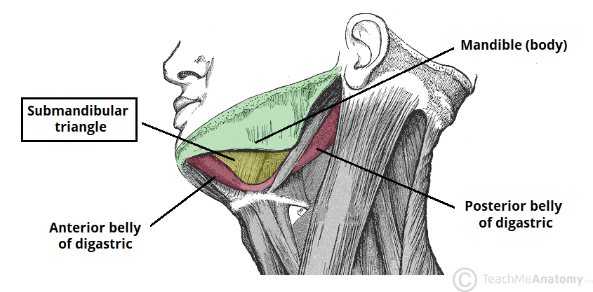
T/F Trigonum submandibulare contains:
A. glandula submandibularis
B. accessory nerve
C. phrenic nerve
D. facial artery
E. lingual nerve
T/F Trigonum submandibulare contains:
T - A. glandula submandibularis
F - B. accessory nerve
The accessory nerve (cranial nerve XI) passes more posteriorly
F - C. phrenic nerve
phrenic nerve runs in the neck but not in the submandibular triangle
T - D. facial artery
curves around the lower border of the mandible within this triangle
supplies blood to many structures of the face including the skin and muscles of facial expression, the palate, tonsils, submandibular gland, and nasal area
T - E. lingual nerve
lingual nerve passes near the submandibular gland in this area
provides general sensory (touch, pain, temperature) innervation to the anterior two-thirds of the tongue, the floor of the mouth, and the lingual gingiva
T/F The following elements are part of trigonum submandibulare:
A. n. mylohyoideus
B. n. hypoglossus
C. glandula thyroidea
D. trigonum Pirogovi
E. a. thyroidea inferior
T/F The following elements are part of trigonum submandibulare:
T - A. n. mylohyoideus
T - B. n. hypoglossus
F - C. glandula thyroidea
T - D. trigonum Pirogovi
F - E. a. thyroidea inferior
MATCH EACH NUMBERED TERM WITH THE MOST PROPER LETTERED ONE
A. M. latissimus dorsi
B. M. levator scapulae
C. M. platysma
D. M. trapezius
E. M. erector spinae
1. N. facialis
2. Rr. dorsales of nn. spinales
3. N. dorsalis scapulae
4. N. thoracodorsalis
5. N. accessories
A. M. latissimus dorsi - 4. N. thoracodorsalis
B. M. levator scapulae - 3. N. dorsalis scapulae
C. M. platysma - 1. N. facialis
D. M. trapezius - 5. N. accessories
E. M. erector spinae - 2. Rr. dorsales of nn. spinales (dorsal rami of the spinal nerves)
MATCH EACH NUMBERED TERM WITH THE MOST PROPER LETTERED ONE
Which of the following A to F supplies the muscles 1 to 6?
A. Cervical plexus
B. Spinal accessory nerve
C. Cranial accessory nerve
D. Facial nerve
E. None of these
1 Platysma
2 Infrahyoid
3 Sternocleidomastoid
4 Levator veli palatini
5 Orbicularis oculi
Which of the following A to F supplies the muscles 1 to 6?
A. Cervical plexus - 2 Infrahyoid
B. Spinal accessory nerve - 3 Sternocleidomastoid
C. Cranial accessory nerve - 1 Platysma
D. Facial nerve - 5 Orbicularis oculi
E. None of these - 4 Levator veli palatini
MATCH EACH NUMBERED TERM WITH THE MOST PROPER LETTERED ONE
A. Anterior cervical triangle
B. Posterior(lateral) cervical triangle
1. Omotrapezoid (Subclavian triangle)
2. Carotid
3. Omoclavicular (Occipital triangle)
4. Digastric triangle
A. Anterior cervical triangle - 2. Carotid, 4. Digastric triangle
B. Posterior(lateral) cervical triangle - 1. Omotrapezoid (Subclavian triangle), 3. Omoclavicular (Occipital triangle)
Erector spinae muscle comprises three muscle columns
A.
B.
C.
Erector spinae muscle comprises three muscle columns
A. spinalis
B. longissimus
C. iliocostalis

List the boundaries of lateral cervical triangle:
A.
B.
C.
List the boundaries of lateral cervical triangle:
A. trapezius m.
B. sternocleidomastoid m.
C. clavicle
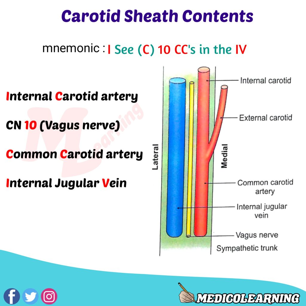
Vagina carotica in the neck contains:
A.
B.
C.
Vagina carotica in the neck contains:
A. A. carotis communis (interna)
B. v. jugularis interna
C. n. vagus
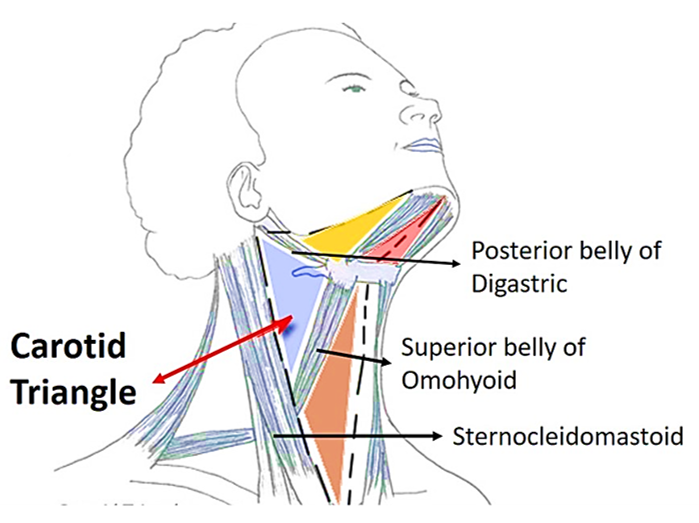
List the boundaries of carotid triangle:
A.
B.
C.
List the boundaries of carotid triangle:
A. venter posterior of m. digastricus
B. m. sternocleidomastoideus
C. m. omohyoideus - venter superior
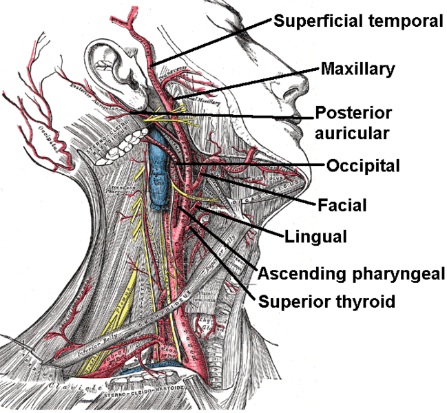
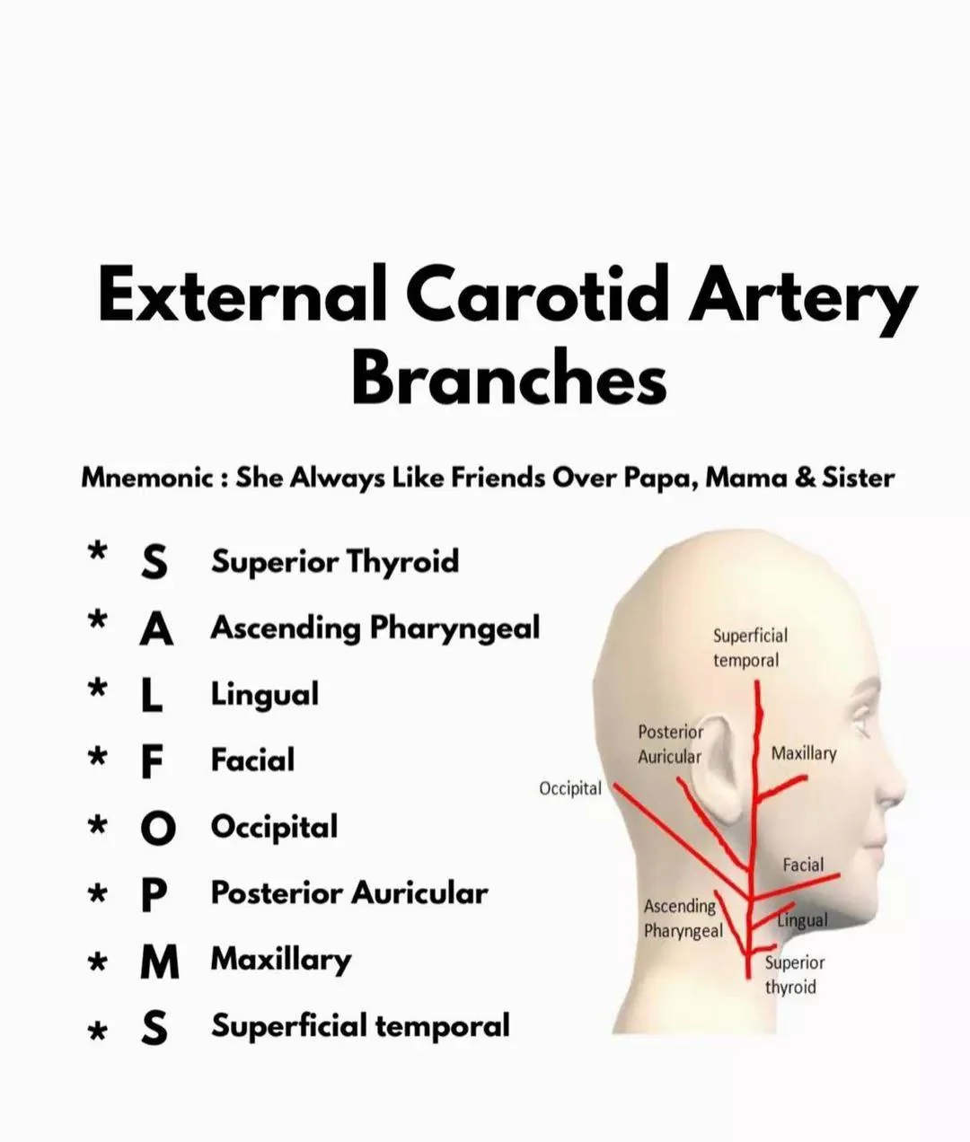
External carotid artery gives off the following branches from its anterior surface:
A.
B.
C.
External carotid artery gives off the following branches from its anterior surface:
A. superior thyroid a.
B. lingual aa.
C. facial aa.
The thyroid gland is drained by the following three pairs of veins:
A.
B.
C.
The thyroid gland is drained by the following three pairs of veins:
A. superior thyroid vv.
B. middle thyroid vv.
C. inferior thyroid vv.
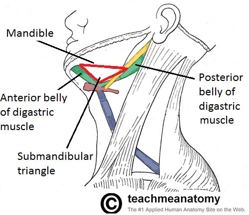
Define the boundaries of trigonum submandibulare:
A.
B.
C.
Define the boundaries of trigonum submandibulare:
A. posterior bellies of digastric m.
B. anterior bellies of digastric m.
C. mandible
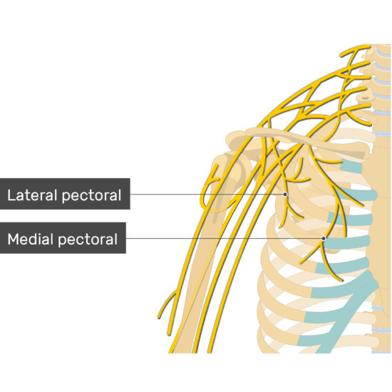
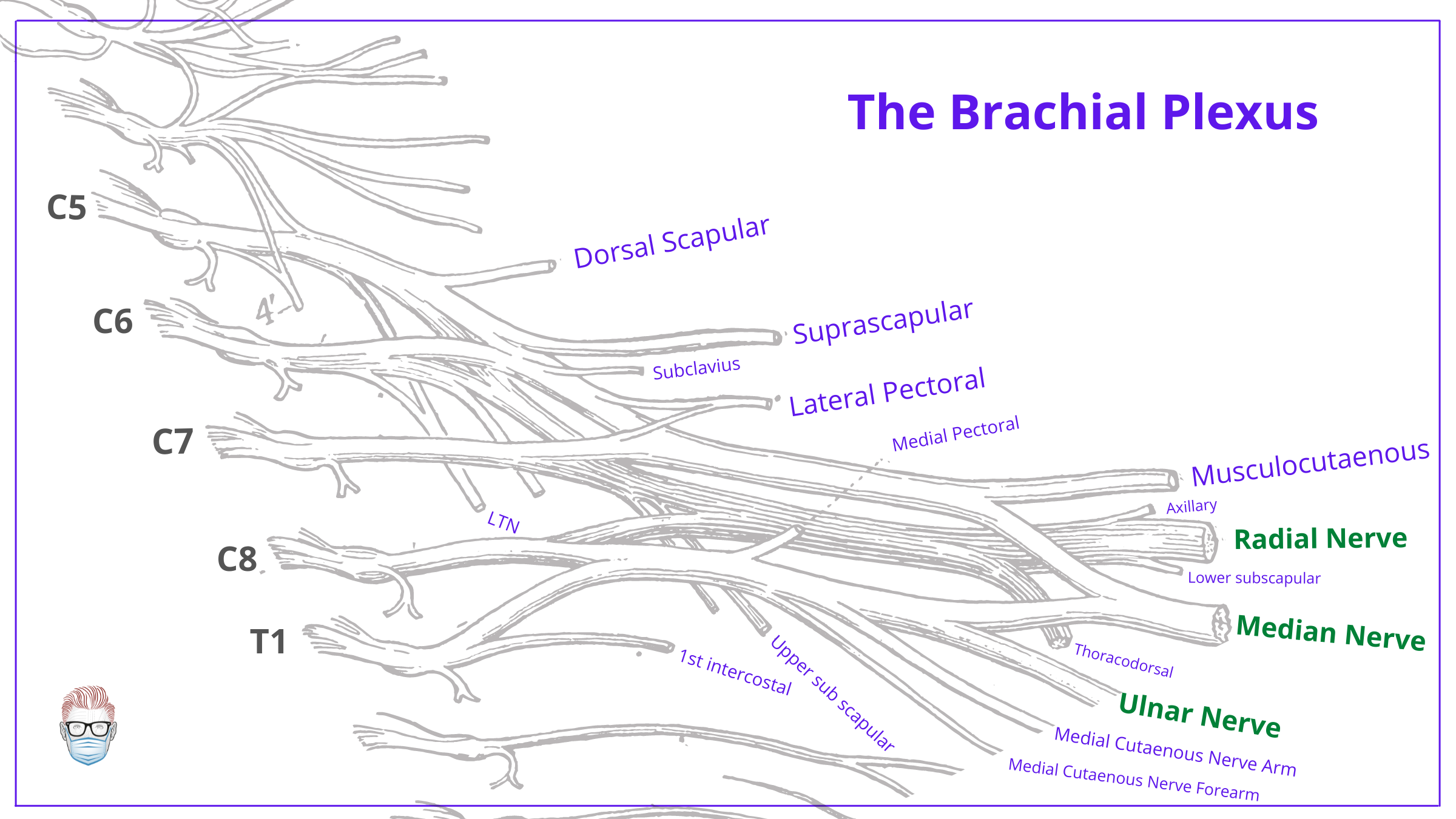
Pectoral nerves are branches of the medial and lateral cords of the brachial plexus.
A. Yes
B. No
Pectoral nerves are branches of the medial and lateral cords of the brachial plexus.
A. Yes
B. No
The lateral pectoral nerve arises from the lateral cord (C5–C7).
The medial pectoral nerve arises from the medial cord (C8–T1)
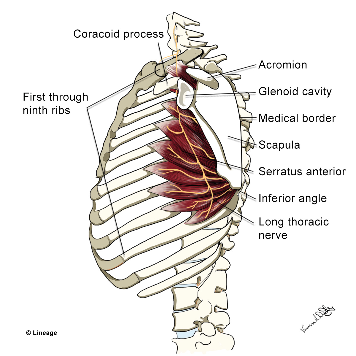
Long thoracic nerve is a direct branch of the roots of the brachial plexus.
A. Yes
B. No
Long thoracic nerve is a direct branch of the roots of the brachial plexus.
A. Yes
long thoracic nerve arises directly from the C5, C6, and C7 roots of the brachial plexus.
B. No
dorsal scapular nerve arises from the C5 root of the brachial plexus. It innervates the rhomboid major, rhomboid minor, and levator scapulae muscles
Dorsal scapular nerve is a direct branch of the roots of the brachial plexus.
A. Yes
B. No
Dorsal scapular nerve is a direct branch of the roots of the brachial plexus.
A. Yes
dorsal scapular nerve typically arises directly from the C5 root of the brachial plexus.
B. No
Superior trunk of the brachial plexus gives off n. suprascapularis and nerve to the subclavius.
A. Yes
B. No
Superior trunk of the brachial plexus gives off n. suprascapularis and nerve to the subclavius.
A. Yes
"S" theme:
Superior trunk → Suprascapular nerve and Subclavius nerve
B. No
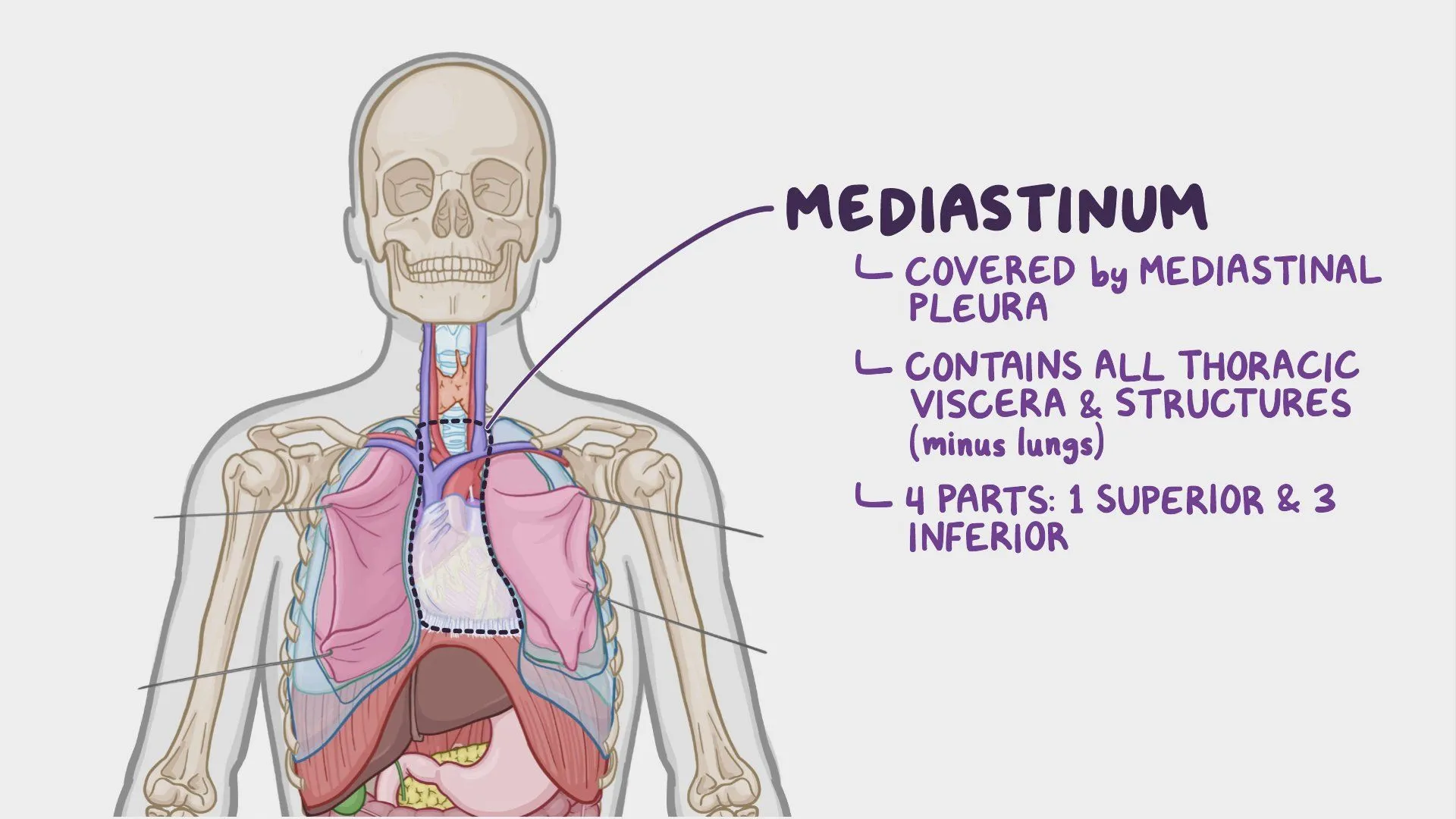
Mediastinum is the space in thoracic cavity where the lungs are located.
A. Yes
B. No
Mediastinum is the space in thoracic cavity where the lungs are located.
A. Yes
B. No
mediastinum is the central compartment of the thoracic cavity between the two lungs, containing the heart, great vessels, trachea, esophagus, thymus, and other structures — but not the lungs themselves.
lungs are located in the pleural cavities on either side of the mediastinum
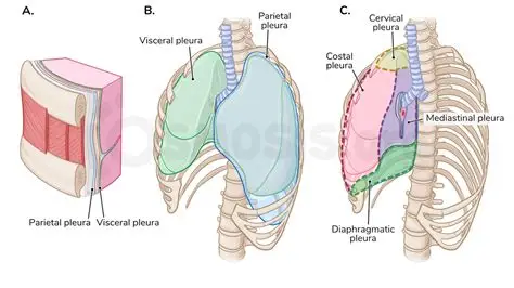
The lower border of parietal pleura is projected at the level of 12th rib on the back of the thorax.
A. Yes
B. No
The lower border of parietal pleura is projected at the level of 12th rib on the back of the thorax.
A. Yes
B. No
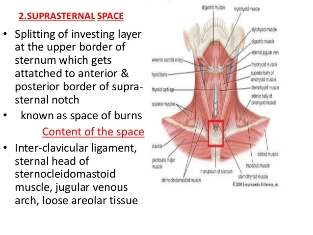
The suprasternal space is between the superficial (investing) and middle lamina of deep cervical fascia.
A. Yes
B. No
The suprasternal space is between the superficial (investing) and middle lamina of deep cervical fascia.
A. Yes
B. No
The phrenic nerve is a part of the neurovascular bundle of the neck.
A. Yes
B. No
The phrenic nerve is a part of the neurovascular bundle of the neck.
A. Yes
B. No
neurovascular bundle = carotid sheath
Common carotid artery
Internal jugular vein
Vagus nerve
The cervical parietal pleura forms the dome of each pleural cavity.
A. Yes
B. No
The cervical parietal pleura forms the dome of each pleural cavity.
A. Yes
B. No
The parietal and visceral pericardium are separated by a thin film of fluid.
A. Yes
B. No
The parietal and visceral pericardium are separated by a thin film of fluid.
A. Yes
B. No
The subclavian vessels arch over the anterior surface of the dome of the pleura.
A. Yes
B. No
The subclavian vessels arch over the anterior surface of the dome of the pleura.
A. Yes
B. No
The costomediastinal recess lies along the inferior margin of the pleura.
A. Yes
B. No
The costomediastinal recess lies along the inferior margin of the pleura.
A. Yes
B. No
inferior margin of the pleura refers to the costodiaphragmatic recess
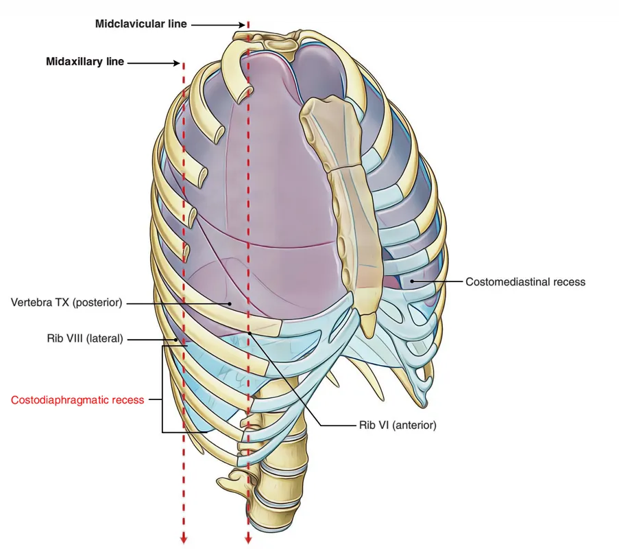
The costodiaphragmatic recess extends between the thoracic wall and the vertical part of the diaphragm.
A. Yes
B. No
The costodiaphragmatic recess extends between the thoracic wall and the vertical part of the diaphragm.
A. Yes
B. No
The phrenic nerve and its accompanying vessels pass anterior to the lung root.
A. Yes
B. No
The phrenic nerve and its accompanying vessels pass anterior to the lung root.
A. Yes
B. No
The vagus nerve descends posterior to the lung root.
A. Yes
B. No
The vagus nerve descends posterior to the lung root.
A. Yes
B. No
The phrenic and vagus nerves descend between the pericardium and sternum.
A. Yes
B. No
The phrenic and vagus nerves descend between the pericardium and sternum.
A. Yes
B. No
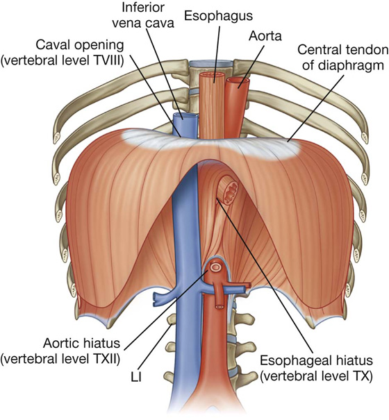
The thoracic duct enters the thoracic cavity through hiatus aorticus.
A. Yes
B. No
The thoracic duct enters the thoracic cavity through hiatus aorticus.
A. Yes
B. No
Diaphragm hiatuses:
Caval hiatus (T8): inferior vena cava + right phrenic n.
Esophageal hiatus (T10): Esophagus + vagus nerves + esophageal branches of left gastric artery and vein
Aortic Hiatus (T12): Aorta + thoracic duct + azygos vein
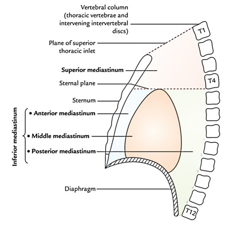
The lower boundary of mediastinum superius is the plane between angulus sterni and Th4/Th5.
A. Yes
B. No
The lower boundary of mediastinum superius is the plane between angulus sterni and Th4/Th5.
A. Yes
B. No
The upper boundary of mediastinum superius is the plane between the sternal notch and Th1.
A. Yes
B. No
The upper boundary of mediastinum superius is the plane between the sternal notch and Th1.
A. Yes
“thoracic inlet”
Inferior - Transverse thoracic plane
from the manubriosternal joint to the intervertebral disc between T4 and T5
Anterior - Manubrium of the sternum
Posterior - Upper thoracic vertebrae → T1 to T4
Lateral - Mediastinal pleura (covering the lungs)
B. No
Phrenic nerve passes posteriorly to the root of the lungs.
A. Yes
B. No
Phrenic nerve passes posteriorly to the root of the lungs.
A. Yes
B. No
Vagus nerve passes anteriorly to the root of the lungs.
A. Yes
B. No
Vagus nerve passes anteriorly to the root of the lungs.
A. Yes
B. No
Phrenic and vagus nerves pass together posteriorly to the root of the lungs.
A. Yes
B. No
Phrenic and vagus nerves pass together posteriorly to the root of the lungs.
A. Yes
B. No
In the thorax sympathetic trunk is intimately related to the esophagus.
A. Yes
B. No
In the thorax sympathetic trunk is intimately related to the esophagus.
A. Yes
B. No
sympathetic trunk runs along the vertebral column, posterior and lateral in position.
The esophagus lies anterior to the vertebral column, in the posterior mediastinum.
The nerves intimately related to the esophagus are:
Vagus nerves (especially the esophageal plexus formed by branches of the vagus)
Thoracic splanchnic nerves pass more anteromedially from the sympathetic trunk toward the abdomen, but the trunk itself remains posterior.
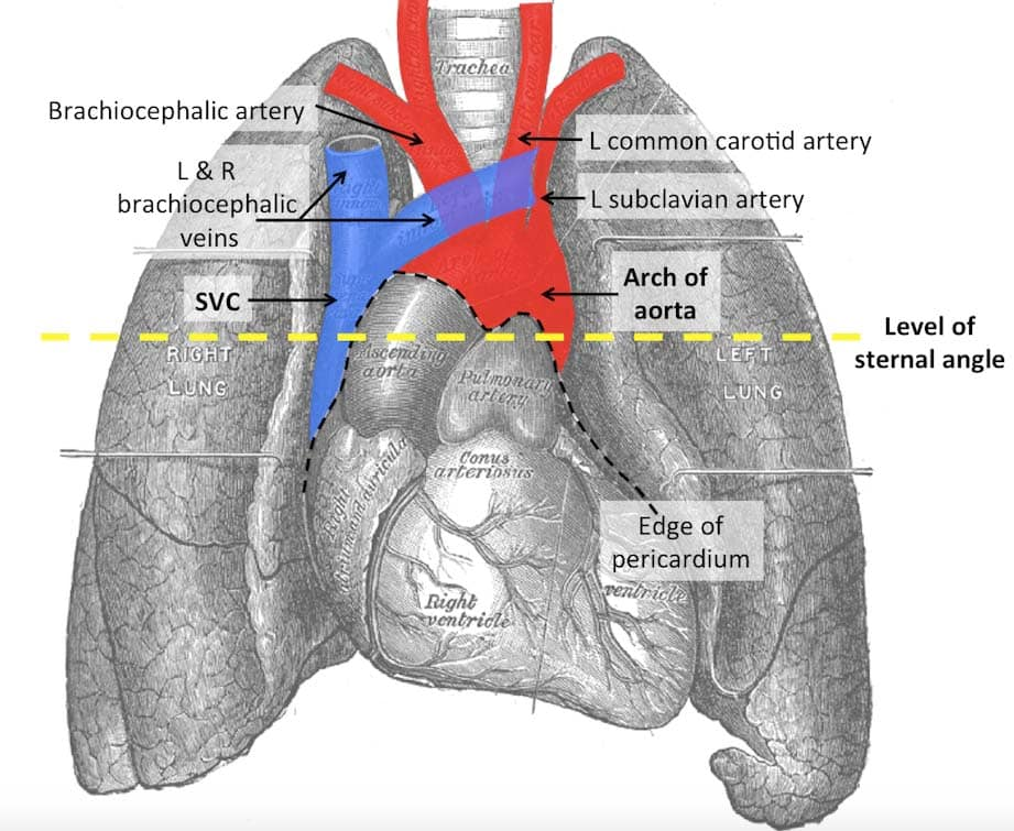
Aortic arch may have from 1 to 6 branches.
A. Yes
B. No
Aortic arch may have from 1 to 6 branches.
A. Yes
B. No
While most individuals have three classic branches off the aortic arch —
Brachiocephalic trunk
Left common carotid artery
Left subclavian artery —
there can indeed be anatomical variations, and embryologically, the aortic arch system develops from six pairs of aortic arches.
So, when your book says the aortic arch may have from 1 to 6 branches, it may be referring to embryological development or rare anatomical variants, such as:
separate origins of the carotid arteries,
vertebral arteries arising directly from the arch,
retroesophageal right subclavian artery (aberrant right subclavian),
thyroidea ima artery (occasionally from the arch),
or a persistent ductus arteriosus.
The pleural cavity contains:
A. Blood
B. Mucosal fluid
C. Serous fluid
D. Air
The pleural cavity contains:
A. Blood
B. Mucosal fluid
C. Serous fluid
D. Air
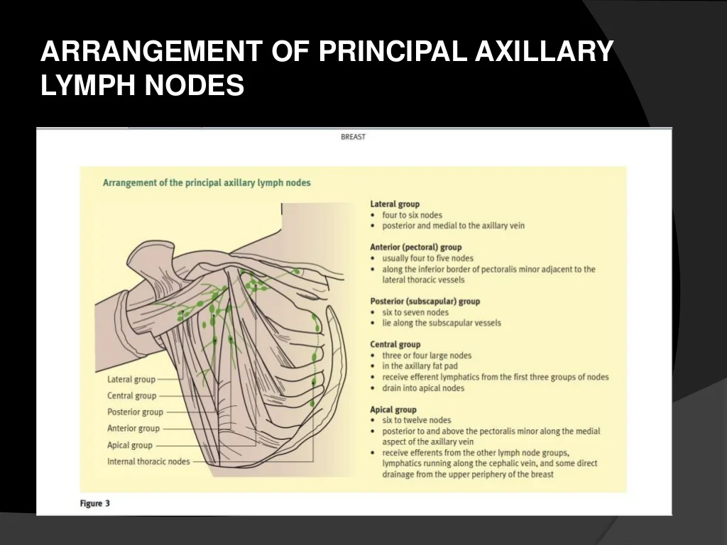
In lymphatic drainage of the breast, the major portion (about 75%) enters eventually into which group of nodes?
A. Central axillary
B. Deltopectoral
C. Lateral axillary
D. Parasternal
E. Subscapular
In lymphatic drainage of the breast, the major portion (about 75%) enters eventually into which group of nodes?
A. Central axillary
About 75% of the lymph from the breast drains into the central axillary lymph nodes, which then drain into the apical nodes and further into the subclavian lymphatic trunk
B. Deltopectoral
C. Lateral axillary
D. Parasternal
E. Subscapular
BUT ALSO PECTORAL AXILLARY
Pectoral (anterior) axillary nodes = first stop for lymph from the breast.
Central axillary nodes = major collecting nodes receiving lymph from pectoral and other axillary groups
Here’s the typical flow:
Lymph from the breast first drains into pectoral (anterior) axillary nodes.
Then, lymph passes to the central axillary nodes.
Finally, it drains into the apical (subclavian) nodes.
A woman with breast cancer subsequently develops metastases in her vertebral column. The most direct route for spread of the tumor to the vertebral column was via:
A. branches of the cephalic vein
B. branches of the lateral thoracic vein
C. branches of the thoracoacromial veins
D. lymphatic vessels draining into the axilla
E. branches of the intercostal veins
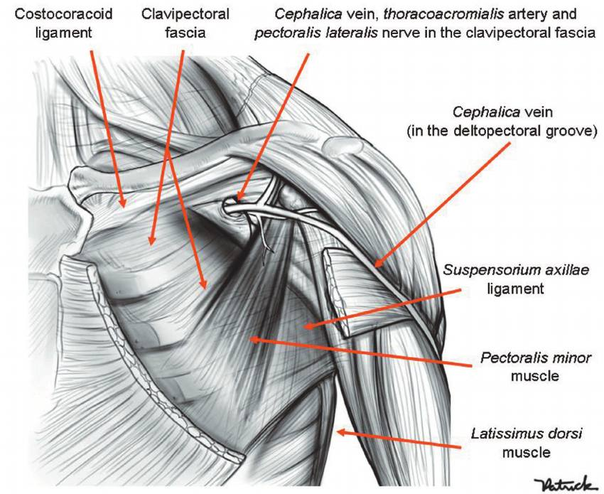
The clavipectoral fascia is penetrated by which artery?
A. Anterior circumflex humeral
B. Axillary
C. Subscapular
D. Thoracoacromial
E. Thoracodorsal
The clavipectoral fascia is penetrated by which artery?
A. Anterior circumflex humeral
B. Axillary
runs beneath the fascia
C. Subscapular
D. Thoracoacromial
branch of axillary artery
Also penetrated by:
cephalic vein
lateral pectoral n.
lymphatic vessels
E. Thoracodorsal
supplies latissimus dorsi