A&P Lecture 8-9 (Ch. 6)
 Call Kai
 Learn
Learn Practice Test
 Spaced Repetition
 Match
Match Flashcards
 Knowt Play
1/97

There's no tags or description
Looks like no tags are added yet.
Name | Mastery | Learn | Test | Matching | Spaced | Call with Kai | Chat |
|---|
No analytics yet
Send a link to your students to track their progress
98 Terms
Skeletal system
Comprised of bone and cartilage
Bone function
Forms rigid scaffolding for protection support and movement. Important for mineral storage, triglyceride storage, and homatopoiesis. Classified as organs
Cartilage function
Forms joint structures, growth plates, and templates for future bones during embryonic development
Hematopoiesis
Red blood cell and platelet synthesis
Bone as an organ
Includes bone, cartilage, nerves, vessels, connective tissue, and marrow
Hyaline cartilage
Lines articular surfaces of joints, form growth plates near the ends of long bones, form cartilage models that many bones develop from

Fibrocartilage
Present in areas subject to both stretch and compression (ex: intervertebral discs, menisci in knee, pubic symphysis between pelvic bones)
Elastic cartilage
Forms the external ear and epiglottis
Cartilage growth
Includes interstitial growth and appositional growth
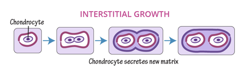
Interstitial growth
Cartilage grows from within- chondrocytes in lacunae divide and secrete new matrix
Appositional growth
Cartilage is surrounded by a layer of dense irregular connective tissue called perichondrium. Chondroblasts differentiate from cells in perichondrium and secrete new matrix on outer surface of cartilage

Perichondrium
Layer of dense irregular connective tissue surrounding cartilage
Axial bone classification
Vertebral column, rib cage, sternum, skull
Appendicular skeleton
Limbs and limb girdles (scapula/clavicle and pelvic bones)
Long bones
Bones made up of a shaft with distinct ends (most limb bones)
Short bones
Cube or round shaped bones (wrist, ankle, sesamoid bones)
Sesamoid bones
Bones that form in a tendon (ex: patella)
Flat bones
Thin, flat, and usually curved bones (ex: cranial bones of skull, ribs, sternum)
Irregular bones
Bones with shapes more complex than other categories (ex: vertebra, pelvic bones)
Gross bone structure
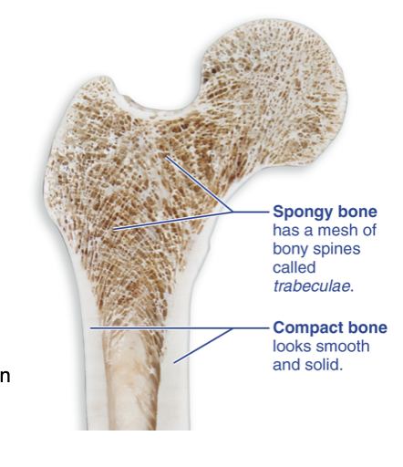
Macroscopic, includes cortical bone, trabecular bone, skeletal features, and marrow cavity
Compact (cortical) bone
Dense bone tissue lining the bone’s outer surface

Spongy (trabecular) bone
Made up of many trabeculae (small beams/spines of bone tissue that form a network on the interior of a bone). Arranged along lines of stress from mechanical forces
Microscopic bone structure
Includes osteons, canals, and lamellae
Chemical organization of bone structure
Includes organic (osteoid, bone cells) and inorganic (hydroxyapatite) components
Bone cell function
Create, maintain, and recycle bone
Osteoid
Unmineralized extracellular matrix made of collagen fibers, calcium-binding proteins, and ground substance
Collagen fibers function in organic component of bone
Give bone strength under tension and allow it to bend slightly without breaking
Hydroxyapatite crystals
Inorganic component of bone: mineral salts, mainly calcium phosphate. Give bone its rigidity and strength under compression
Mineralized bone
Composite of collagen encased in hydroxyapatite crystals
2 categories of bone tissue organization
Compact (cortical) bone
Spongy (trabecular) bone
Trabeculae
Small beams or spines of bone tissue that form a network in spongy bone. Arranged along lines of stress from mechanical forces
Periosteum
Connective tissue membrane that lines the outer surface of bone. Two layers- outer: fibrous; inner: osteogenic
Fibrous layer of periosteum
Outer layer, made of dense irregular connective tissue
Osteogenic layer of periosteum
Inner layer, contains osteoprogenitor cells
Endosteum
Connective tissue membrane that lines all interior surfaces of bone, including surface of trabecular bone and canals within cortical bone. Single layer, also contains osetoprogenitor cells
Organization of short, flat, and irregular bone
Outer compact bone (on both sides) surrounds inner spongy bone (called diploe in flat bones)
Diploe
Spongy bone inner layer found in flat bones
Organization of long bones
Diaphysis: shaft; epiphysis: ends
Diaphysis
Shaft of a long bone. Cylindrical arrangement of compact bone, surrounds medullary cavity

Epiphyses
Ends of long bone. Outer layer of compact bones, inner layer of spongy bone. Has epiphyseal line

Epiphyseal line
Remnant of the growth plate that forms the boundary between the diaphysis and epiphysis
Red marrow
Hematopoietic tissue that forms blood cells. Makes up marrow cavity and spaces between spongy bone in infants. In adults, limited to axial skeleton and proximal ends of humerus and femur
Yellow marrow
Mainly adipose tissue that replaces red marrow in the marrow cavity during growth. Can revert to red in certain conditions (sever, chronic anemia)
Osteoprogenitor cells
Mitotic stem cells found on bone-facing surfaces of the periosteum and endosteum (some can become osteoblasts

Osteoblasts
Mitotic bone-forming cells that secrete osteoid. Some become flat, inactive bone lining cells that line and maintain bone surface. Some become osteocytes

Osteocytes
Mature osteoblasts that maintain bone matrix and sense mechanical strain. Situated in lacunae

Osteoclasts
Large multinucleated cells that resorb or degrade bone extracellular matrix

Osteon
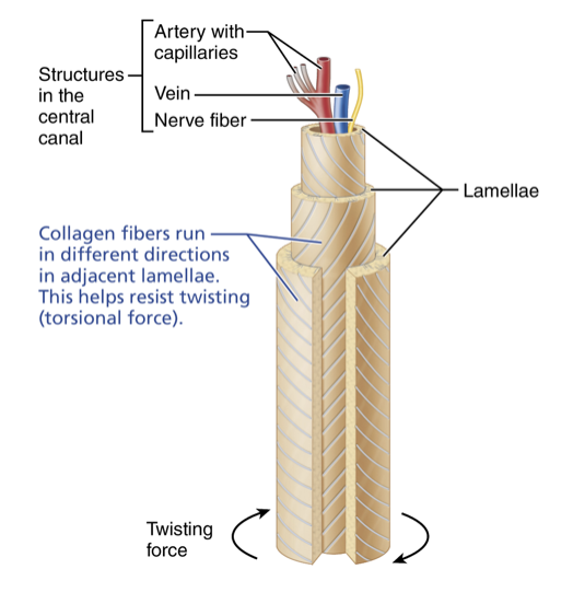
Structural unit of compact bone. Tiny pillars of bone made up of concentric bone layers arranged parallel to ling axis of bone

Osteon structure
Central (haversian) canal, lamellae, lacunae, canaliculi
Central (Haversian) canal
Neurovascular canal located at the center of an osteon
Perforating (Volkmann) canal
Perpendicular connecting channel between adjacent central canals
Lamellae
Concentric layers of bone surrounding the central canal
Lacunae in osteons
Spaces between lamellae that contain osteocytes
Canaliculi
Small channels radiating from lacunae that allow osteocyte-osteocyte communication

Spongy bone microanatomy
Has no proper osteons and no central canals, irregularly arranged lamellae, with osteocytes connected by canaliculi. Nutrients accessed and wastes removed through canaliculi that open into marrow space

Lamellar arrangement
Different directions of collagen fibers differs between neighboring lamellae. Resists torsional (twisting) forces as one lamella reinforces its neighbor
Bone develops from a hyaline cartilage model (majority of bones form this way).
Bone develops directly from mesenchyme. Flat bones of skull and parts of clavicle form this way
Endochondral ossification steps
Bone collar forms around diaphyseal margins of cartilage model
Hypertrophied chondrocytes in center of shaft calcify surrounding cartilage matrix, cavities develop
Periosteal bud invades internal cavities, forming early spongy bone
Medullary cavity formed, diaphysis elongate, and secondary ossification centers form at epiphyses
Epiphyses ossify, articular cartilage and growth plate are remaining cartilage
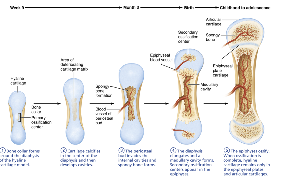
Bone collar forms around the diaphyseal margins of the cartilage model. Perichondrium turns into periosteum, osteoblasts start forming bone on outside of shaft. Chondrocytes in center of shaft hypertrophy (grow bigger)


Hypertrophied chondrocytes in the center of the shaft calcify the surrounding cartilage matrix, and cavities develop. Calcified matrix prevents diffusion of nutrients, chondrocytes die and matrix begins to deteriorate, creating cavities

The periosteal bud invades internal cavities and forms early spongy bone. Osteoclasts break down calcified cartilage, and osteoprogentior cells create osteoblasts, which secrete osteoid around remaining cartilage fragments. Early spongy bone is formed as the primary ossification center
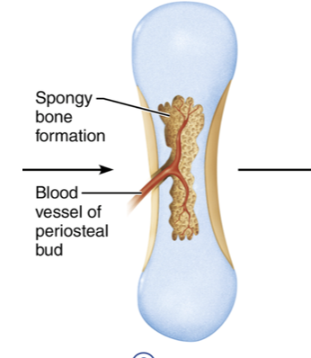
Starter kit containing nerves, vessels, red marrow elements, osteoprogenitor cells, and osteoclasts
Medullary cavity forms, the diaphysis elongates, and secondary ossification centers form at the epiphyses. Epiphyseal cartilage grows at its ends, lengthening model. Primary ossification center expands with growth of overall model. Osteoclasts resorb early trabecular bone at center of diaphysis, forming marrow cavity. Secondary ossification centers established in epiphyses by periosteal buds
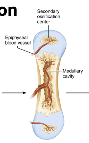
Endochondral ossification step 5
Epiphyses ossify, articular cartilage and growth plate are remaining cartilage. Secondary ossification centers follow same sequence as primary, except trabecular bone is maintained (no medullary cavity in epiphyses). Remaining hyaline cartilage makes up growth plate and articular cartilage on epiphyses

Hyaline cartilage located between the epiphysis and diaphysis
Intramembranous ossification steps
Mesenchymal stem cells become osteoblasts and form an ossification center
Osteoid deposited and calcified around blood vessels, forming early spongy bone
Mesenchyme condenses on outer face of bone, forms periosteum
Compact bone replaces early spongy bone deep to periosteum
Red marrow fills spaces between trabeculae of spongy bone

Occurs at the epiphyseal growth plate. Involves 5 zones: resting, proliferation, hypertrophic, calcification, and ossification
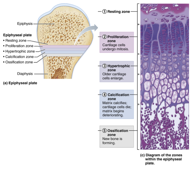
Resting zone of bone growth
Proliferation zone of bone growth
Chondrocytes line up in columns and divide, pushing the epiphysis away from the diaphysis
Hypertrophic zone of bone growth
Calcification zone of bone growth
Cartilage calcifies ,chondrocytes die, and the matrix deteriorates. Former lacunae provide spaces that are invaded by blood vessels
Ossification zone of bone growth
Remnants of calcified matrix are eroded by osteoclasts, and new bone replaces them through osteoblast activity, and marrow elements are introduced
Osteoblasts from osteogenic layer of periosteum secrete osteoid on outer surface of bone, while osteoclasts resorb bone from the inner surface
Bone growth in width during early development
More bone is deposited than lost
Bone growth in width with aging
More bone is lost than gained
Bone tissue becomes more mineralized with time (more brittle) and accumulates microdamage. Remodeling isthe balanced resorption of older bone and replacement by new bone
Bone remodeling process
Osteoclasts move along the bone surface and create depressions as acids and enzymes break down the matrix. Osteoblasts follow osteoclasts, depositing osteoid. Osteoid calcification occurs
Deposited proteins bind Ca 2+
Increase in local Ca 2+ concentration causes osteoblasts to release alkaline phosphatase (ALP)
ALP cleaves phosphate ions off osteoid proteins
Ca 2+ and phosphate forms hydroxyapatite crystal formation
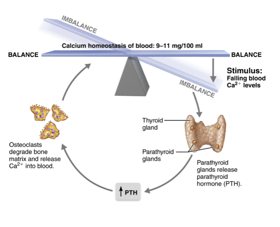
Ca 2+ is constantly deposited into and withdrawn from bone to maintain extracellular calcium homeostasis
PTH negative feedback loop
Low Ca 2+ causes PTH to be released
PTH stimulates release of signaling protein RANK-L from osteoblasts, which increases osteoclast formation and resorption activity
Resorption release Ca 2+ from bone
As Ca 2+ levels rise, PTH release ends and resorption returns to normal
RANK-L
Protein from osteoblasts which increases osetoclast formation and resorption activity
PTH
Parathyroid hormone, released by low Ca 2+ levels and stimulate RANK-L to increase osteoclast activity and resorption
Remodeling and physical activity process
Mechanical stress causes bone to bend slightly
Causes ion-containing fluid to move through canaliculi
Osteocytes sense fluid movement and signal to osteoblasts/clasts to remodel accordingly
Aging shifts remodeling toward net resorption, mainly due to hormonal decline, particularly estrogen
Role of estrogen in bone
Has protective effects on endosteal bone and keeps osteoclasts in check
Estrogen and bone loss
Estrogen decline leads to increased resorption, causing lower bone density
Bone density loss determined by sex
Post-menopausal decline in estrogen results in increased rate of bone density loss in females compared to males.
Weight-bearing exercise, hormone replacement therapy, and RANK-L inhibitors
Bone fracture repair process
Inflammation: hematoma forms from torn vessels and clots, bone cells die and tissue is inflamed
Fibrocartilaginous (soft) callus forms from nearby fibroblasts and chondroblasts
Fibrocartilaginous callus replaced by bony (hard) callus (disorganized spongy bone) from osteoblast activity
Bony callus is continually remodeled until fracture completely repaired
Soft callus formed after a bone fracture from nearby fibroblasts and chondroblasts
Hard callus formed after bone fracture made of disorganized spongy bone, formed through osteoblast activity