Looks like no one added any tags here yet for you.
CNS
Organs: brain and spinal cord
Function: responsible for integrating and processing information
PNS
includes: cranial nerves and spinal nerves
Function: communication between CNS and rest of the body
Sensory (afferent) division
Somatic and visceral sensory nerve fibers
Conducts impulses from receptors to the CNS
Motor (efferent) division
Motor nerve fibers
Conducts impulses from the CNS to effectors (muscles and glands)
Two divisions (SNS and ANS)
Somatic Nervous System
Controls voluntary motor nerve fibers
Conducts impulses from the CNS to skeletal muscles
Autonomic Nervous System
Regulates involuntary motor nerve fibers
Conducts impulses from the CNS to cardiac/smooth muscle and glands
Two further divisions (Sympathetic and Parasympathetic)
Sympathetic Division
“fight-or-flight” response, increases alertness and metabolic activity, mobilizes body systems during activity
releases adrenaline
Parasympathetic Division
“Rest-and-digest” response, consereves energy & promotes house-keeping functions furing rest/sleep
Gray Matter
Contains neuron cell bodies, dendrites, and nonmyelinated fibers
Found in outer cortex of the brain, deep nuclei, and the central region of the spinal cord
Primarily responsible for processsing and integration of information
White Matter
Composed of myelinated axons that transmit signals between different parts of the CNS
Found in deep regions of the brain and spinal cord
Facilitates communication between gray matter regions
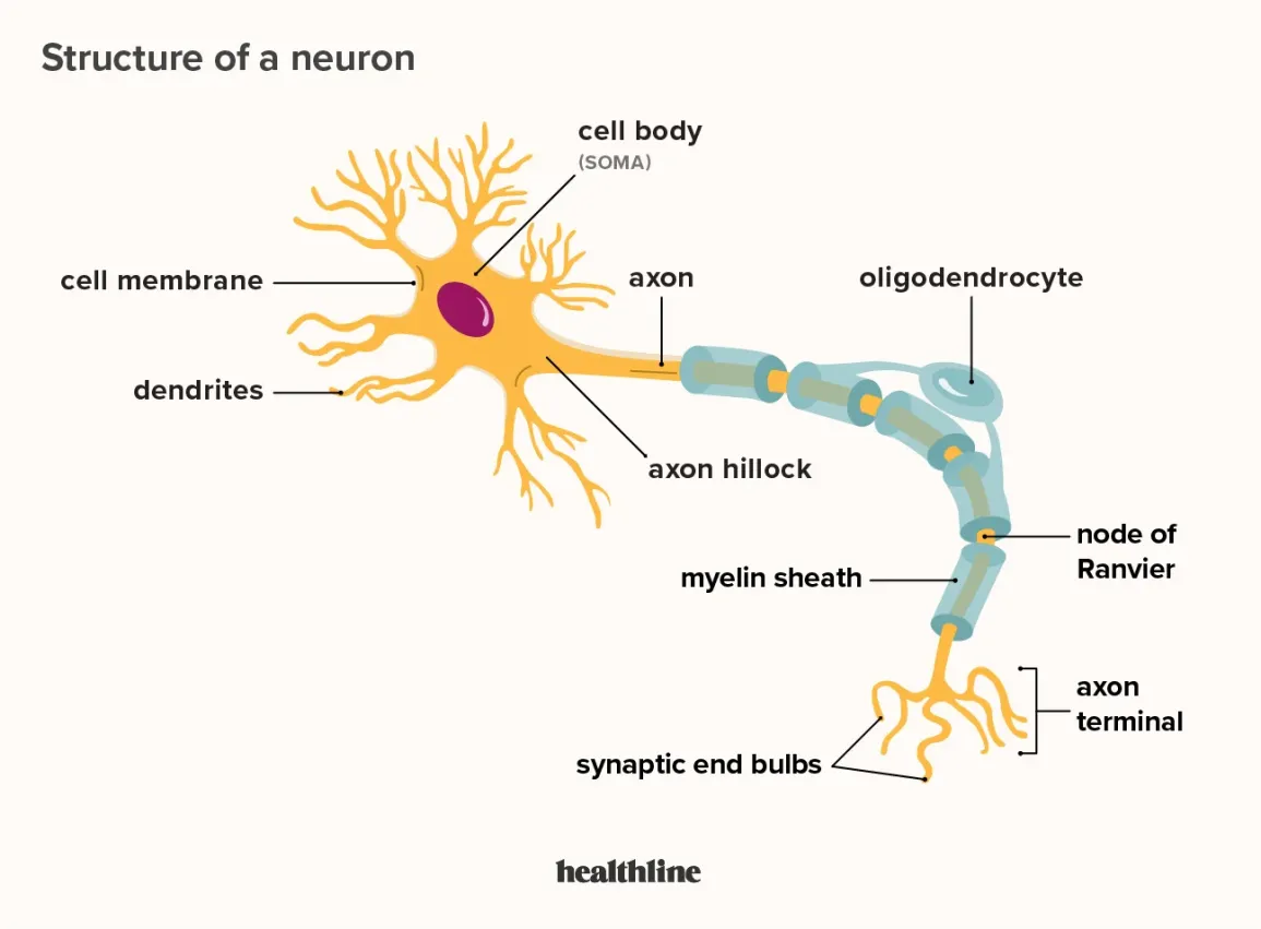
Multipolar Neuron Parts (in order of polarity)
Dendrites: recieve signals from other neurons
Cell Body (Soma): contains the nucleus and organelles; processes incoming signals and is responsible for all biosynthetic activity
Axon Hillock: Initiates/generates action potentials
Axon: transmits electrical signals away from the cell body
Myelin Sheath: increases signal conduction speed
Nodes of Ranvier: gaps in myelin that enable saltatory conduction
Axon Terminals: release neurotransmitters to communicate with other neurons or effectors
CNS Glial Cells - LIST THEM
astrocytes, microglia, ependymal, oligodendrocytes
Astrocytes
Maintain the blood-brain barrier, regulate ions/nutrients, recycle neurotransmitters, and form scar tissue
Oligodendrocytes
Myelinate CNS axons and provide structural support
Microglia
Remove debris, waste, and pathogens via phagocytosis
Act as immune cells
Ependymal Cells
Line brain ventricles and spinal canal, producing, circulating, and monitoring cerebrospinal fluid (CSF)
PNS Glial Cells - LIST
Schwann Cells and Satellite Cells
Schwann Cells
Myelinate axons in the PNS and aid in repair after injury
Satellite Cells
Surround neuron cell bodies in ganglia for support and protection, regulating their environment
Sensation
Detects stimuli from the environment or internal body conditions via sensory receptors
EX: general senses (like touch, pain, temperature, and proprioception) and special senses (like vision, hearing, taste, and smell).
Integration
Processes and interprets sensory information to determine appropriate responses
Occurs within CNS in Phe brain and spinal cord
Response
Produces motor output to muscles or glands, either voluntarily (somatic) or involuntary (autonomic)
Ganglia
Found in the PNS
Nuclei
found in the grey matter of the CNS
Tract
a bundle of axons, or fibers, found in the CNS
Nerve
a bundle of axons, or fibers found in the PNS
Myelin Sheath
A white lipoprotein that insulates and protects axons, increasing nerve impulse speed
How is myelin formed in the PNS?
Schwann cells wrap around a single axon, squeezing out cytoplasm to form a tightly layered sheath
How is myelin formed in the CNS?
Oligodendrocytes extend processes to myelinate multiple axons (up to 60 at once)
What is resting membrane potential?
-70mV
Sodium-Potassium Pump (Na/K +ATPase)
Pumps 3 Na+ out, 2 K+ in, using ATP
Leak Channels
K+ leaks out more than Na+ enters, making the inside more negative
Negatively Charged Proteins
Large anions inside the cell further contribute to negativity
Parts of an Action Potential
Resting State
Depolarization
Rising phase of AP
Falling phase of AP
Undershoot
Depolarization
Voltage-gated Na+ channels open, allowing Na+ influx, making the inside more positive
Peak of AP
(~+30mV): Na+ channels close, and K+ channels open
Repolarization of AP
K+ exits, restoring negativity
Hyperpolarization
Excessive K+ efflux causes a brief dip below resting potential
Absolute Refractory
The time from the opening of Na⁺ channels until they reset - AP cannot occur
Refractory Period
The time in which a neuron cannot trigger another action potential (AP)
Relative Refractory
The period following the absolute refractory period when another AP can only be triggered by an exceptionally strong stimulus
EPSP
Depolarization of the postsynaptic membrane - brings neuron closer to threshold
caused by Na+ or Ca²⁺ influx
IPSP
hyperpolarization of the neuron - moves it away from threshhold
Caused by: K+ efflux or Cl⁻ influx
Cause of EPSP and IPSP
The synapse—a structure that allows a neuron to pass an electrical or chemical signal to another cell
Presynaptic neuron
neuron conducting impulses toward synapse (sends info).
Postsynaptic Neuron
neuron transmitting electrical signal away from synapse (recieves info)
In the PNS, the postsynaptic cell can be a neuron, muscle cell, or gland cell
Chemical Synapse
Chemical signal - neurotransmitter is released from presynaptic neuron
Electrical Synapse
uses gap junctions to send signals from pre to post synaptic neuron
Synaptic Delay
The time required for neurotransmitter release, diffusion, and receptor binding (0.3–5.0 ms). It is the rate-limiting step of neural transmission
Effect of Synaptic Delay
Action potential transmission down an axon is fast, but the synapse slows transmission to the postsynaptic neuron
This delay is not noticeable because it is still very quick
Neurotransmitters
Effects can be excitatory (depolarizing) and/or inhibitory (hyper polarizing)
Acetylcholine
excitatory to vertebeate skeletal muscles; excitatory or inhibitory everywhere elese
Secretion sites: CNS, PNS; vertebrate neuromuscular junction
Norepineohrine
Excitatory or inhibitory - biogenic amines
Secretion sites: CNS; PNS
Dopamine
Generally excitatory; may be inhibitory at some sites - biogenic amines
Sites: CNS;PNS
Serotonin
Generally inhibitory - biogenic amines
Sites: CNS
GABA
inhibitory Amino Acid
Site: CNS; invertebrate
Glycine
Inhibitory Amino Acid
Site: CNS
Glutamate
Excitatory Amino Acid
Site: CNS; invertebrate neuromuscular junction
Aspartate
Excitatory Amino Acid
Site: CNS
Substance P
Excitatory - neuropeptide
Site: CNS; PNS
Met-enkephalin
Inhibitory - Neuropeptide
Site: CNS
Where does the embryonic nervous system develop?
The nervous system originates from the ectoderm, specifically the neuroectoderm
Neural plate
develops from part of the ectoderm and then turns into neural groove
Neural groove
develops from neural plate - these neural folds come together to form the neural tube and crest
What does the neural crest become?
PNS
What does the neural tube become?
CNS
Embryonic Structures —> Adult
Forebrain —> Telencephalon & Diencephalon
Midbrain —> Mesencephalon
Hindbrain —> Metencephalon & Myelencephalon
Telencephalon
Cerebrum (includes cerebral cortex, white matter, basal nuclei)
Diencephalon
Diencephalon (thalamus, hypothalamus, epithalamus)
Mesencephalon
Midbrain (part of brainstem)
Metencephalon
Pons (part of brainstem), cerebellum
Myelencephalon
Medulla oblongata (part of brainstem)
Major Regions of the Adult NS
Cerebrum (including cerebral cortex, white matter, basal nuclei)
Diencephalon (thalamus, hypothalamus, epithalamus)
Brainstem (midbrain, pons, medulla oblongata)
Cerebellum
Spinal cord
Frontal Lobe
Located at the front of the brain (behind the forehead)
Controls movement, thinking, planning, and decision-making
Broca’s area (left side) helps with speech
Parietal Lobe
Located behind the frontal lobe, at the top of the brain
Processes touch, pain, and temperature
Helps with spatial awareness and movement coordination
Temporal Lobe
Located on the sides of the brain (near the ears)
Processes sounds (hearing)
Helps with memory and learning
Wernicke’s area (left side) helps understand language
Occipital Lobe
Located at the back of the brain
Processes vision (color, shape, motion)
Central Sulcus
Deep groove separating the frontal and parietal lobes
Divides motor control (front) and sensory processing (back)
Lateral Sulcus
Separates the frontal and parietal lobes from the temporal lobe
Divides auditory areas from motor and sensory regions
Corpus Callosum
Thick nerve band deep in the brain
Connects the left and right hemispheres for communication
Commissural fibers
WHITE MATTER
horizontal fibers that connect gray matter of two hemispheres
Association fibers
WHITE MATTER
horizontal running fibers that connect different parts of same hemisphere
Projection fibers
WHITE MATTER
vertical fibers that connect hemispheres with lower brain or spinal cord
Internal capsule
projection fibers on each side of brain stem form compact band
- Passes between thalamus and some of basal nuclei
Corona radiata
WHITE MATTER
projection fibers that radiate through cerebral white matter to cortex
basal ganglia function
movement, decision making and reward/addiction
Diencephalon Location
Between brainstem and cerebrum
Diencephalon Function
memory processing and emotional response, relay and control center
Midbrain
BRAIN STEM
coordinates sensory representations of the visual, auditory, and somatosensory perceptual spaces
Pons
BRAIN STEM
main connection with the cerebellum
Medulla Oblongata
signals nerves to and from your body
Controls heart rate, blood pressure, respiration
Reflex centers for vomiting, swallowing, coughing
BRAIN STEM
Cerebellum Structure
FILL IN
Brain Stem
connects the cerebrum of the brain to the spinal cord and cerebellum
Limbic System Location
Encircles the upper brainstem, includes parts of the cerebrum (hippocampus, amygdaloid body, cingulate gyrus) and connects to the hypothalamus
Limbic System Function
Emotional responses to odors and memory
Amygdaloid body
Recognizes angry or fearful expressions
Assesses danger and triggers fear response
Cingulate Gyrus
Helps express emotions through gestures
Aids in resolving mental conflict
Where is the limbic system output?
Hypothalamus
What does the hypothalamus play a role in?
Influences psychosomatic illnesses (stress-related disorders)
What helps form and store memories?
Hippocampus & amygdaloid body
Storage site of neural stem cells