Muscular System
 Call Kai
 Learn
Learn Practice Test
 Spaced Repetition
 Match
Match Flashcards
 Knowt Play
1/29

There's no tags or description
Looks like no tags are added yet.
Name | Mastery | Learn | Test | Matching | Spaced | Call with Kai | Chat |
|---|
No analytics yet
Send a link to your students to track their progress
30 Terms
Sagittal Plane - Flexion
Decreases the angles of two body parts (e.g. bending elbows)
Sagittal Plane - Extension
Increases the angles of two body parts (e.g. straightening elbows)
Sagittal plane - Dorsiflexion
Ankle flexion - bring toes toward shin (toward up of the body)

Sagittal plane - Plantar flexion
Ankle (toes) pointing toward the ground.

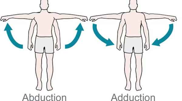
Frontal Plane - Abduction
Move away from the midline

Frontal Plane - Adduction
Move toward the midline
Frontal Plane - Elevation
move upward, superior (e.g. closing mouth - mandible goes up)
Frontal Plane - Depression
move downward, inferior (e.g. shoulder shrug)
Frontal plane - Inversion
lifting medial border of the foot (put weight inside the foot)
Front plane - Eversion
lifting lateral border of the foot (put weight outside of foot)
Transverse plane - Pronation
palm face down to floor; rotate inward/ medially 向內
Transverse plane - Supination
palm and wrist face up; rotate outward/ laterally 向外
Transverse plane - Horizontal Adduction
Angle between two joints decreases

Transverse plane - Rotation
Pivoting on the axis (rotating head, thighs)
Skeletal Muscle structures
Multi-nucleated; Striated; somatic voluntary control; tubular and cylinder shaped
Smooth muscle
Autonomic control; can use myogenic mechanism to contract; non-striated; uninucleate; spindle-shaped

Cardiac msucle
Autonomic control; myogenic; striated and branched; uninucleate BUT may have two nuclei; connected by intercalated discs with gap junctions
Myogenic mechanism
muscle can contract on its own without nerve signals

Label the t-tubule system from the top
Sarcolemma
Sarcoplasmic Reticulum
Terminal cisternae (two chambers that sandwich T-tubules)
Transverse (T) Tubules
Triad (the sandwich structure of T-tubules and terminal cisternae)
Where is the AP impulse going in skeletal muscle fibers for contraction?
AP propagated to sarcolemma, AP impulse send to the t-tubles and depolarize to SR, Ca2+ channels on SR open to release Ca2+ ions. Muscle contraction. ATP-Calcium pumps within the SR membrane pump Ca2+ back into SR and muscle relax.
Sarcomere
Functional contractile unit of muscle between two Z-lines
What forms myofibril?
Many sarcomere lie end to end.
How does sarcomere contract?
Myosin head attach to actin binding site and form cross bridges. Pulling actin toward the mid line of sarcomere, and myosin is close to Z-line (power stroke).
Thin filament structure
Actin molecules form double helical strands.
Troponin complex (the one inside - connect to G-actin; middle - hold tropomyosin in place; top one - link to calcium)
Tropomyosin - block actin binding site for link up myosin when Ca2+ is not presented.

Thick filament structure
Two myosin molecules (looooonggg) form double strands.
Myosin head have Actin-binding site and ATPase site
What is the role of ATP in slide filament contraction?
ATP binds to the myosin head ATPase site; ATP is hydrolyzed into ADP + Pi. This breakdown causes myosin to release from actin and reset myosin to bind to another actin site.
Three types of skeletal muscle fibers
Slow Oxidative (SO Type I); Red
Fast Oxidative (FOG Type IIa); Red
Fast Glycolytic (FG Type IIb); White
The characteristics of Slow Oxidative Type I Fibers
Small diameter, high myoglobin = Red, high capillary density, many mitochondria, rely primarily on aerobic oxidative phosphorylation (low glycolytic enzyme content)
For prolonged, low-intensity aerobic activities, maintain posture
The characteristics of Fast Glycolytic Type II Fibers
Fast twitch, Larger diameter, low myoglobin and low capillary density = White, few mitochondria = forced to rely on glycolysis, high glycolytic enzyme content
For short bursts, e.g. sprinting/ heavy lifting
Fast Oxidative Type IIa Fibers’ characteristics are all moderate/ between type I and IIb
For moderate intensity activities, running
Pacemaker cell pathways
Sinoatrial (SA) node
Atrioventricular (AV) node