HIstology of Early Tooth Development
 Call Kai
 Learn
Learn Practice Test
 Spaced Repetition
 Match
Match Flashcards
 Knowt Play
1/77

There's no tags or description
Looks like no tags are added yet.
Name | Mastery | Learn | Test | Matching | Spaced | Call with Kai | Chat |
|---|
No analytics yet
Send a link to your students to track their progress
78 Terms
T/F Each tooth is considered an organ
true
define odontogenesis
tooth development
what are the 2 basic tissues required to start odontogenesis
oral epithelium and ectomesenchyme
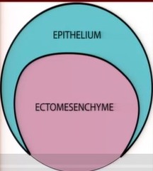
what is oral epithelium derived from
the oral ectoderm
what does the oral epithelium form
the enamel organ
where is the ectomesenchyme coming from
the neural crest
what does the ectomesenchyme form
dentin and pulp in the crown and root
what is the neural crest
transient vertebrate embryonic cells
what is the first sign of tooth development
formation of dental lamina (initiation)
what occurs in the initiation step of tooth development
the formation of the dental lamina
when does the formation of the dental lamina occur
week 6 in utero
where can we see the dental lamina
looking down on developing arch in a jaw
the dental lamina is _________ into the ectomesenchyme
invaginating
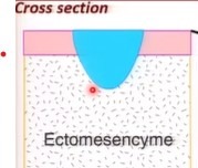
the horseshoe shape describes how-
the oral epithelium is thickening to form the developing arch
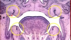
pig dental lamina
circles show deep invaginations of dental lamina
what is the first tooth stage
the formation of the tooth bud
the formation of the dental placode is the start of what stage
the tooth bug stage
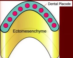
what is the dental placode made of
epithelial thickening (further thickening of the dental lamina)
what does epithelial thickening mean, cellularly
increased division of cells
how many dental placode points are made in EACH arch
10 for the 10 primary teeth on each arch
what shape does the tooth bud resemble
a boot (bulbous)
why does a concavity form in the tooth bud stage
MITOSIS takes place on the superior (top) surface and the distal tip
this forms the boot shape
is the dental lamina still connected to the oral epithelium in the bud stage
yes
at which stage do cells become functionally different
cap stage
what are all the functionally different areas in the cap stage
outer enamel epithelium
inner enamel epithelium
stallate reticulum
dental papilla
dental sac/follicle
cervical loop
what is the outer enamel epithelium made of
simple cuboidal epithelium
where is the outer enamel epithelium found
on the convexity of the cap (the sides that curve OUTWARD)
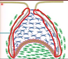
where it the inner enamel epithelium found
on the concavity of the cap (the side that curve inward)
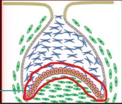
what is the inner enamel epithelium made of
simple low columnar cells
where is the stellate reticulum found
INSIDE the cap
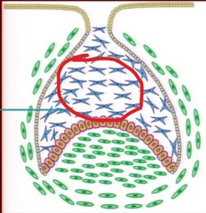
what is between the stellate shaped cells
glycosaminoglycans (GAGS)
why do stellate reticulum have a star shaped
the desmosomes pull on the cells to connect them
what does the space of the stellate reticulum give space for
the formation of the crown
what is the ectomesenchyme below the inner enamel epithelium called
dental papilla
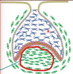
what does the dental papilla eventually form
the dentin and pulp of the crown
what is the ectomesenchyme around the developing enamel organ and dental papilla called
dental sac/follicle
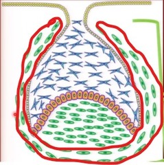
what does the dental sac/follicle form
the cementum, PDL, and alveolar process
what is the cervical loop in the cap stage
the junction of the outer and inner enamel epithelium
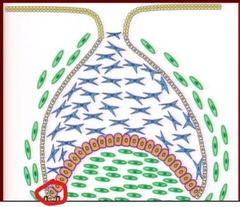
what is the cervical loop used to form
the root
what makes up the enamel epithelium
- outer enamel epithelium
- inner enamel epithelium
- stellate reticulum
define morphodifferentiation
significant changes in shape, ultimately determining the final shape and structure of the tooth crown
what stage can we see the shape of the tooth histologically
the bell stage
Define Histodifferentiation
cells differentiate into their final "phenotype" which determines what they make
what does the inner enamel epithelium differentiate into
ameloblasts
what does the dental papilla differentiate into
odontoblast layer under the inner epithelium
in what direction does differentiation move
cuspal tip to cervical area (neck)
is epithelial tissue vascular?
no
is the outer enamel epithelium vascular?
yes
what stage is the enamel knot found
the bell stage
why does the enamel knot form
the epithelial cells have been told that they need to differentiate into ameloblasts cells, SO they stop dividing
where can the enamel knot be found
near the tip of the inner enamel epithelium
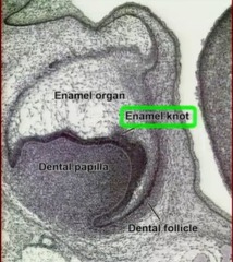
which cells are the first to differentiate
the ones near the enamel knot
why do the sister cells near the enamel knot, not differentiate
they need to keep growing
what is the purpose of the enamel knot
orchestrate cuspal morphogenesis (by differentiating some, but not all cells into ameloblasts)
what stage is the stratum intermedium formed in
the bell stage
where can the stratum intermedium be found
on the inner layer of the inner enamel epithelium
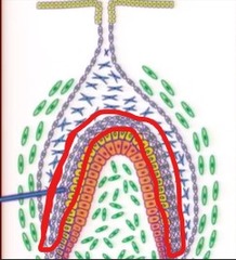
what is the purpose of the stratum intermedium
nutritional multilayer for the formation of enamel (feed ameloblasts)
the remaining cells from the dental papilla that did not differentiate into odontoblasts, form what?
the dental pulp
what is the inner enamel epithelium constrained between
the cervical loop and cuspal tip
why does the cuspal outline form
differential proliferation (mitosis)
at each cusp, what is happening as far as cells
there is an enamel knot of cells that are being differentiated into ameloblasts
in what direction is the future cusp being pushed
up, towards the outer enamel epithelium
what type of tissue is ectomesenchyme
Connective tissue
how is the vestibular lamina formed
thickening of oral epithelium lateral (next to) to the dental lamina
which direction do cells grow in the vestibular lamina
downward (to invaginate the ectomesenchyme)
how is a cavity formed from the vestibular lamina
the cells degenerate (via apoptosis)
what is the vestibule forming
the space between the gingiva and the inside of the cheek
what is the successional lamina and what does it ultimately form
- it branches from the parent lamina that was used for the formation of the primary tooth
- it will ultimately form the successional tooth
how many teeth, IN TOTAL, does the the successional lamina create
20
does the permanent tooth use the same placode as the primary tooth
yes since they are coming from the same parent dental lamina
what is the lateral lamina
connects the developing primary tooth to the parent lamina
the extension of the dental lamina (successional lamina) is always _________ to the primary tooth
lingual
during which stage does the enamel organ lose its connection with the oral epithelium
the bell stage
how does the enamel organ lose its connection to the oral epithelium
the parent lamina degenerates (apoptosis)
when cells degenerate, what could be left behind
islands of epithelial cells (dental lamina) in the connective tissue (ectomesenchyme)
permanent molars are derived direction from the ________ portion of the ________ _________ without a primary tooth precursor
distal; dental lamina
where is the more mineralized enamel found
near the cuspal tip
how is enamel deposited by ameloblasts
in increments