Topic 19- Musculoskeletal System
1/31
There's no tags or description
Looks like no tags are added yet.
Name | Mastery | Learn | Test | Matching | Spaced |
|---|
No study sessions yet.
32 Terms
What are the characteristics of skeletal muscle? (3)
Enables voluntary movement, is stimulated by neurons, and is attached to bones via tendons.
Where does skeletal muscle originate from developmentally? What does this origin also give rise to? (4)
The mesoderm, which also gives rise to cardiac muscle, smooth muscle, kidney cells, and red blood cells.
What makes a muscle fiber unique as a cell? (3)
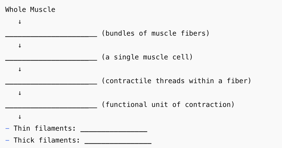
It's multinucleated, does not undergo cytokinesis (elongates instead), and is bundled with others to form whole muscles.
What are myofibrils and what are they made up of?
Long, contractile fibers running the length of muscle fibers, composed of repeating units called sarcomeres.
What is the function of T-tubules in muscle cells and describe them?
They are invaginations of the sarcolemma that carry action potentials deep into the cell, ensuring simultaneous contraction.
What role does the sarcoplasmic reticulum (SR) play in muscle contraction? (2)
It stores calcium ions and releases them into the cytosol to initiate contraction.
What proteins make up the thin filament and what do they do? (2)
Two strands of actin covered by tropomyosin and anchored by troponin; tropomyosin blocks the myosin binding sites at rest.
What is the structure and function of thick filaments?
Composed of ~350 myosin molecules, whose heads have ATPase activity to hydrolyze ATP for contraction.
What is a sarcomere?
The basic unit of contraction in a muscle fiber, bounded by Z lines and centered by an M line.
What happens to the sarcomere during contraction? (2)
It shortens as Z lines move closer together while filament lengths remain constant—this increases filament overlap.
What keeps the muscle at rest and prevents contraction? (2)
Tropomyosin blocks actin’s binding sites, and no Ca²⁺ is present in the cytosol.
How is a muscle contraction initiated? (5)
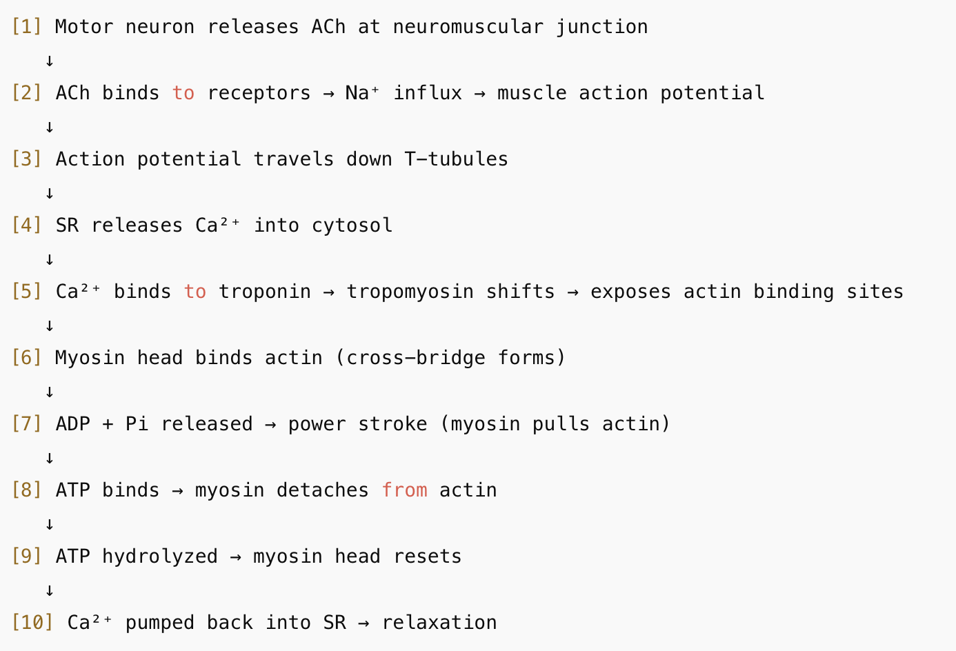
A motor neuron releases ACh → Na⁺ influx → muscle AP → travels down T-tubules → Ca²⁺ released from SR.
What does calcium do once released into the cytosol? (1 → 2)
It binds troponin, causing a conformational change that shifts tropomyosin and exposes actin binding sites.
What is the cross-bridge formation step?
A high-energy myosin head (with ADP + Pi) binds to exposed actin sites.
What happens during the power stroke? (a, b → c)
ADP + Pi are released, and the myosin head pivots, pulling actin toward the M line.
How is the cross-bridge broken?
A new ATP binds to myosin, causing it to release from actin.
How is the myosin head reactivated?
ATP is hydrolyzed into ADP + Pi, returning the head to its high-energy conformation.
When does the cross-bridge cycle repeat?
As long as ATP and Ca²⁺ are available in the muscle fiber.
How does the muscle relax after contraction? (3)
Ca²⁺ is actively pumped back into the SR; tropomyosin re-covers actin; the fiber returns to rest.
What are the energy sources for muscle contraction? (3)
Creatine phosphate transfers phosphate to ADP; glycogen is a glucose store for ATP; cellular respiration produces ATP from glucose and O₂.
What causes rigor mortis? (4)
After death, ATP production stops → myosin can't detach from actin → Ca²⁺ is not reabsorbed → muscles stiffen.
Where is cardiac muscle found and what is its function?
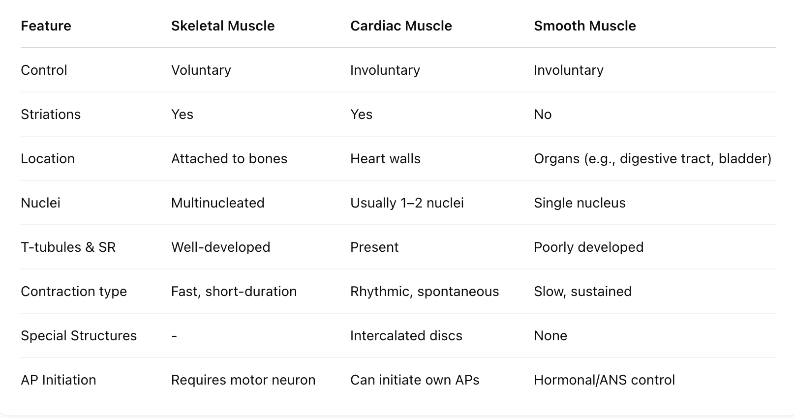
Only in the heart walls; contracts rhythmically to pump blood.
What is the role of pacemaker cells?
They generate spontaneous action potentials without neural input.
What are intercalated discs?
Specialized junctions that allow ion flow between cells, synchronizing contractions.
Can the heart beat without nerve input?
Yes. The heart has an autonomous rhythm and can beat outside the body if oxygen is present.
Where is smooth muscle found? (4)
In the digestive tract, bladder, blood vessels, and reproductive organs.
What distinguishes smooth muscle structurally? (4)
It is involuntary, non-striated, has no sarcomeres or T-tubules, and an underdeveloped SR.
How does smooth muscle contract and what are they controlled by?
Slowly and sustainably, controlled by hormones and the autonomic nervous system.
Comprehend the Levels of Muscle Structure (5 levels)

Sequence the Process of Muscle Contraction (10) from Motor neuron releasing ____ → Ca2+ pumped back into SR

Hypothesize and Diagnose Variability in Contraction when each variable is increased or decreased [Ca2+ in the cytosol, ATP availability, Troponin or Tropomyosin defect, T-tubules or SR damage, Motor Neuron Signal]

Compare and contrast muscle types [voluntary/involuntary, striations, locations, nuclei, T tubules and SR, contraction type, special structures, AP initiation]
