Cardiac Cycle
1/45
There's no tags or description
Looks like no tags are added yet.
Name | Mastery | Learn | Test | Matching | Spaced |
|---|
No study sessions yet.
46 Terms
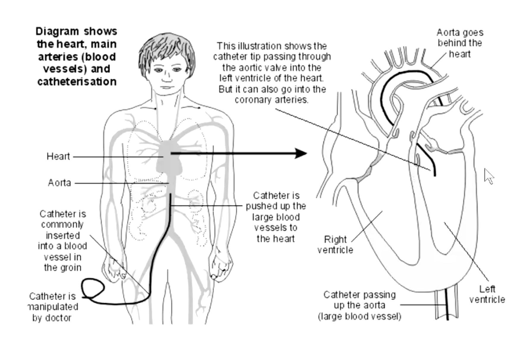
how can we study the mechanical events of the cardiac cycle?
catheters inserted into arteries or veins and passed into chambers of heart
catheters filled with saline and attached to electronic pressure transducers
how is cardiac catheterization done for the left heart?
From the femoral artery, a catheter can advance to aorta, coronary arteries and left ventricle
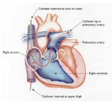
how is cardiac catheterization done for the right heart?
Swan-Ganz catheter
inserted into vein and pushed to entrance to right atrium
balloon at tip is inflated so that flow of blood carries catheter into atrium, right ventricle, and pulmonary artery
catheter tip advanced into pulmonary circulation until it wedges in a small vessel
forward flow of blood in that vessel is blocked and transducer detects “pulmonary wedge pressure” (good indicator of left atrial pressure)
A cardiac cycle consists of precisely timed electrical and mechanical events that are responsible for the rhythmic atrial and ventricular contractions: It includes:
systole- ventricular contraction and ejection of blood
diastole- ventricular relaxation and filling with blood
assuming a resting rate of 72 beats per minutes, one
cardiac cycle takes __ seconds
0.8
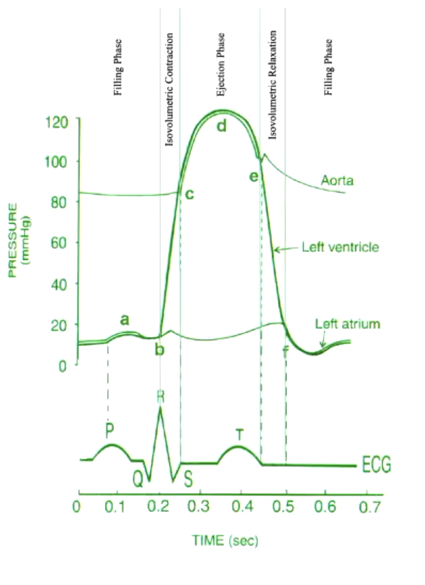
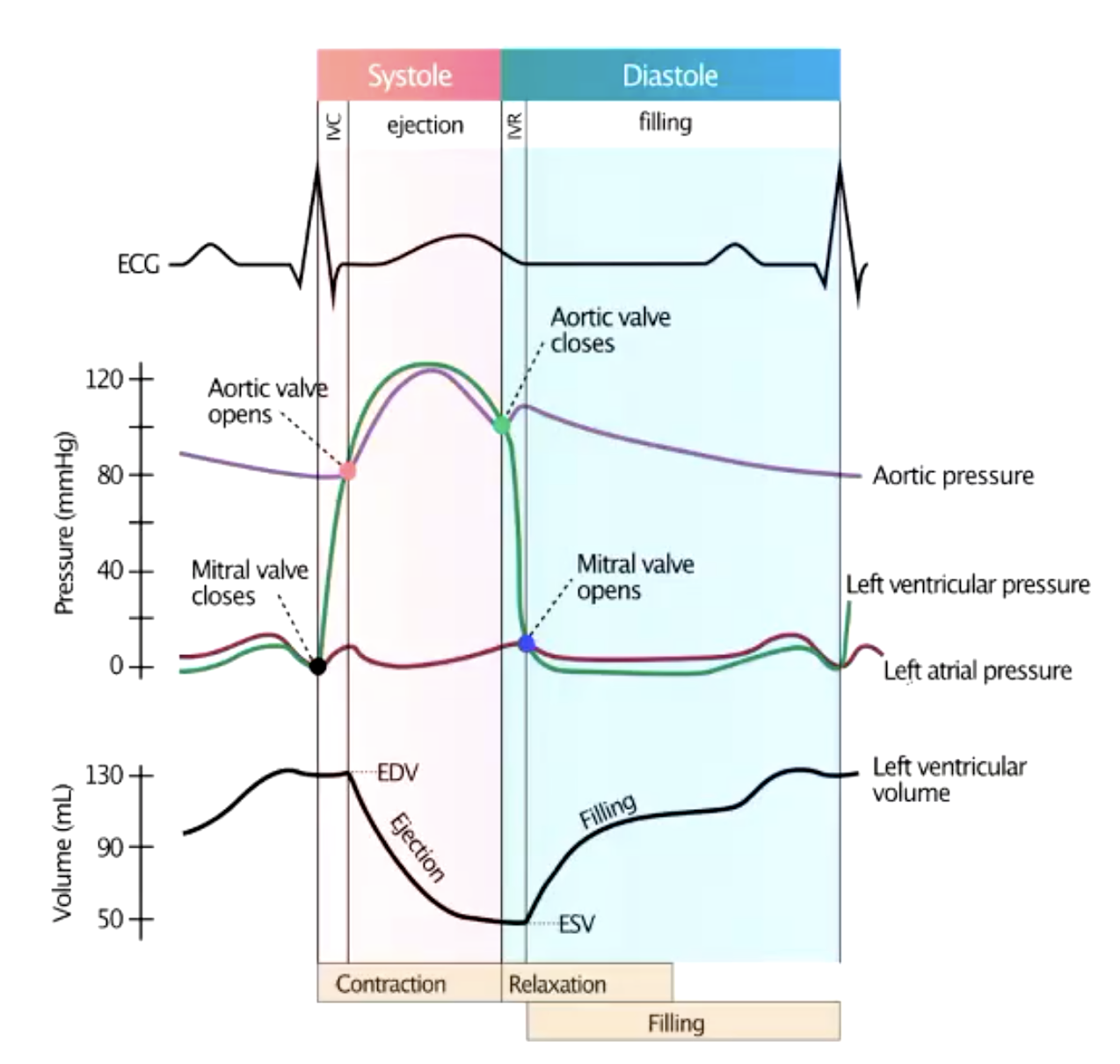
what diagram presents the events of a cardiac cycle for the left-sided cardiac chambers?
Wiggers diagram
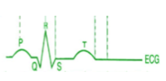
what does the QRS complex indicate on an EKG?
ventricular depolarization (+ atrial repolarization) – triggers ventricular contractions
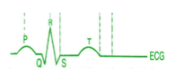
what does the P wave indicate on an EKG?
depolarization of the atria – triggers atrial contraction
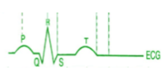
what does the T wave indicate on an EKG?
ventricular repolarization

what happens during atrial systole? (a)
triggered by P wave
small pressure rise as atrium squeezes blood into ventricle
tops off ventricle (already 90-95% full of blood)
what happens during isovolumetric contraction? (b-c)
triggered by QRS complex
mitral valve closes (creates C wave in atrium)
aortic valve is closed
pressure rises rapidly
volume is constant
aortic pressure ~80 mmHg

what is happening at each point of the Wiggers diagram?
a) atrial systole
b-c) isovolumetric contraction
c) opening of aortic valve (ejection)
d) reduction in ejection rate
e-f) isovolumetric relaxation (closing of aortic valve)
f) opening of mitral valve

what happens during opening of aortic valve - ejection? (c)
ventricular pressure exceeds aortic
begins ejection phase
ejection initially rapid
aortic pressure rises

what happens during reduction in ejection rate (d)?
ventricle approaches the end of contraction (T wave occurring)
outward flow declines as kinetic energy of blood decreases
ventricular and aortic pressures begin to fall

what happens during isovolumetric relaxation? (e-f)
forward movement of blood reverses, closing aortic valve
incisura (dichrotic notch)indicates aortic valve closure
ventricular pressure falls precipitously
atrial pressure rises as venous inflow fills the atrium
aortic pressure falls as ejected blood drains away from heart

what happens during opening of mitral valve? (f)
begins rapid filling of ventricle from accumulated blood in atrium
rapid filling followed by reduced filling from pulmonary veins.
when does ventricular systole begin and end?
ventricular systole begins when the mitral valve closes and ends when the aortic valve closes
what are the normal pressure ranges for the left heart?
ventricular:
aortic:
atrial:
ventricular: 0 – 120 mmHg
aortic: 80-120mmHg
atrial: 6-~10 mmHg
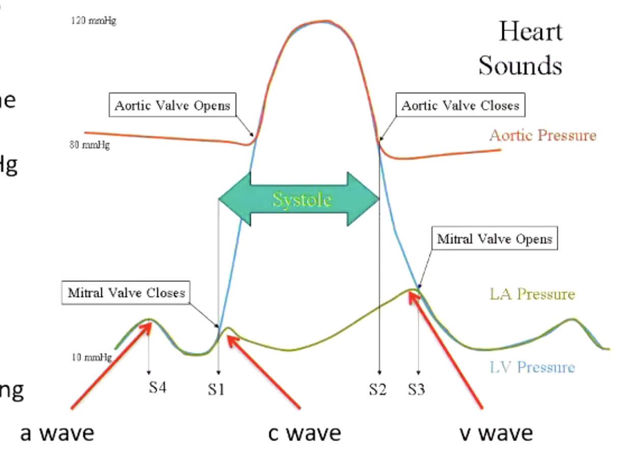
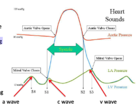
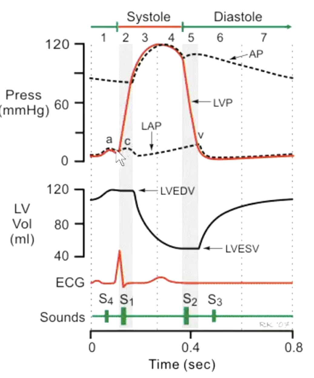
what are the different atrial pressure waves that occur on a Wiggers diagram?
a – atrial systole
c – closure of mitral valve
v – atrial filling and emptying
what is end diastolic volume (EDV)?
atrial systole fills the ventricular to the end diastolic volume (EDV)
what is end systolic volume (ESV)?
at the end of ejection, the volume remaining in the ventricle is the end systolic volume (ESV)
how does volume change during isovolumetric contraction and ejection?
the volume stays constant during isovolumetric contraction, then decreases as blood is ejected into the aorta
how do you calculate stroke volume?
EDV - ESV = stroke volume
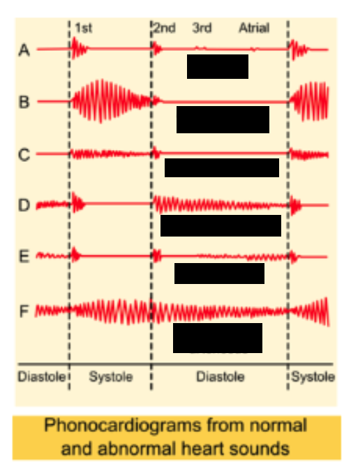
what are the heart sounds? what causes heart sounds?
S1, S2, S3, S4
caused by turbulence or vibrations of blood

what is S1 sounds caused by?
closure of mitral (and tricuspid) valves with the sudden rise in ventricular pressure causes turbulence of blood in ventricles
this is the “lub”

what is S2 sounds caused by?
closure of aortic and pulmonic valves causes vibrations of blood in the high pressure vessels
this is the “dub”

what is S3 sounds caused by?
during rapid filling in some individuals- normal in children (Kentucky)
what is S4 sounds caused by?
vibration of ventricular walls during atrial systole in some individuals (Tennessee)
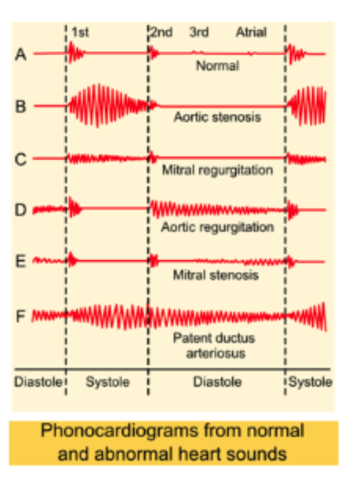
what are abnormal heart sounds that arise from congenital or acquired abnormalities that create turbulence in flow?
Murmurs
murmurs can result from…?
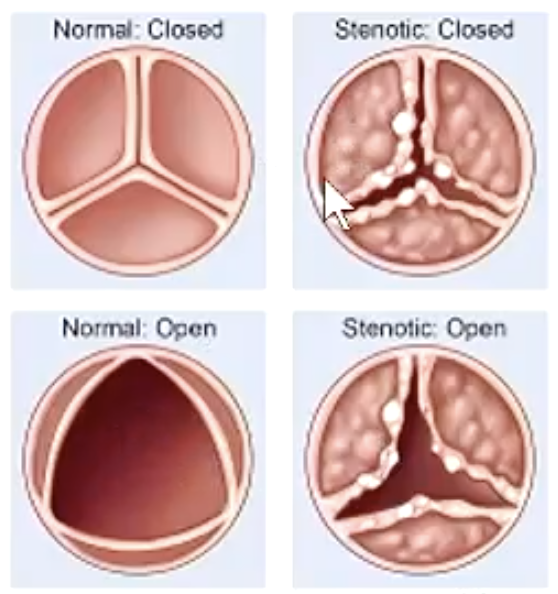
partial obstruction of flow (e.g. aortic stenosis)
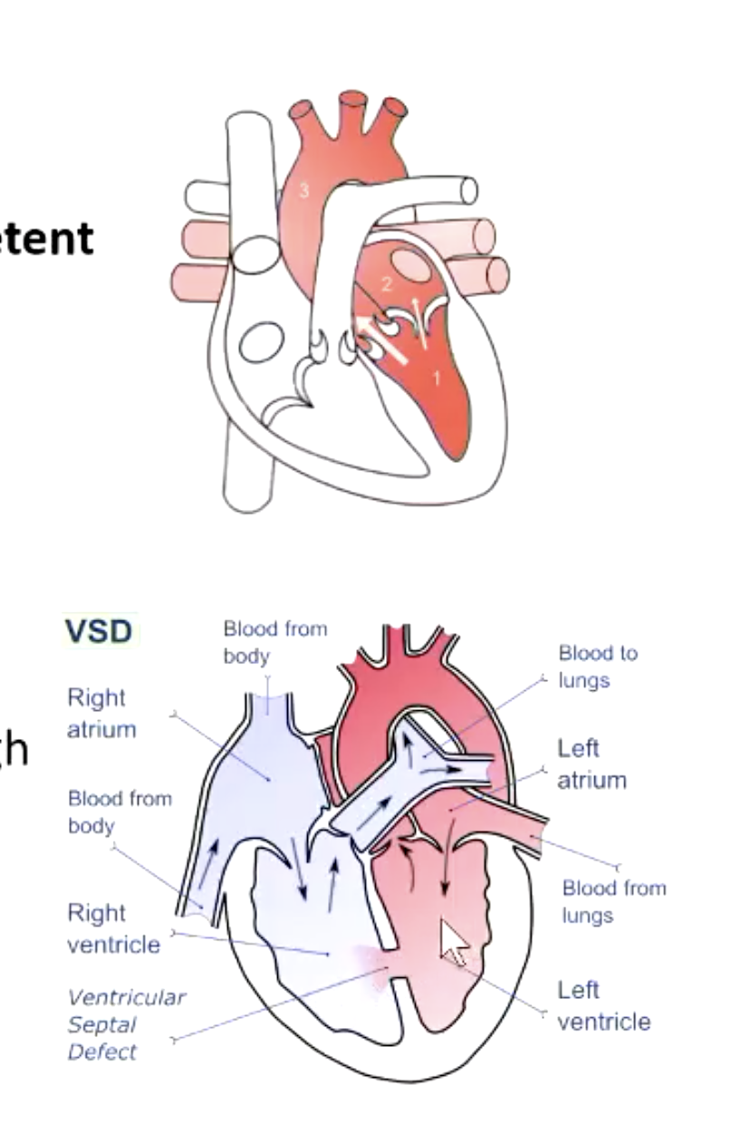
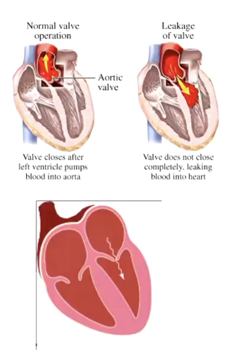
regurgitant flow across an incompetent valve (e.g. mitral regurgitation)
abnormal shunting of blood from one vascular chamber to another lower pressure chamber (ventricular septal defect).
increased flow through normal structures (e.g. high flow rate associated with anemia)
systolic (ejection) murmurs are caused by…?
aortic or pulmonic stenosis
pansystolic murmurs are caused by…?
regurgitant flow through incompetent mitral or tricuspid valve
From left to right ventricle through a ventricular septal defect
diastolic murmurs are caused by…?
regurgitant flow through incompetent aortic or pulmonic valve
mitral or tricuspid stenosis
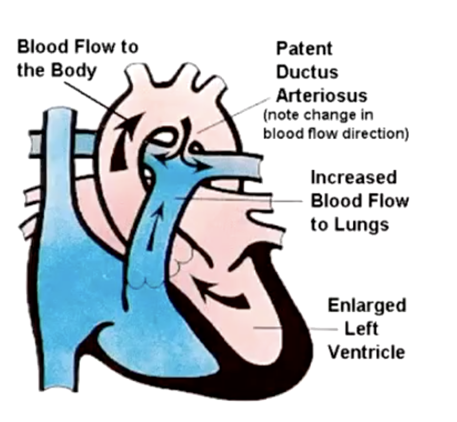
continuous murmurs are caused by…?
Patent ductus arteriosus: pressure gradient between aorta and pulmonary artery persists in both systole and diastole
(heard throughout cardiac cycle)
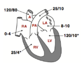
A Wiggers diagram for the right heart has qualitatively all of the features that we saw for the left. However, what’s different?
the pressures in the chambers are lower as shown in the figure
• the right ventricle develops a systolic pressure of only 25 mmHg and drops to 0-4 in diastole
• the pulmonary arterial blood pressures are ~ 25/10
the duration of systole is about ___ of the cycle while diastole is ~__
1/3
2/3
emptying occurs at high/low pressure while filling is mostly passive at high/low pressure
emptying occurs at high pressure while filling is mostly passive at low pressure
t/f: systemic and pulmonary blood flow are equal although SVs can vary from beat to beat
true
how is ejection fraction calculated?
SV/EDV = ejection fraction (55%)
volumes and pressures can change in …?
exercise or disease
all valves are opened/closed during isovolumetric contraction and relaxation
closed
what are the major heart sounds? when do they occur?
S1 and S2 (each occurs when valves close and blood becomes turbulent)
what events occur almost simultaneously with onset of systole?
- QRS complex
- S1
- c wave
- closure of mitral valve
- rise in ventricular pressure
what events occur almost simultaneously with onset of diastole?
- S2
- closure of aortic valve
-incisura
-drop in ventricular pressure
when does the heart pump its stroke volume?
time between S1 and S2