a&p 2 lab practical
 Call Kai
 Learn
Learn Practice Test
 Spaced Repetition
 Match
Match Flashcards
 Knowt Play
1/76

There's no tags or description
Looks like no tags are added yet.
Name | Mastery | Learn | Test | Matching | Spaced | Call with Kai | Chat |
|---|
No analytics yet
Send a link to your students to track their progress
77 Terms
White Matter
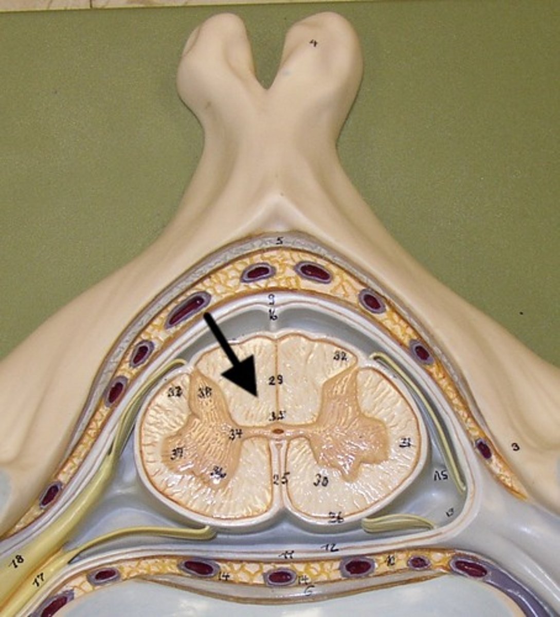
Grey Matter
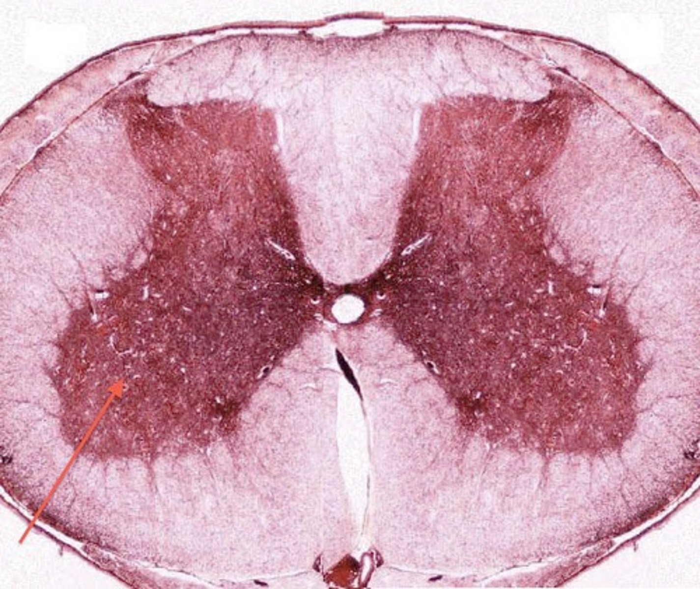
Dorsal Horn
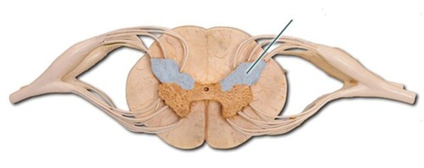
Ventral Horn
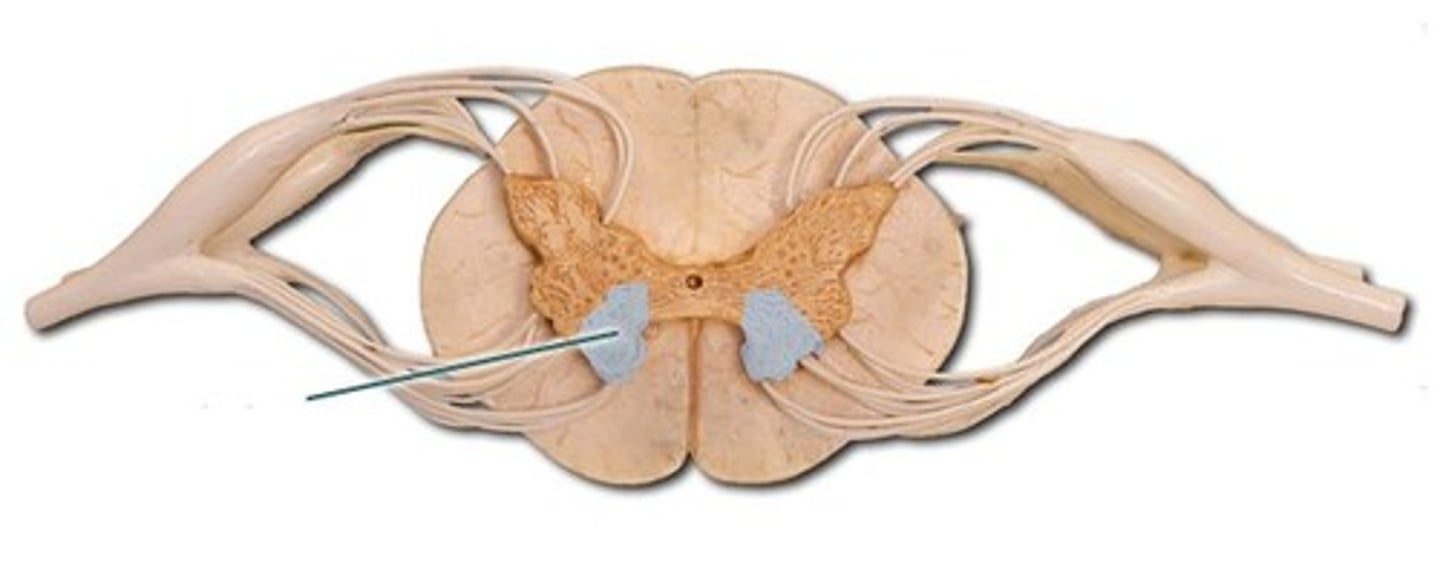
Grey Commissure
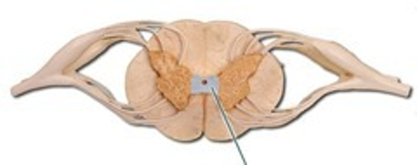
Dura Mater
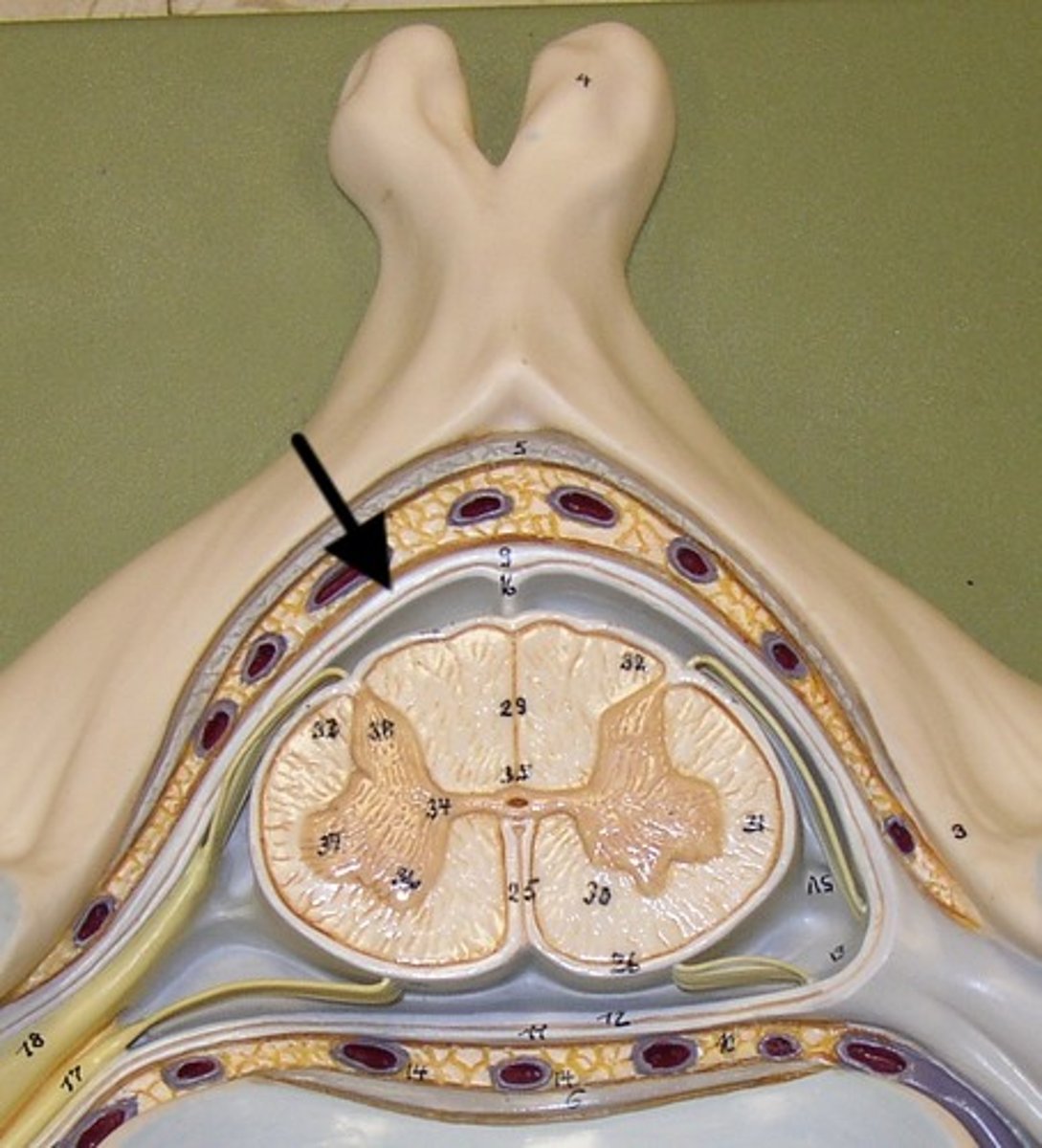
Arachnoid Mater
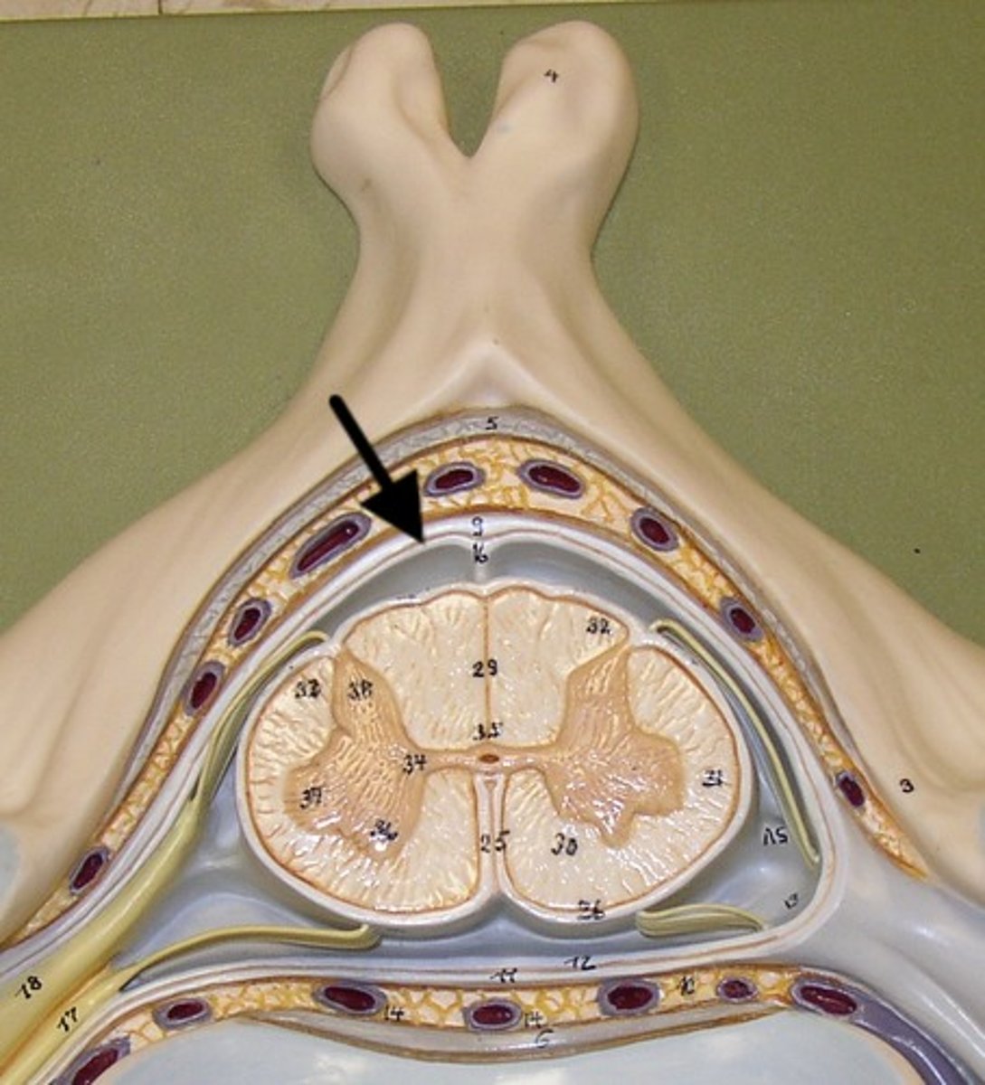
Pia Mater
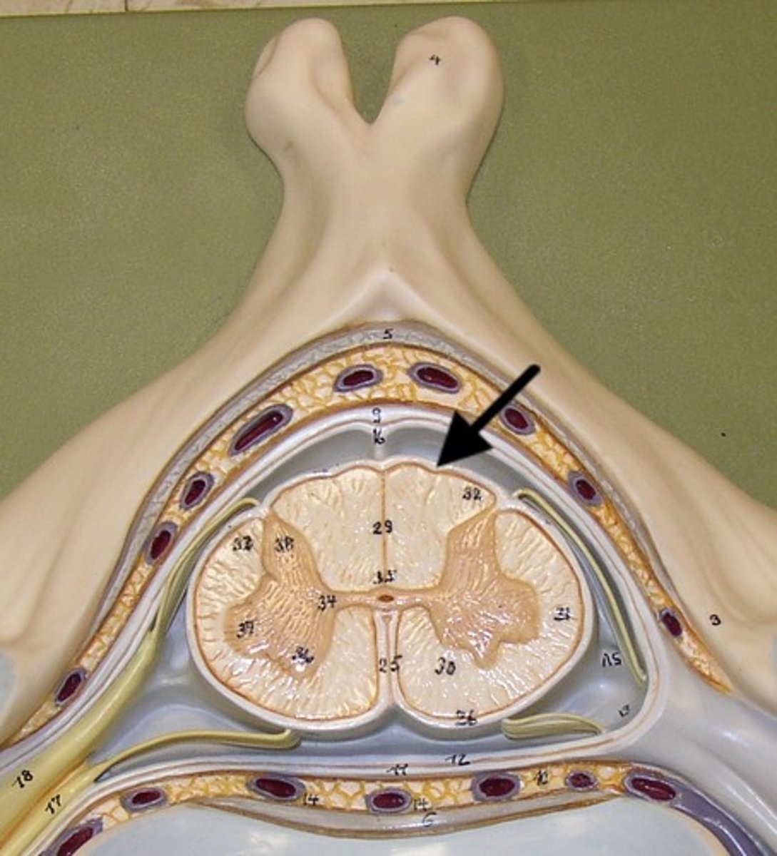
Dorsal Roots
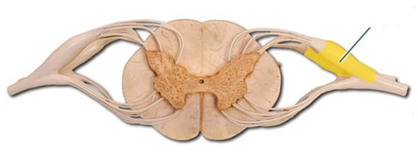
Ventral Roots
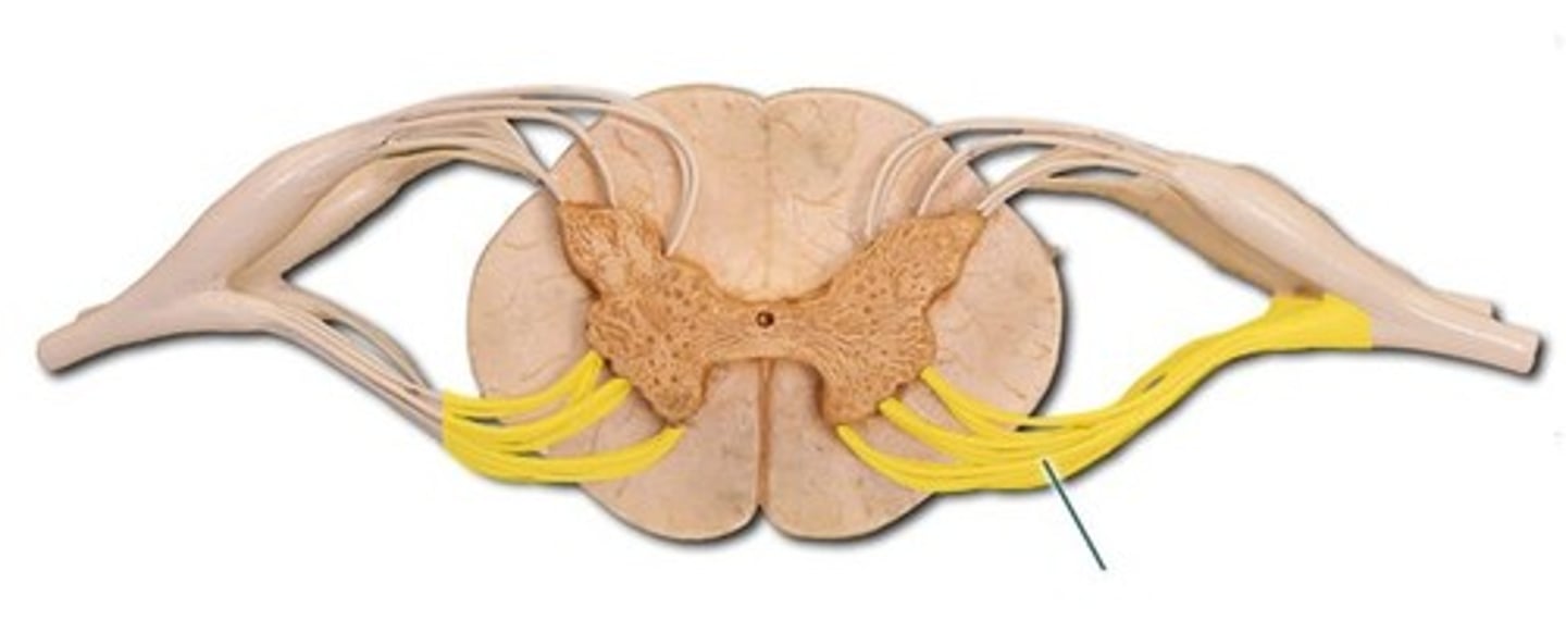
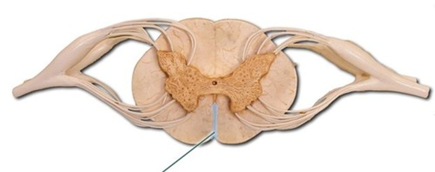
Posterior Median Sulcus
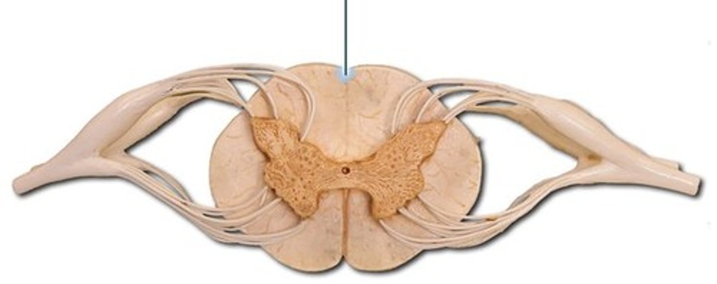
Central Canal
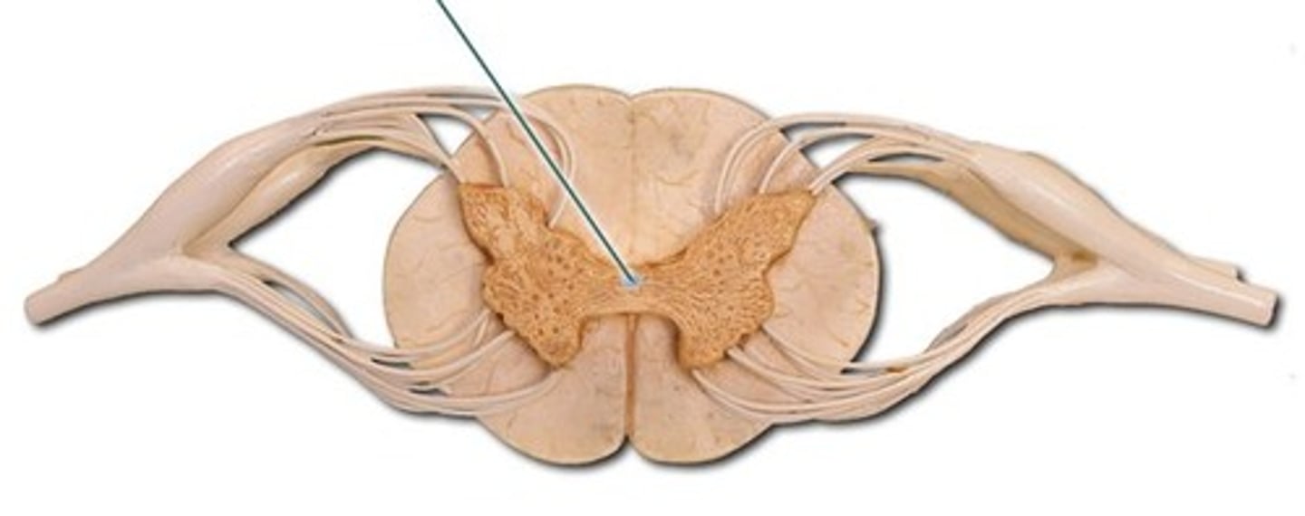
Anterior Median Fissure
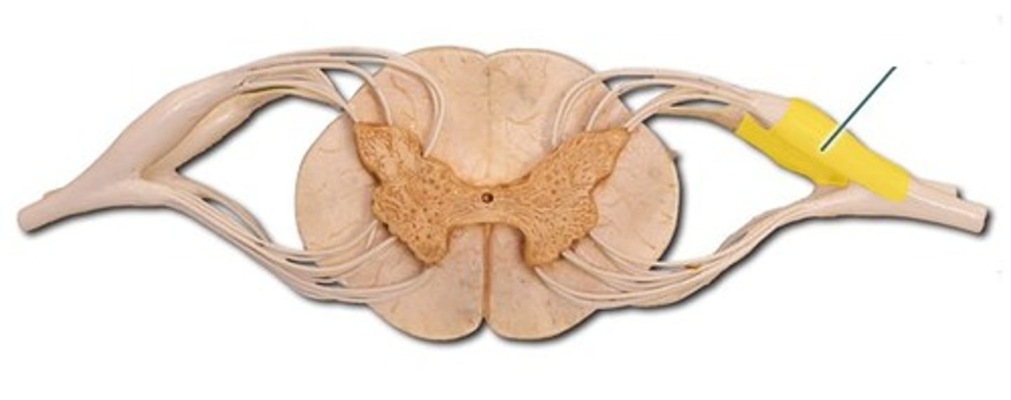
Dorsal Root Ganglion
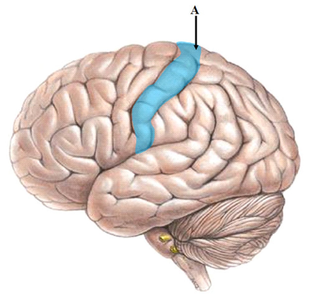
gyrus
ridges
sulcus
grooves
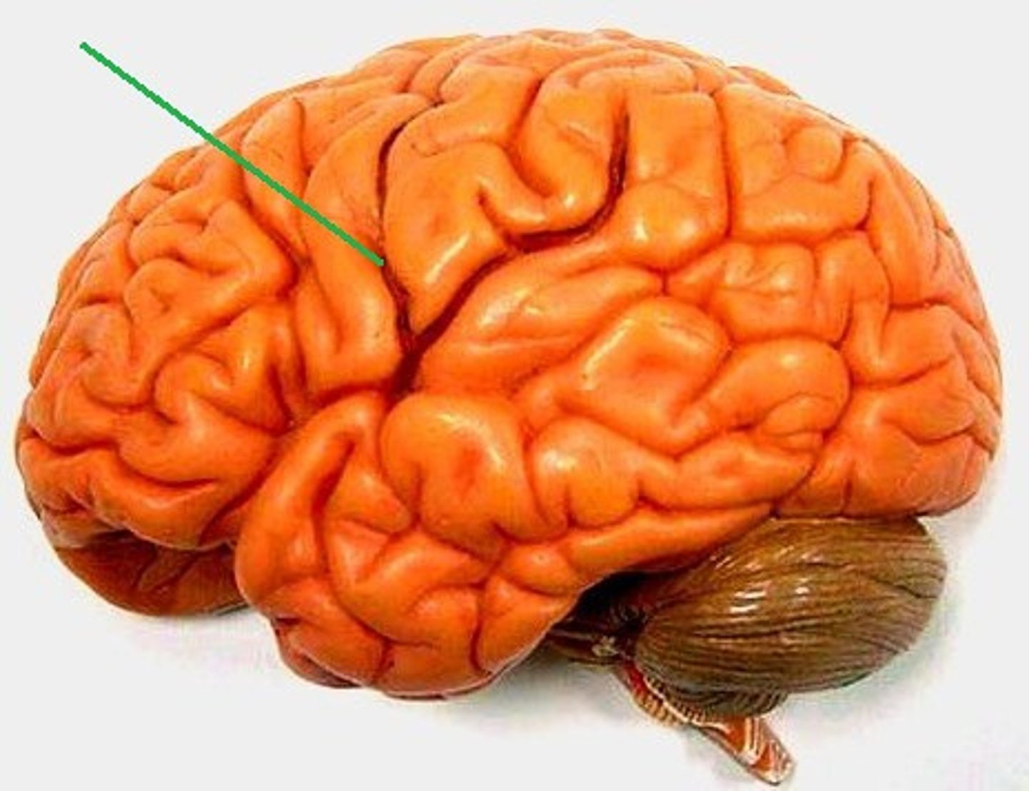
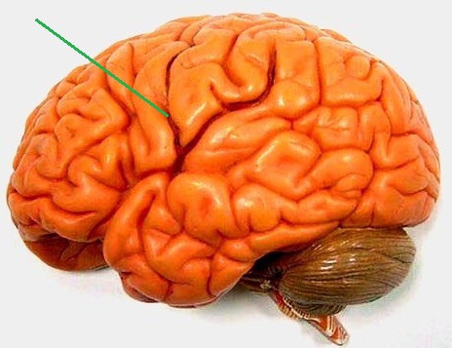
central sulcus
divides the frontal lobe from parietal lobe
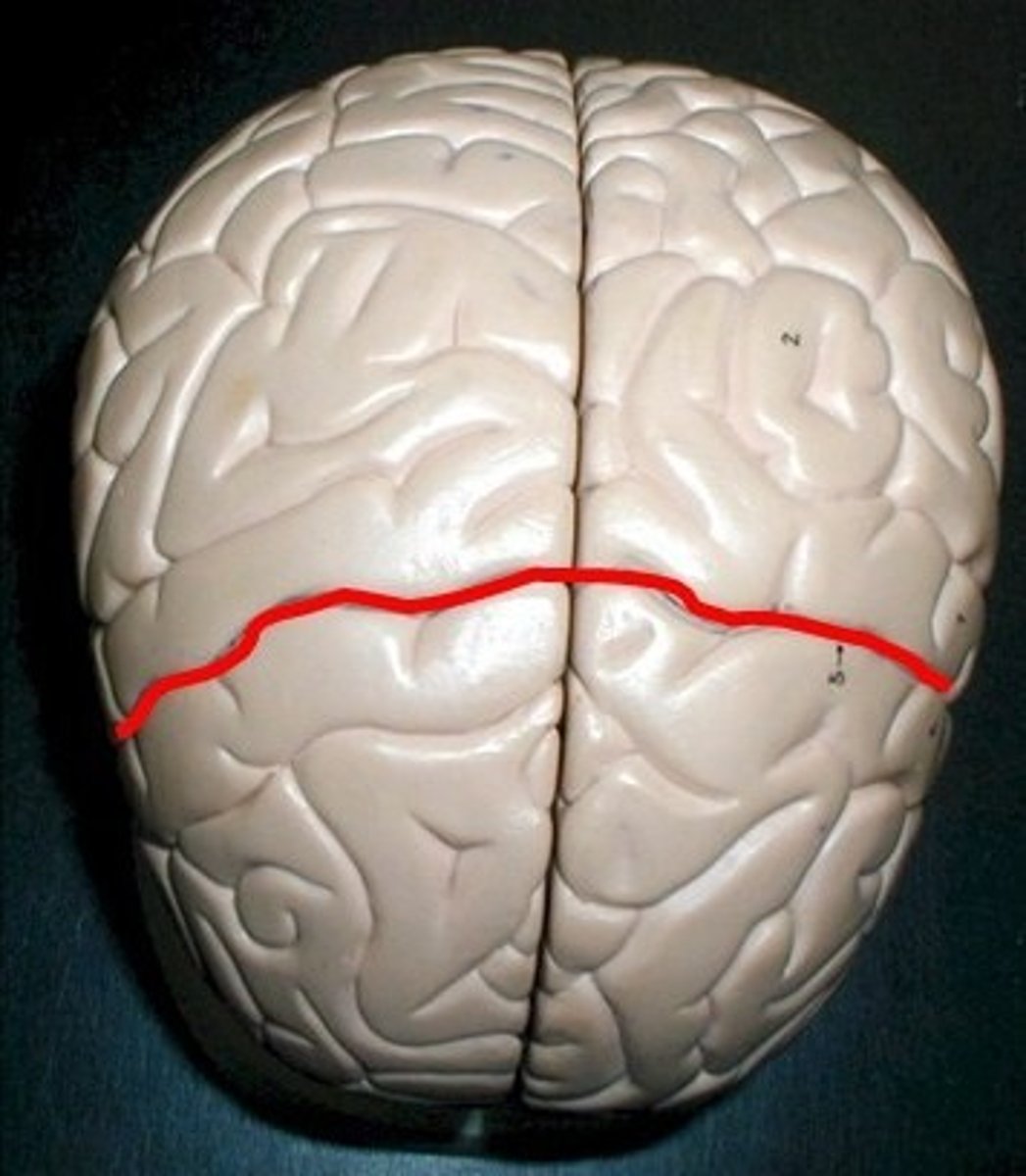
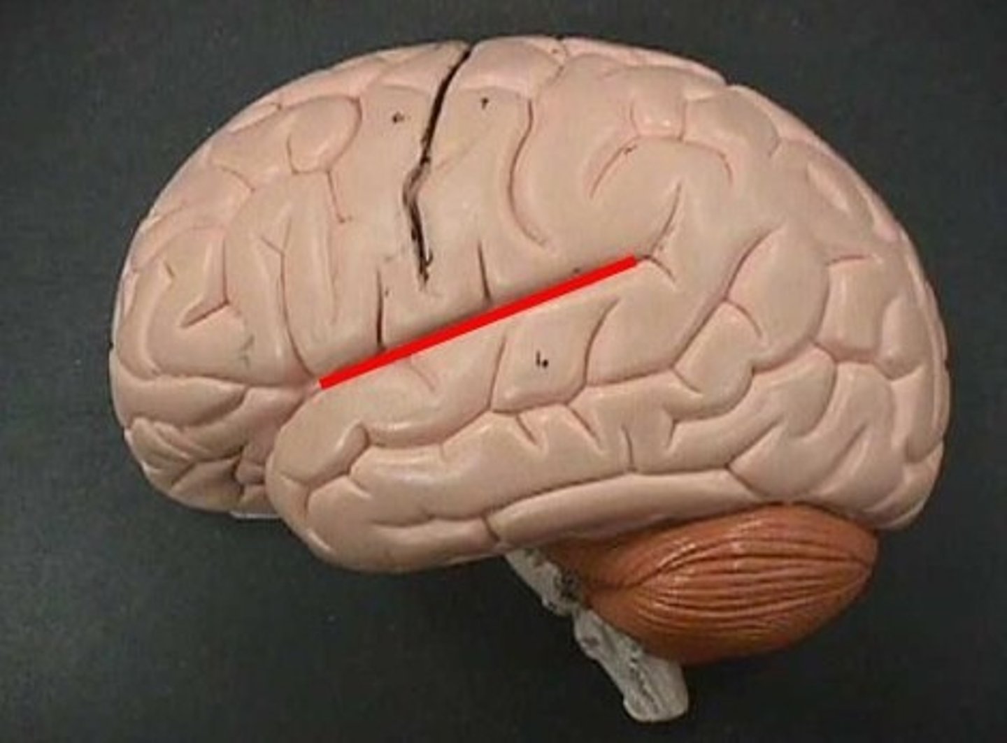
lateral sulcus
divide from temporal
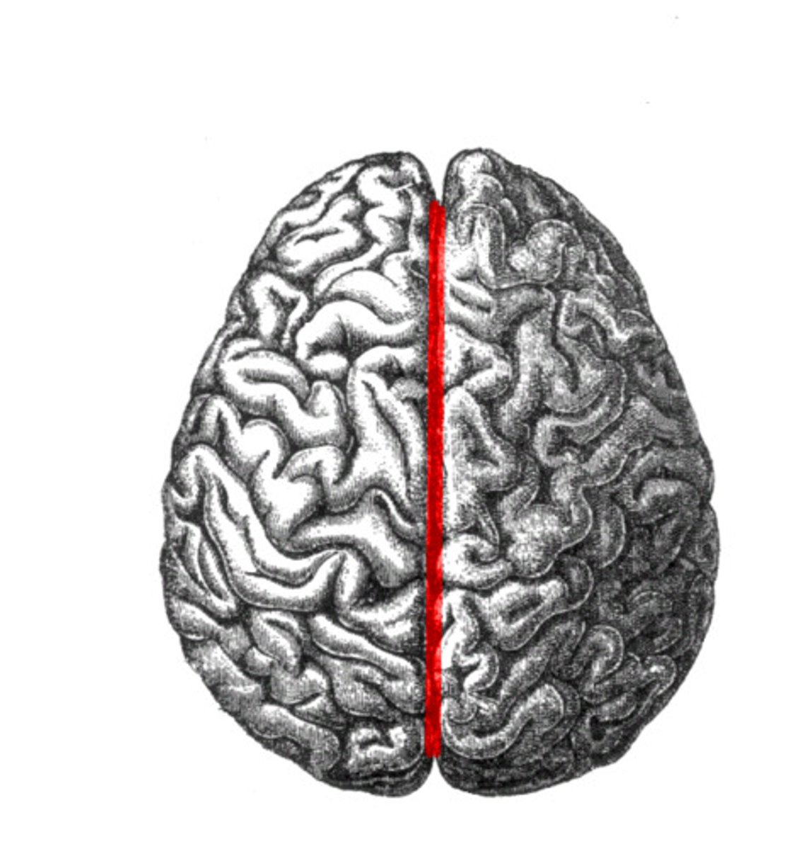
longitudinal sulcus
divides hemi- left and right
pituitary gland
master endocrine gland
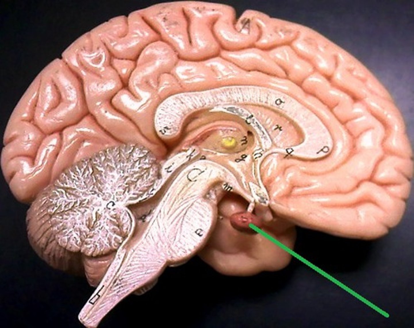
hypothalamus
maintenance function
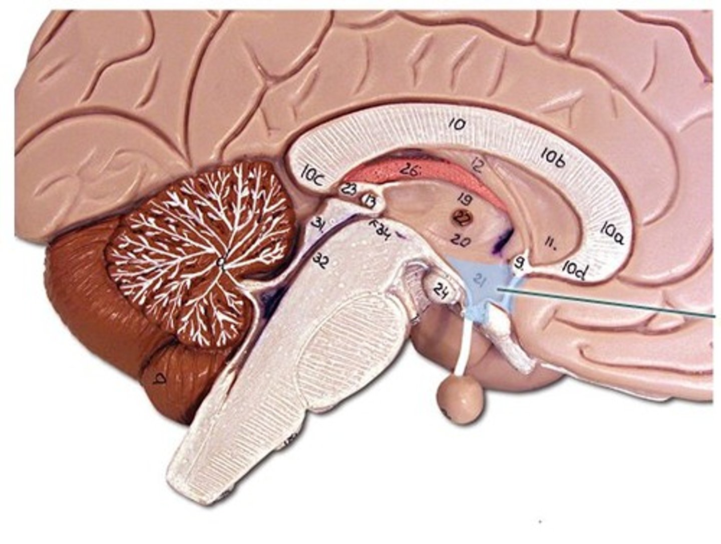
thalamus
relays messages between lower brain and cortex
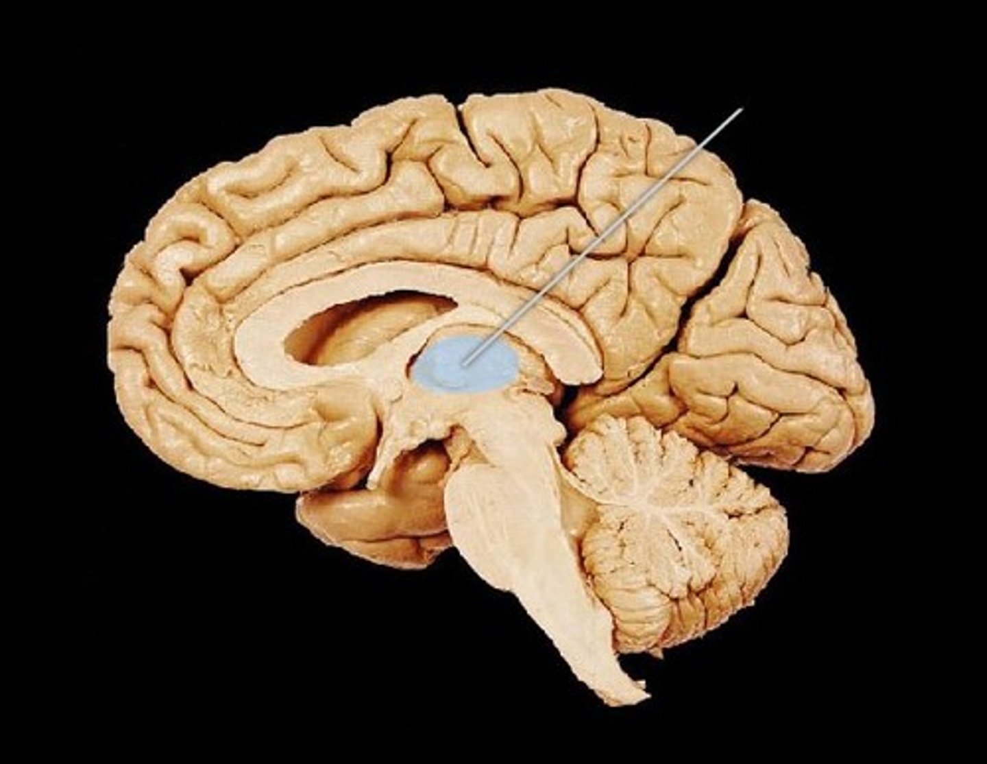
mammillary bodies
used for feeding reflexes
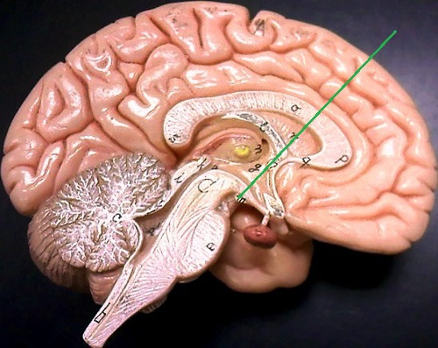
fornix
to limbic system connects mammillary bodies to temporal lobe
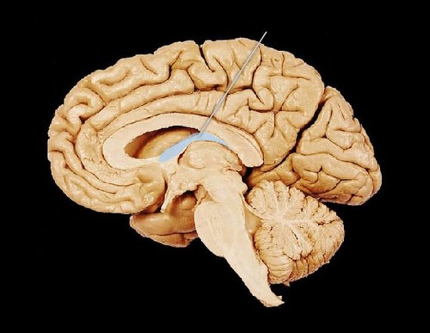
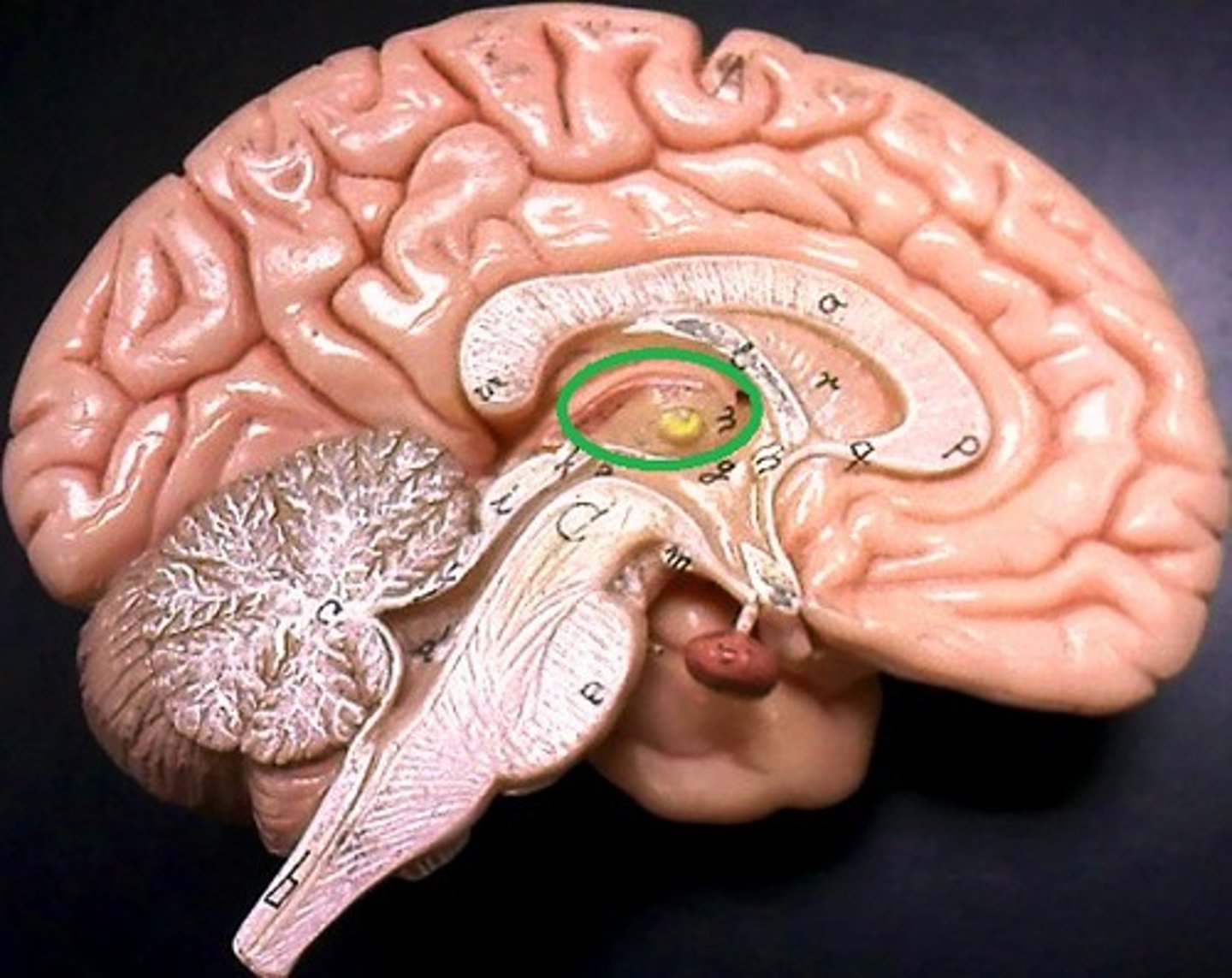
third ventricle
lines the thalamus
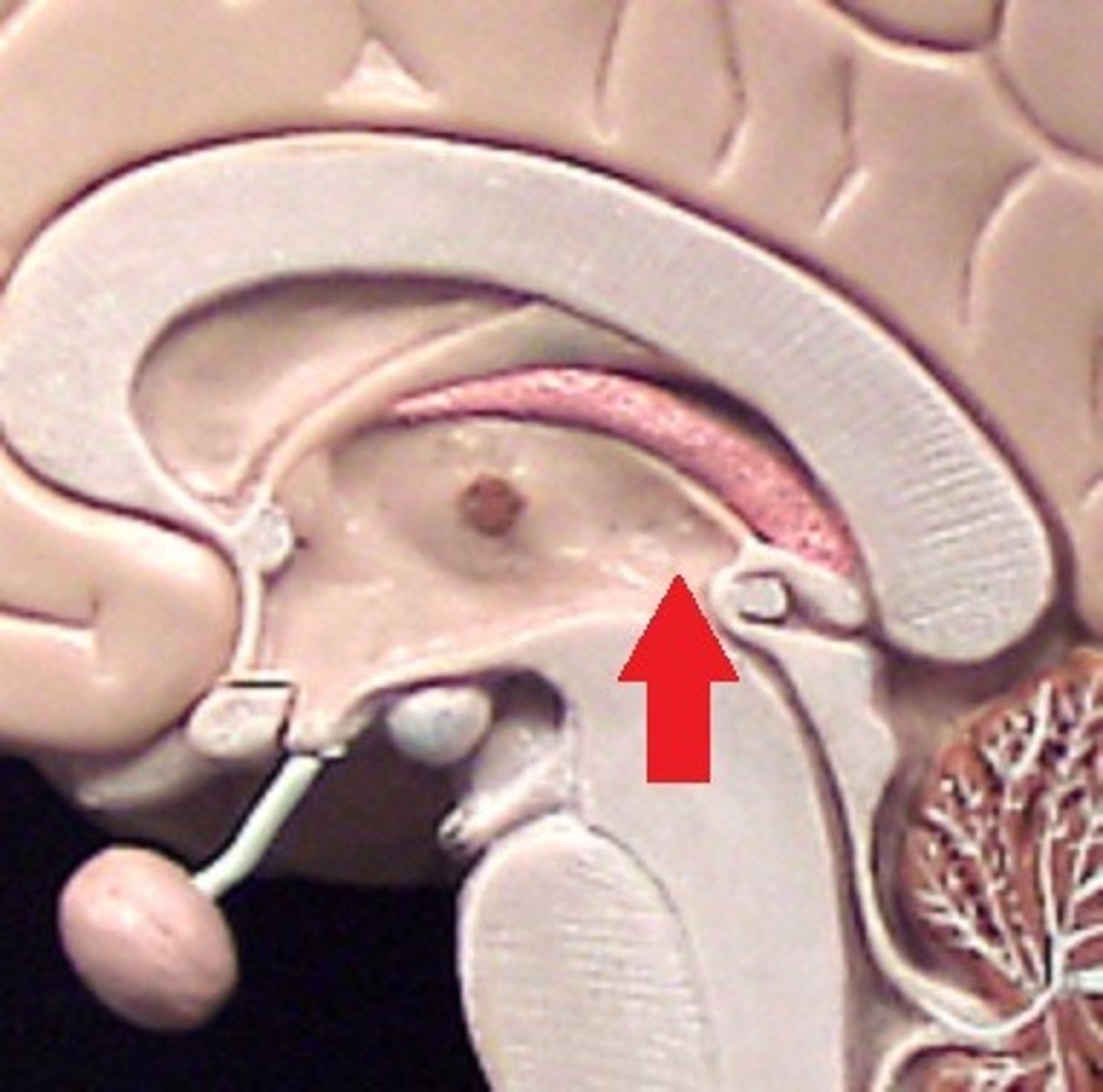
cerebral aqueduct
connects third and fourth ventricles
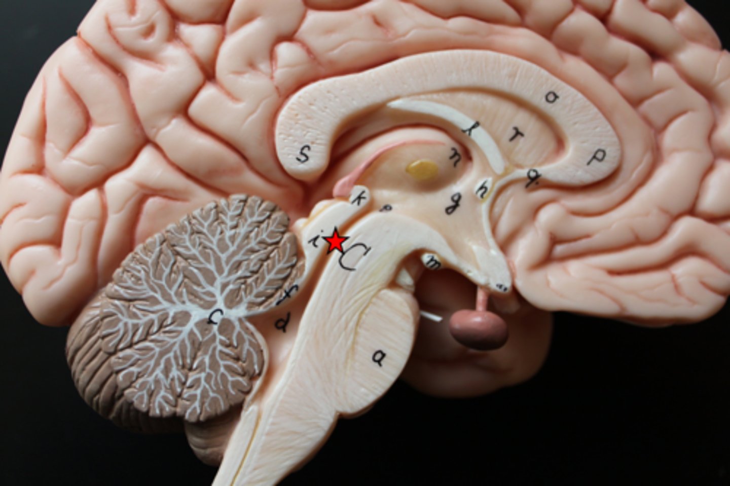
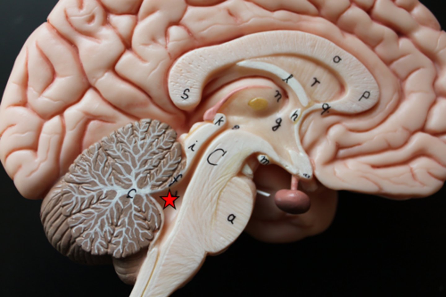
cerebellum
voluntary movement and balance
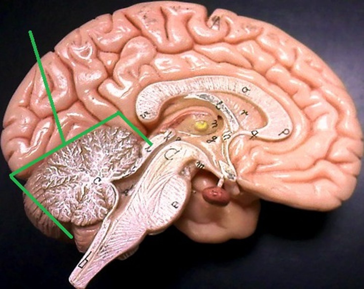
arbor vitae
connects stem to cerebellum
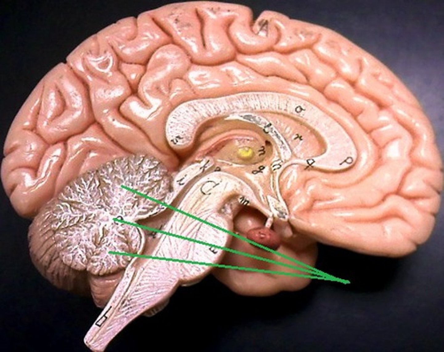
brainstem-midbrain
process visual/ auditory info - arousal
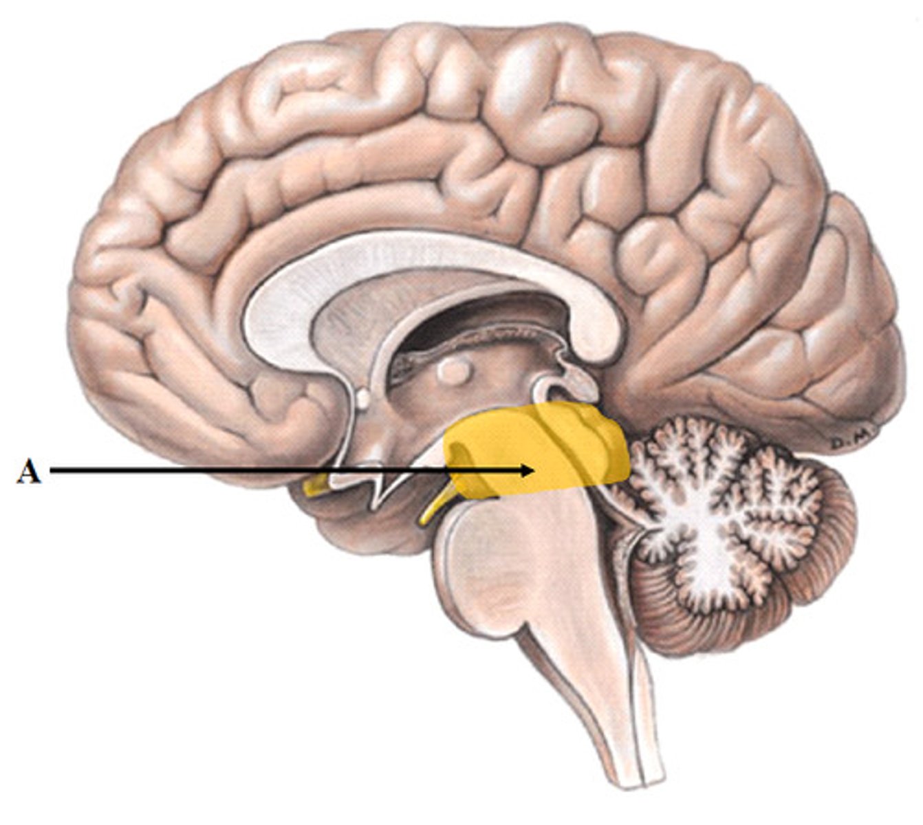
brainstem-pons
respiration control relays info
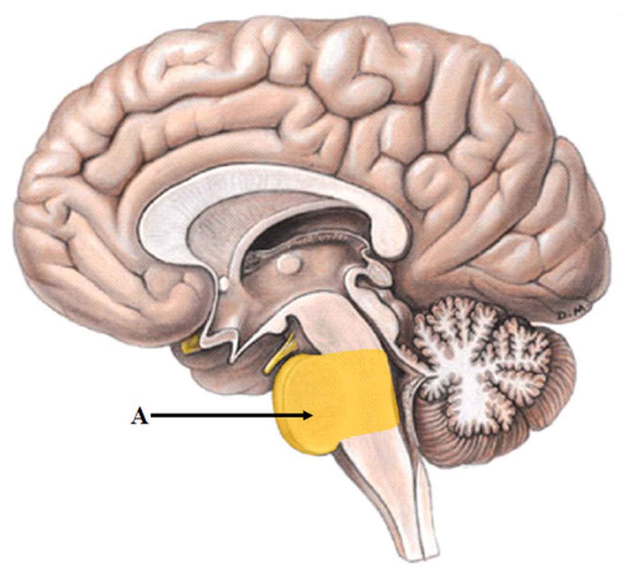
brainstem-medulla
heart beat and breathing
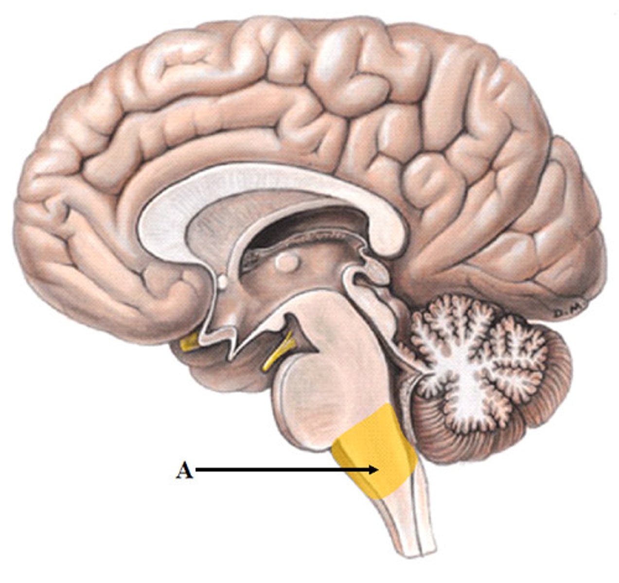
insula
taste
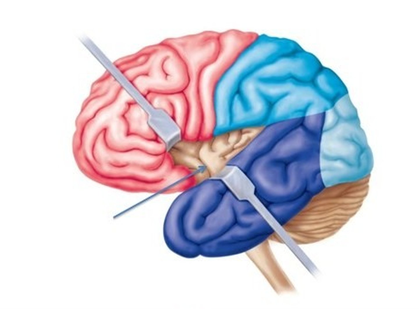
corpus callosum
communicates between two hemispheres
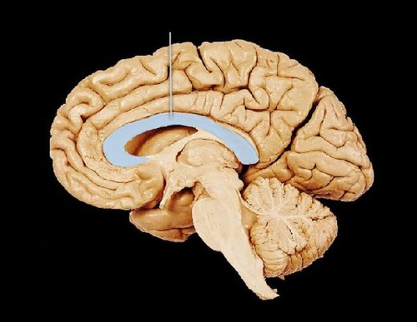
pineal gland
sleep/wake cycle- melatonin
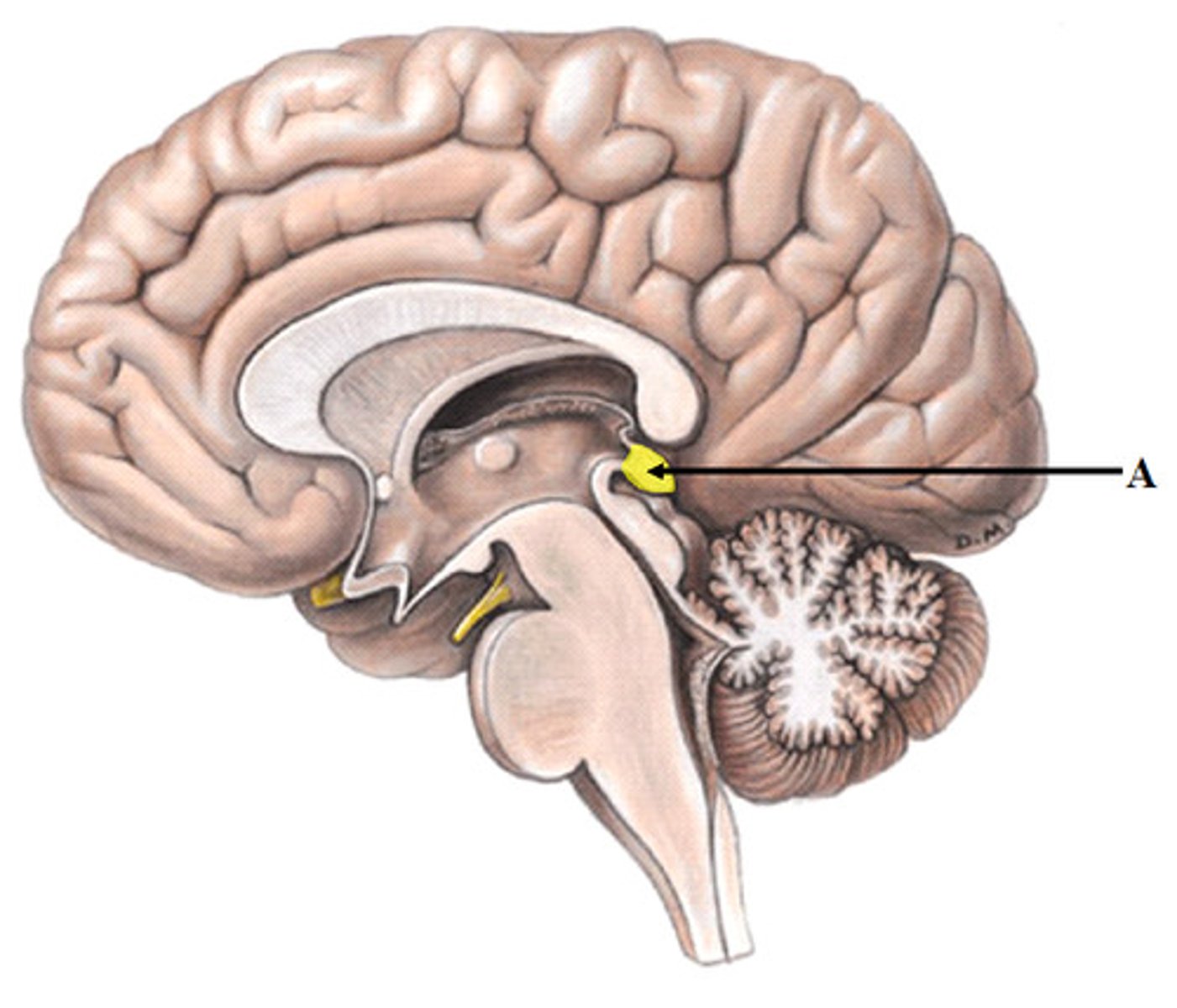
corpora quadrigemina
process info
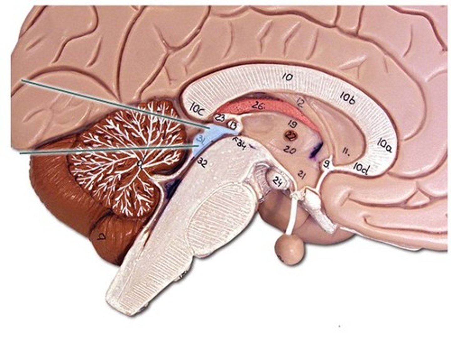
lateral ventricle
contains cerebral spinal fluid
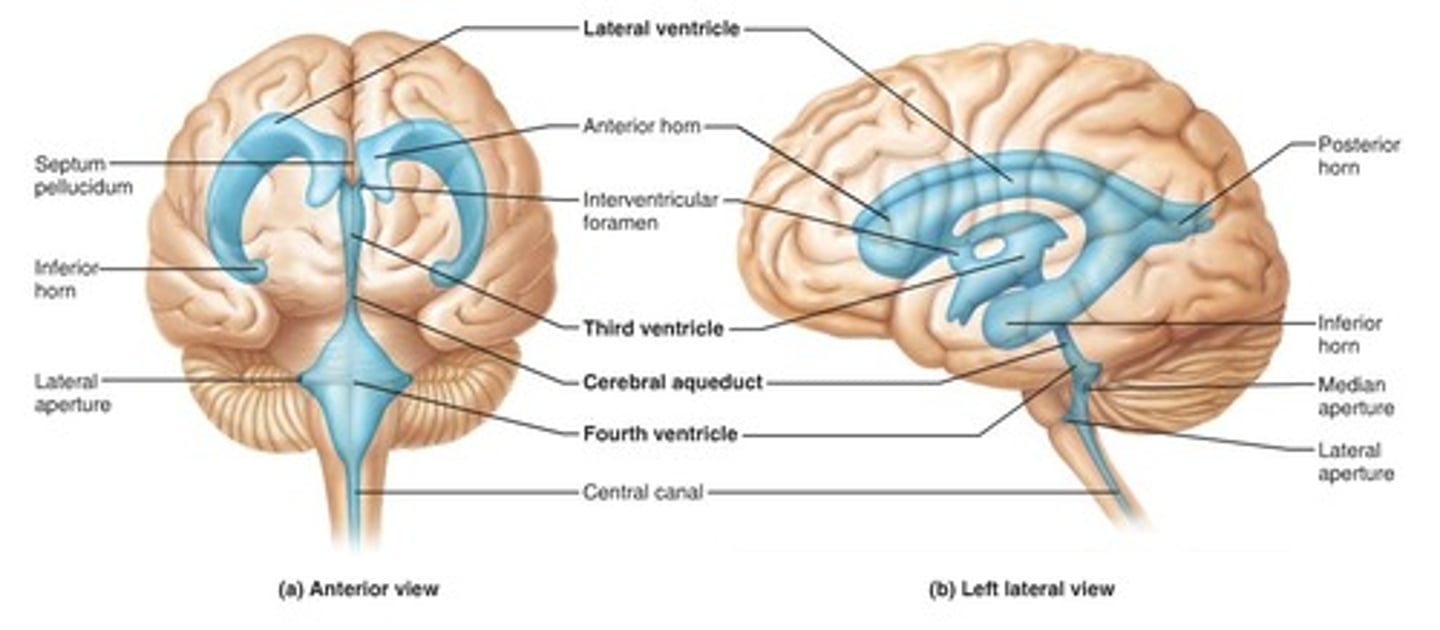
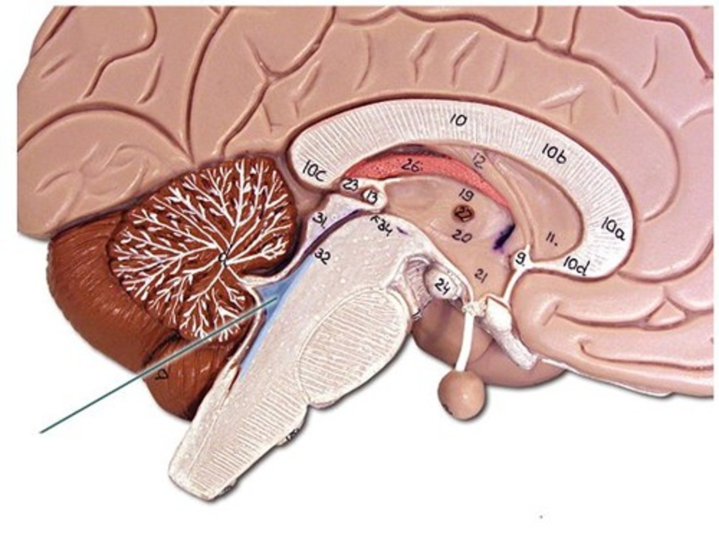
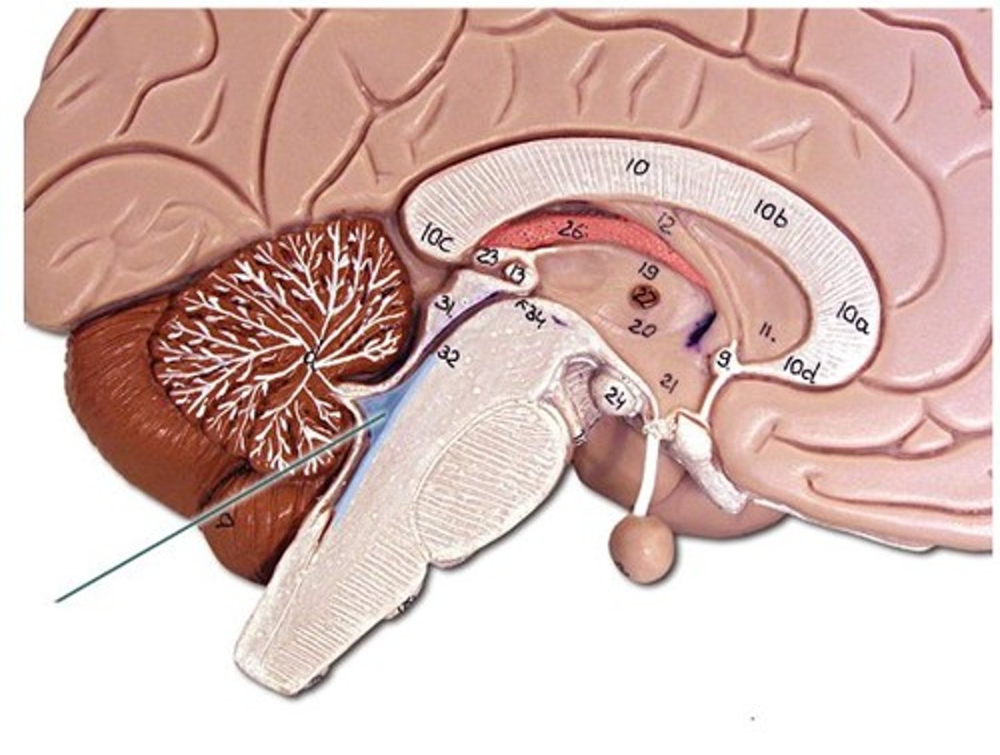
fourth ventricle
connects to central canal
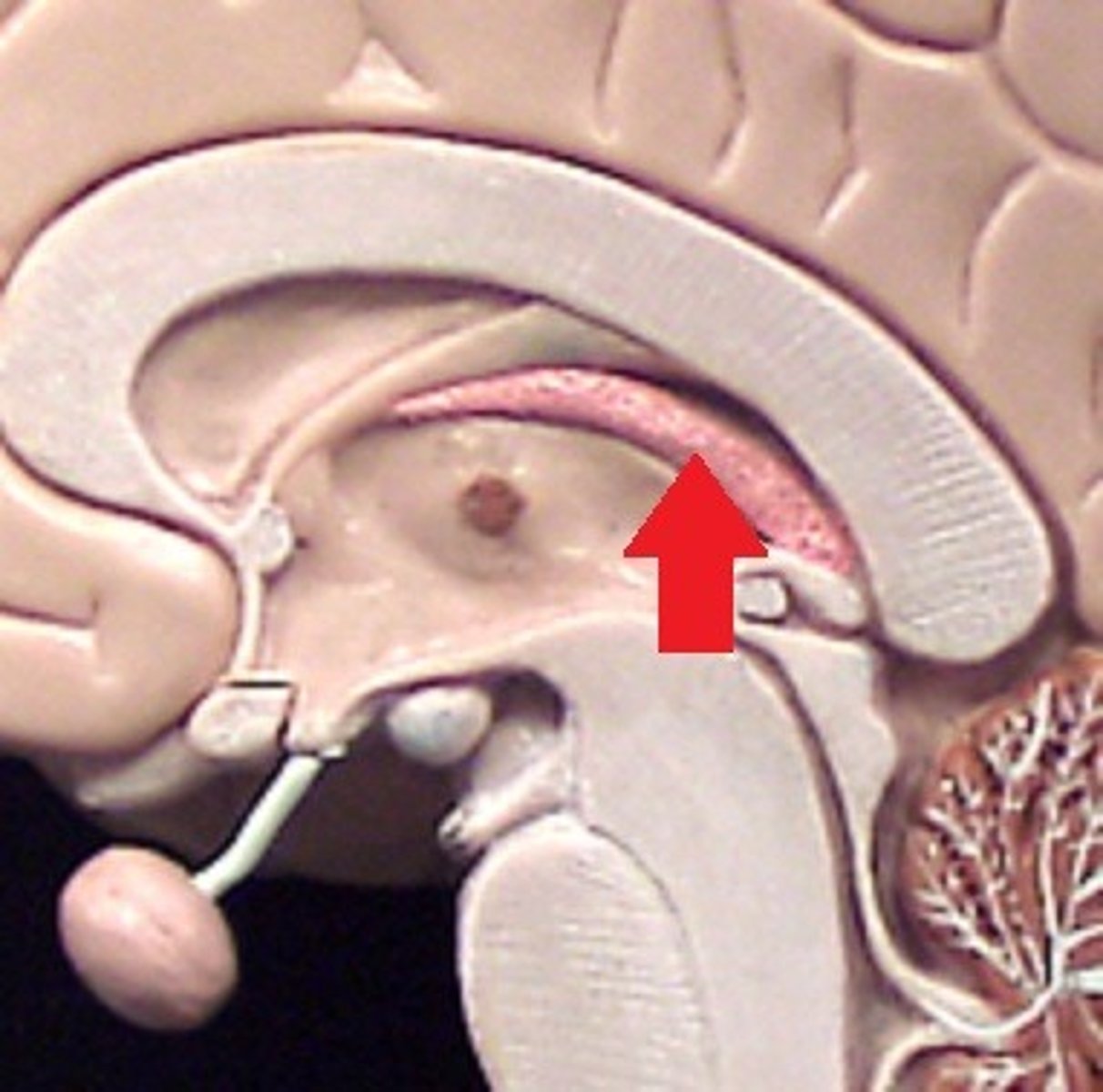
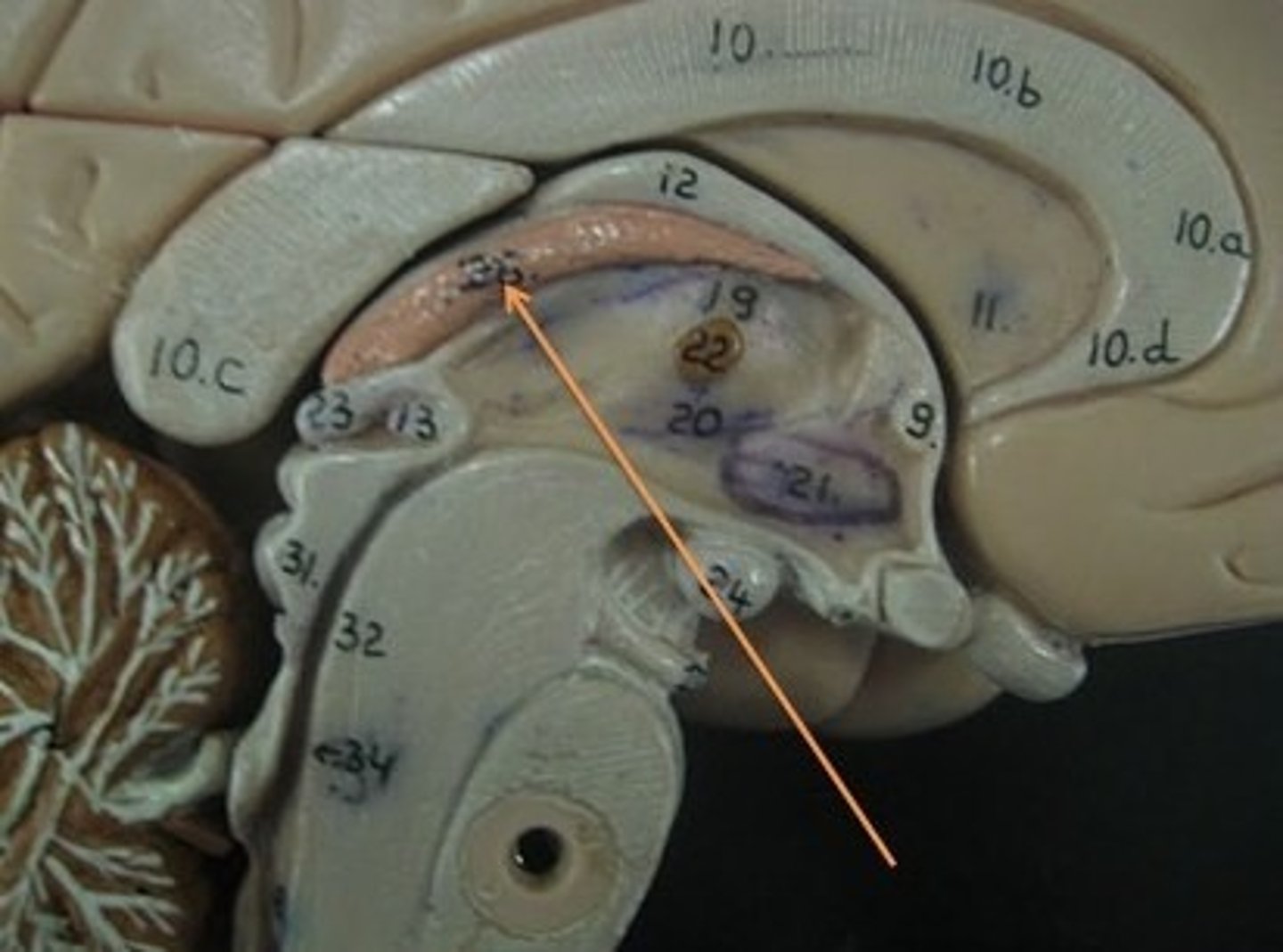
choroid plexus
secretion spinal fluid
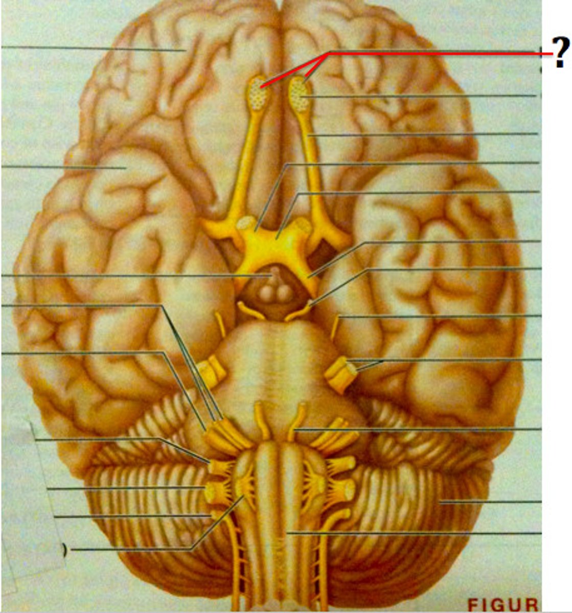
Olfactory Nerve (I) 1
Function: Sensory-Sense of smell
Exit: Cribriform plate of the ethmoid bone
Optic Nerve (II) 2
Function: Sensory-Sense of Sight
Exit: Optic Canal
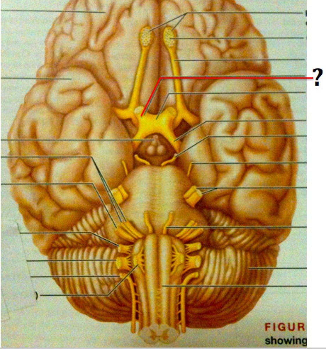
Oculomotor Nerve (III) 3
Function: Motor-Controls eye movement, pupil constriction, & eyelid movement
Exit: Superior orbital fissure
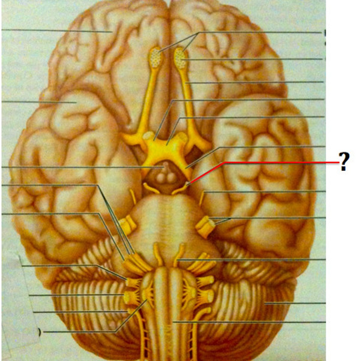
Trochlea Nerve (IV) 4
Function: Motor-Lateral (side to side) eye movement
Exit: Superior orbital fissure
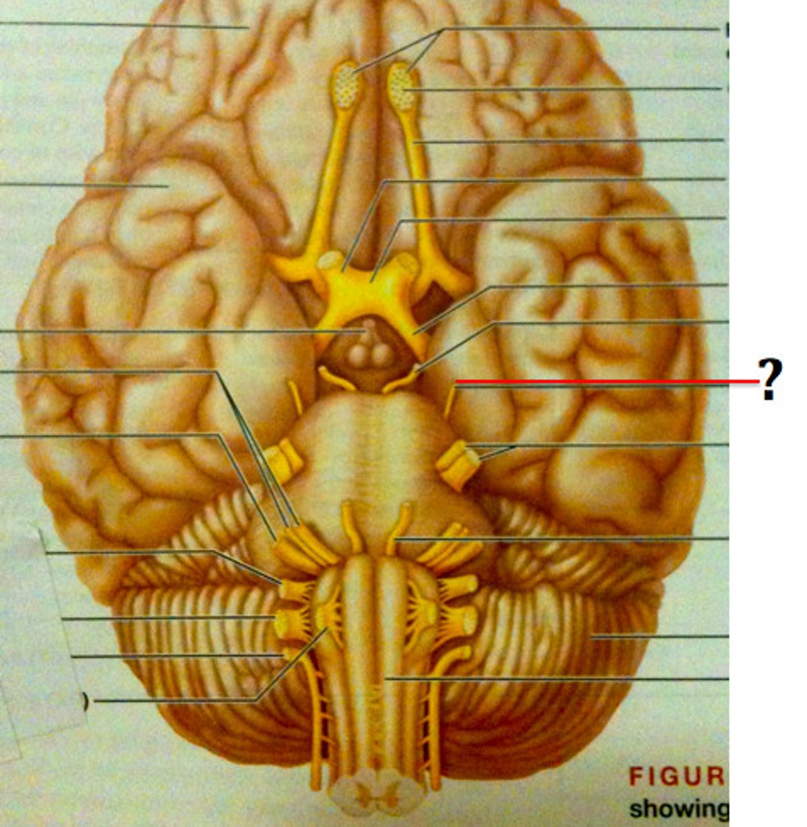
Trigeminal Nerve (V) 5
Function: Both
1. Ohpthalmic-Sensory-orbital
2. Maxillary-Sensory-upper teeth
3. Mandibular-Mixed-
Sensory-muscles for swallowing and anterior 2/3 of tongue
Motor-opening jaw, chewing (mastication)
Exit: Superior orbital fissure, Foramen Rotundum, Foramen Ovale
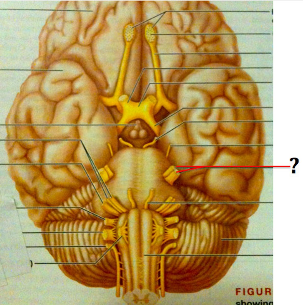
Abducens Nerve (VI) 6
Function: Motor-Eye movement
Exit: Superior orbital fissure
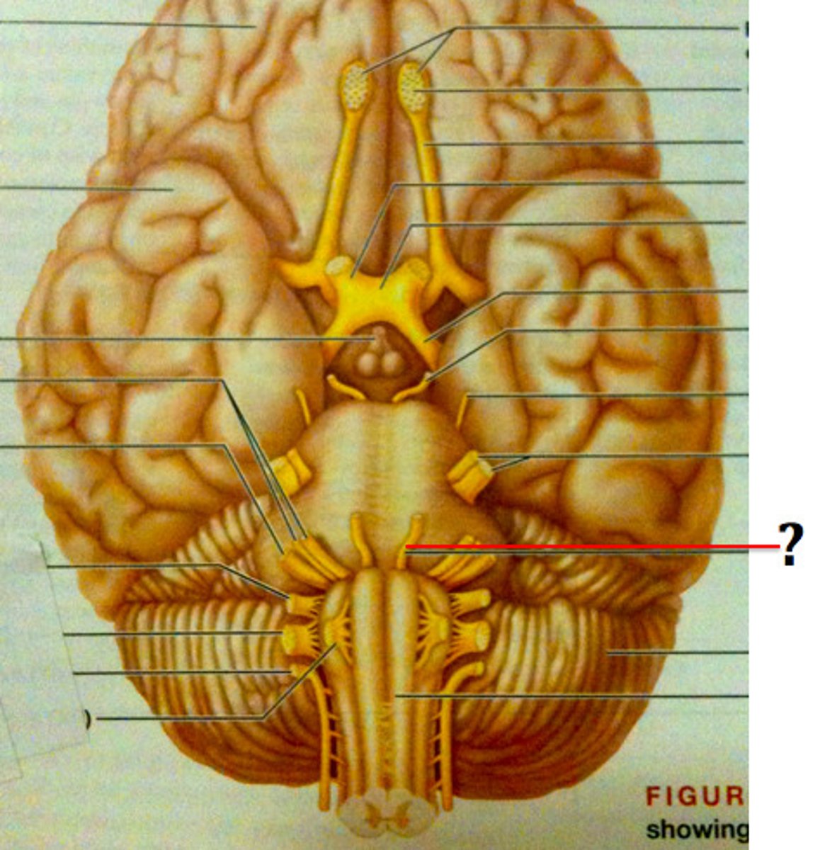
Facial Nerve (VII) 7
Function: Both
Sensory-
Motor-Facial Expressions
Exit: Internal acoustic meatus
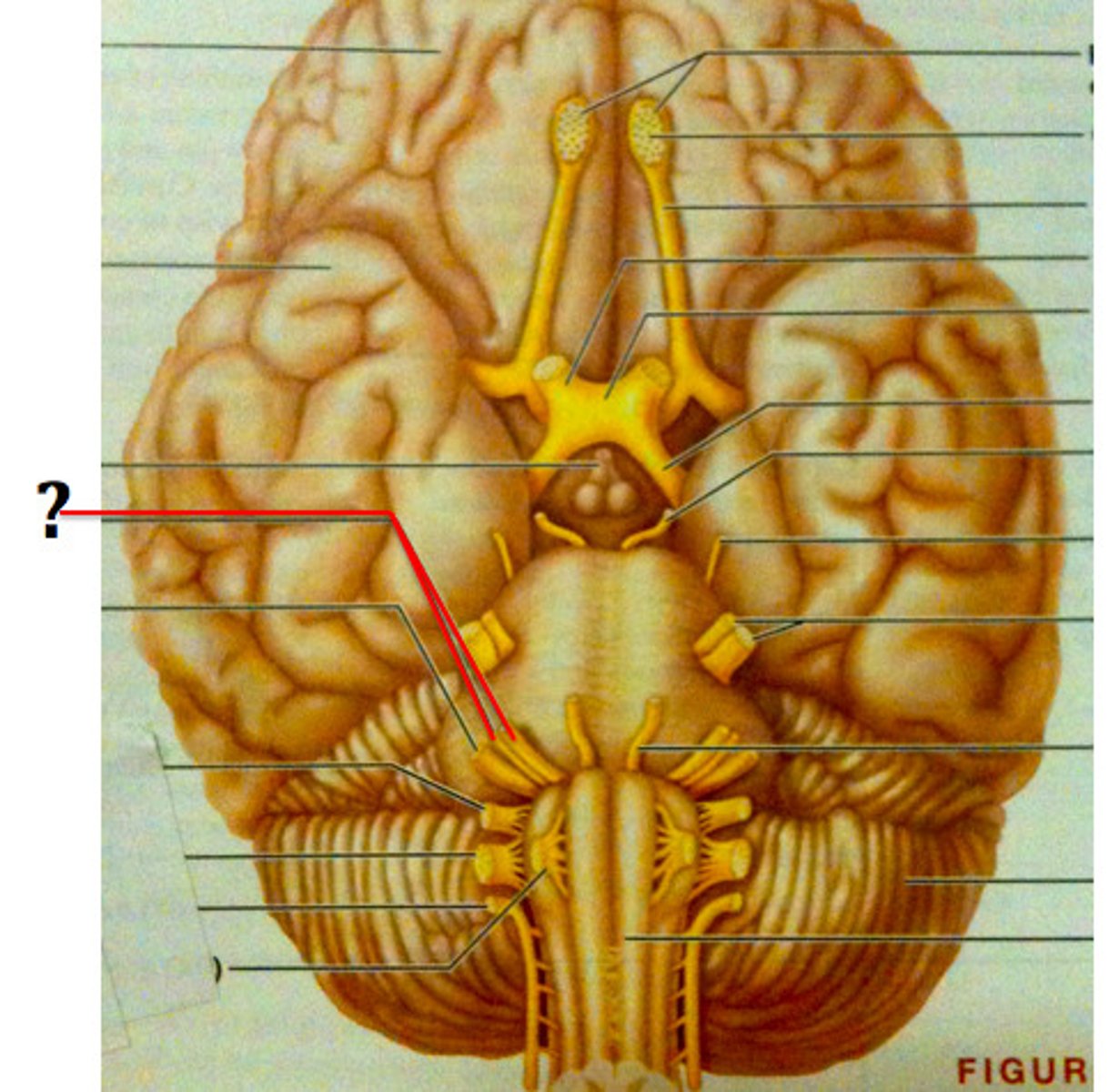
Vestibulocochlear Nerve (VIII) 8
Function: Sensory-Hearing, equillibrium (balance)
Exit: Internal acoustic meatus
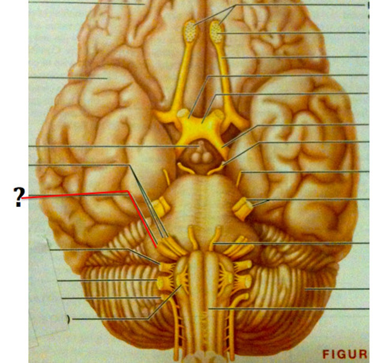
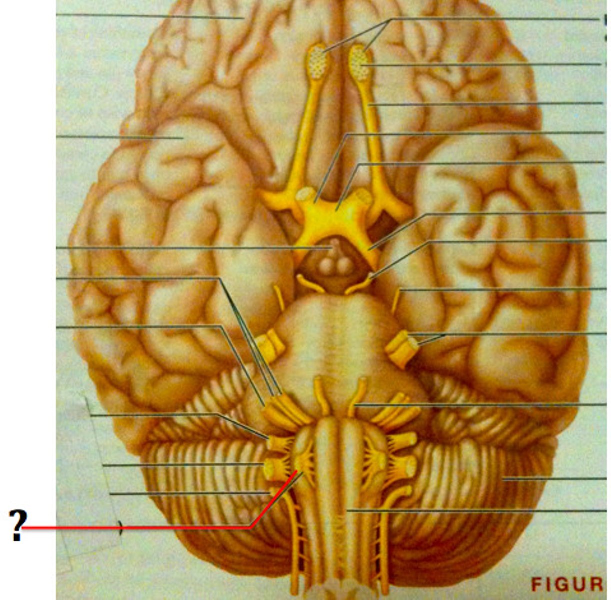
Glossopharyngeal Nerve (IX) 9
Function: Both
Sensory-Taste, respiration, blood pressure
Motor- Swallowing & gagging
Exit: Jugular foramen
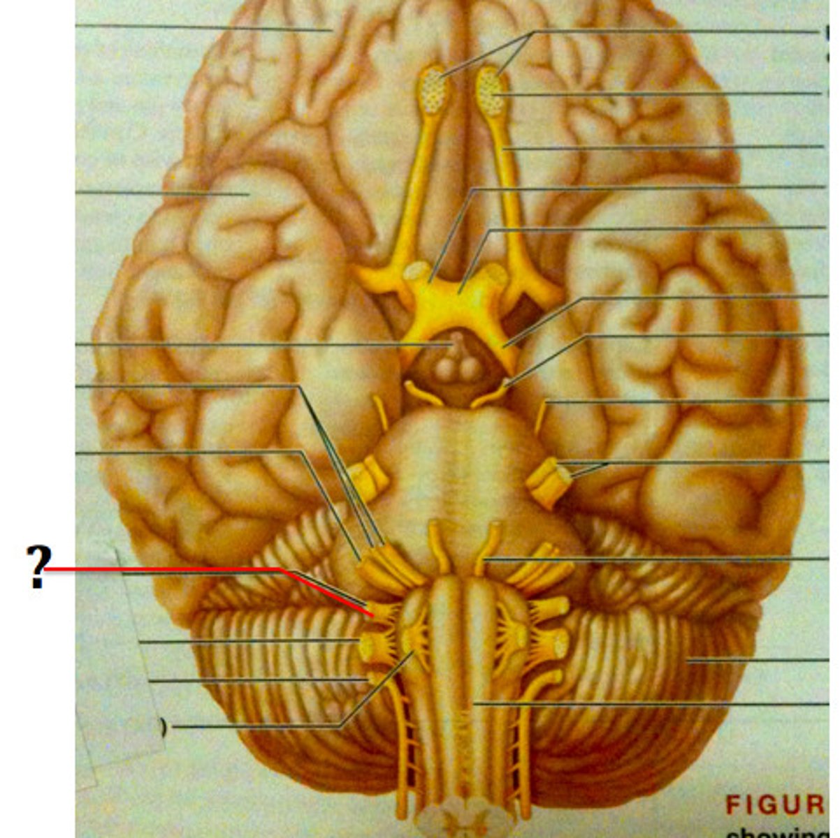
Vagus Nerve (X) 10
Function: Both
Sensory-Taste, hunger, and GI Tract
Motor-Digesting food, plump blood
Exit: Jugular foramen
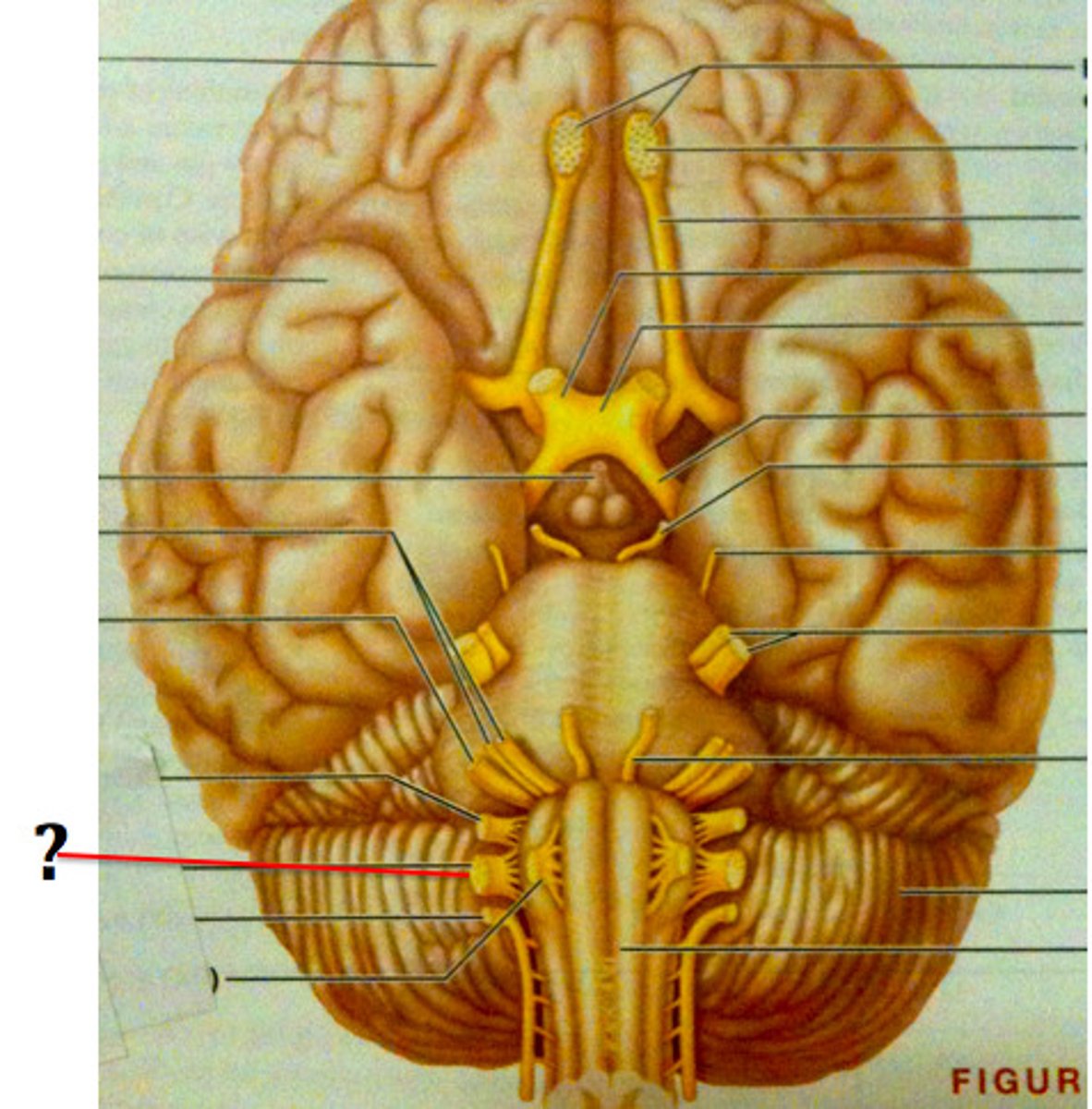
Accessory Nerve (XI) 11
Function: Motor-Moving head and shrugging shoulders
Exit: Jugular foramen
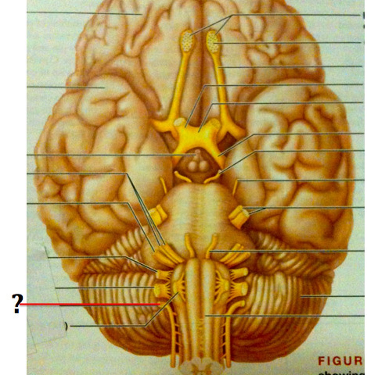
Hypoglossal Nerve (XII) 12
Function: Motor-Movement of the tongue and for speech
Exit: Hypoglossal canal
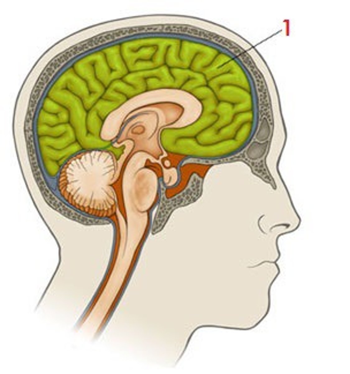
Cerebrum
It is large, is the dominant brain structure, with many folds and crevices enveloping the diencephalon.
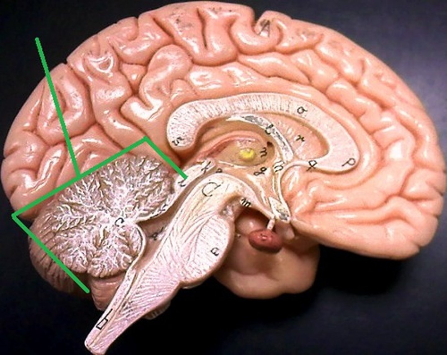
Cerebellum
Posterior to the brain stem.
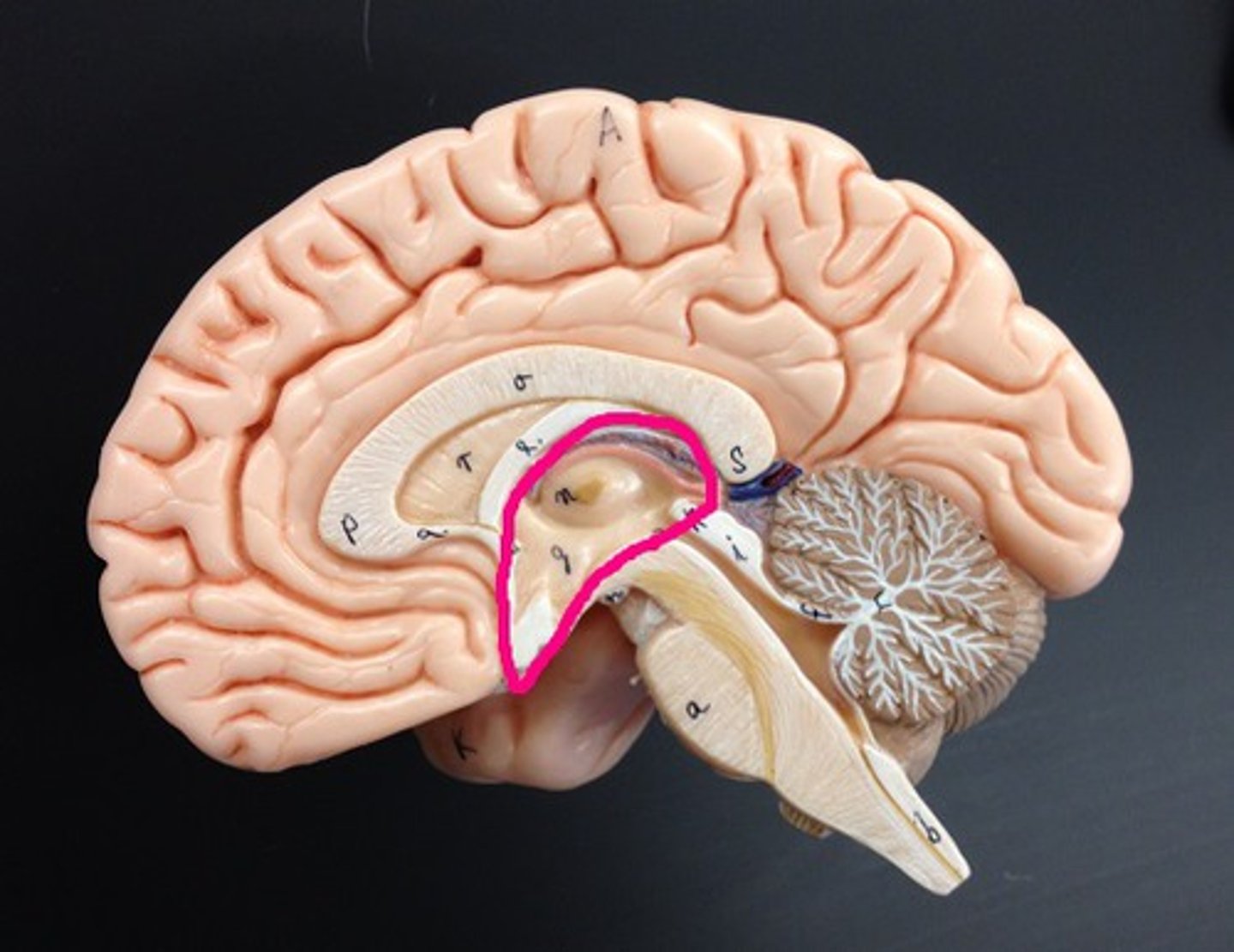
Diencephalon
Superior to the brain stem in the center of the brain.
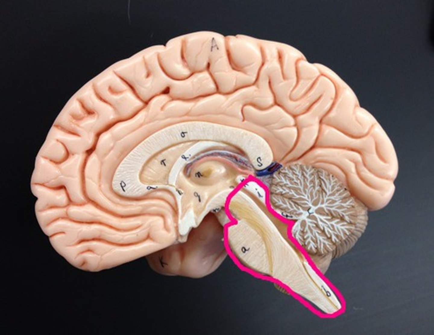
Brain stem
Connected to the superior part of the spinal cord.
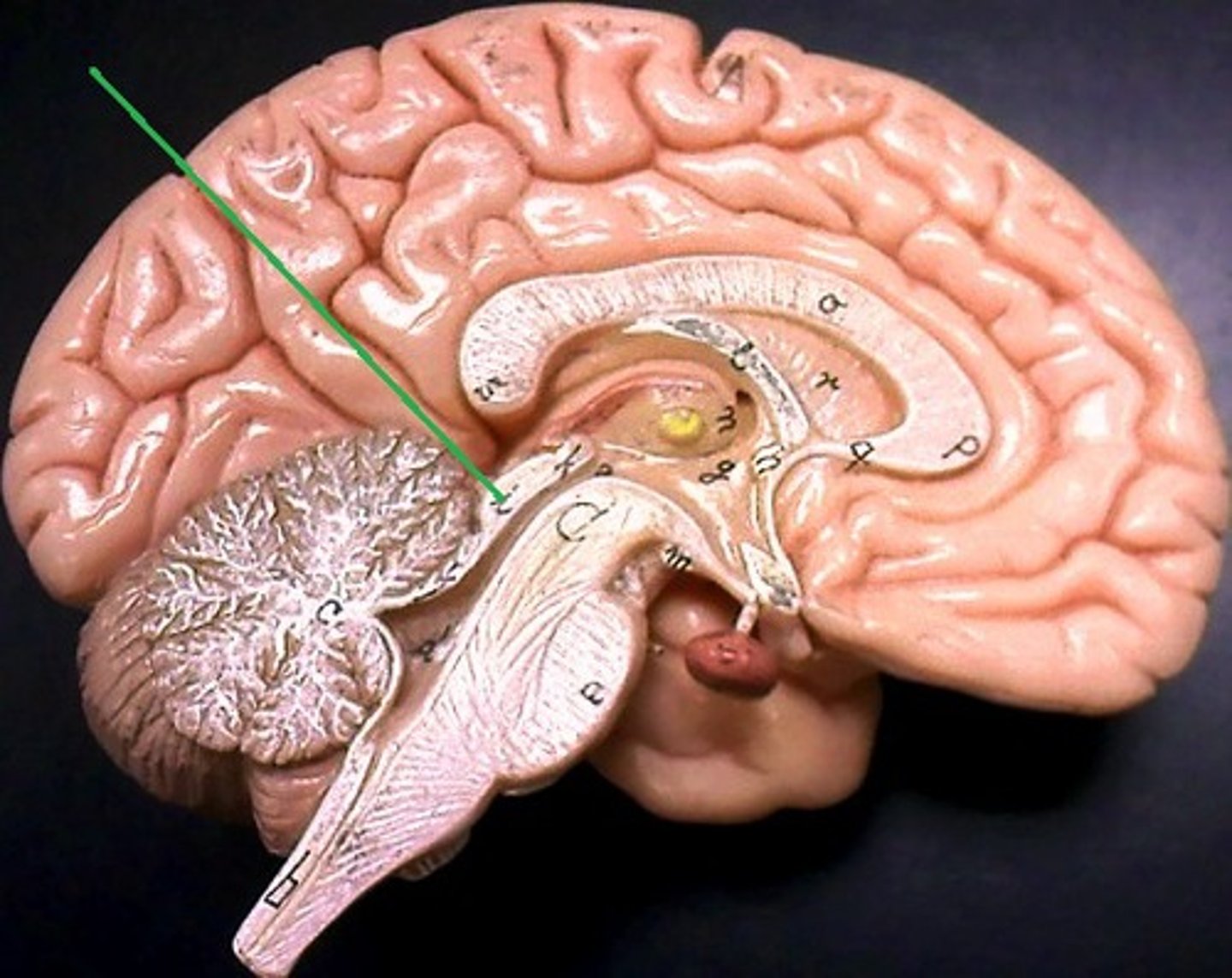
Corpora quadrigemina
Composed of 2 superior colliculi and 2 inferior colliculi.
Midbrain
A smaller area superior to the pons and inferior to the diencephalon, consisting of cerebral peduncles and the corpora quadrigemina.
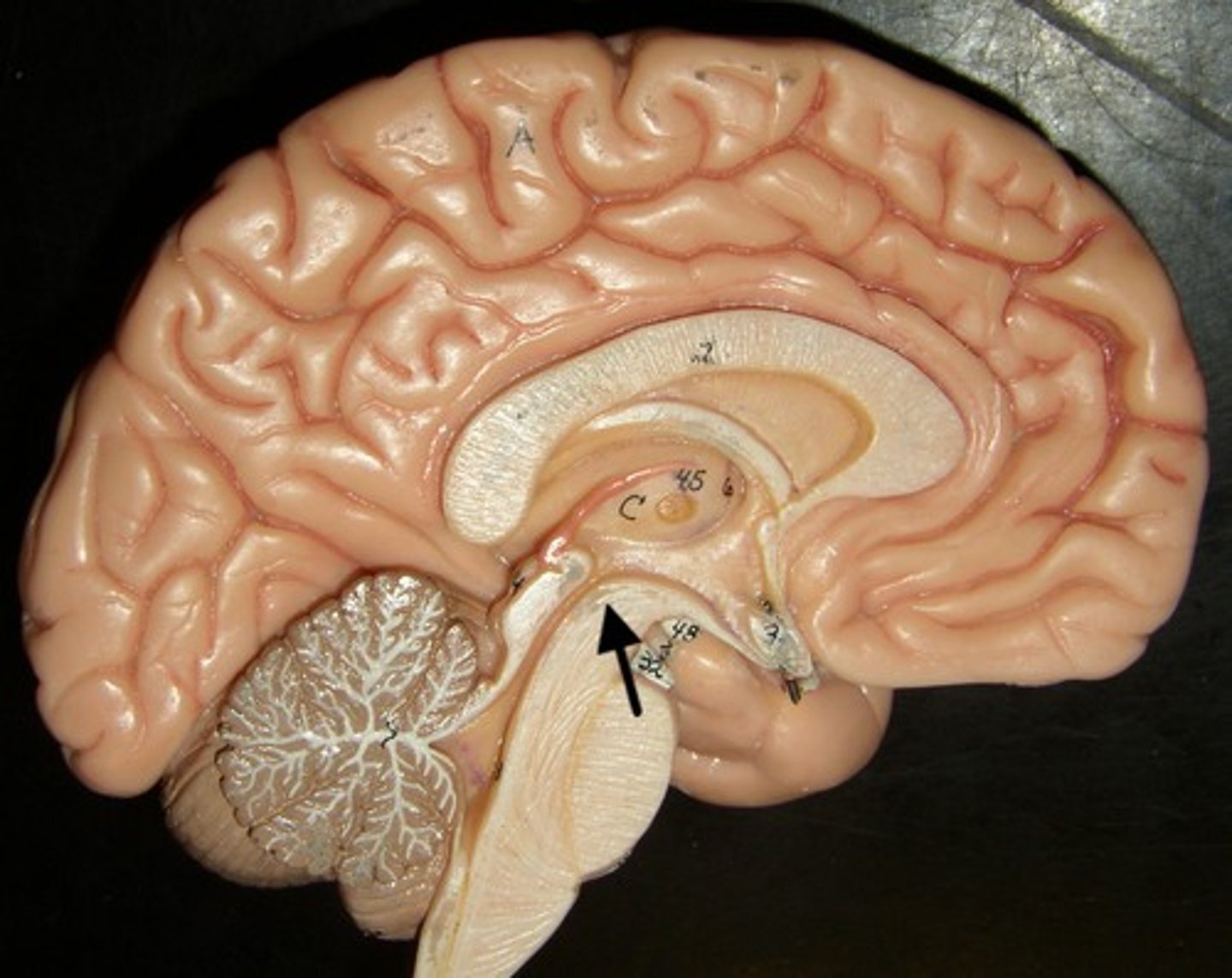
Pons (Bridge)
An expanded structure located superior to the medulla oblongata and anterior to the cerebellum, and has respiratory centers that assist the medulla oblongata in controlling breathing.
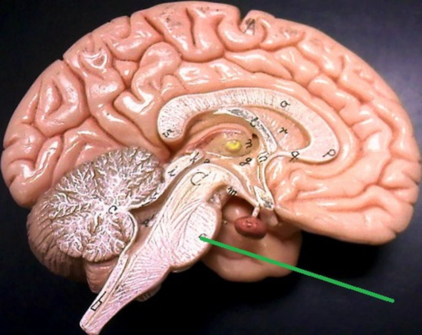
Medulla oblongata
The first brain structure, is immediately superior to the spinal cord and is the most vital part of the brain because it houses the respiratory and cardiovascular control centers.
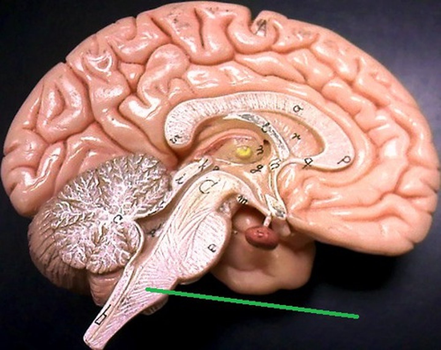
Arbor vitae (Tree living)
When cut in sagittal section, gray matter can be observed on the exterior, with deeper white matter.
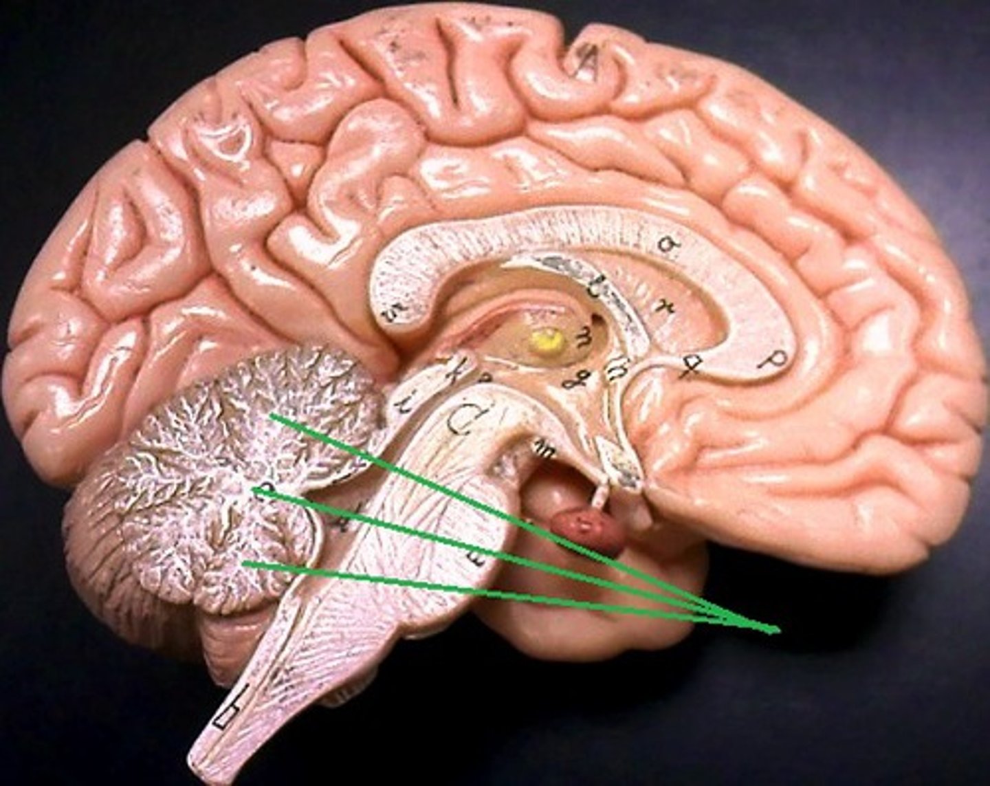
Cerebellar cortex
The outer layer of gray matter.
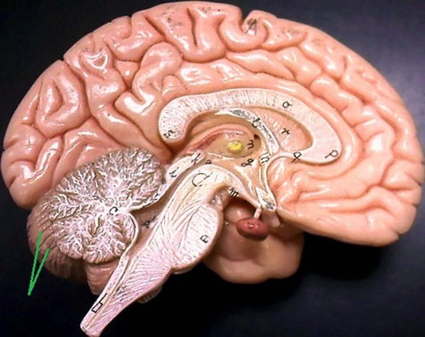
Mammillary body
Two small, round masses located just posterior to the infundibulum are relay stations for smell and taste reflexes.
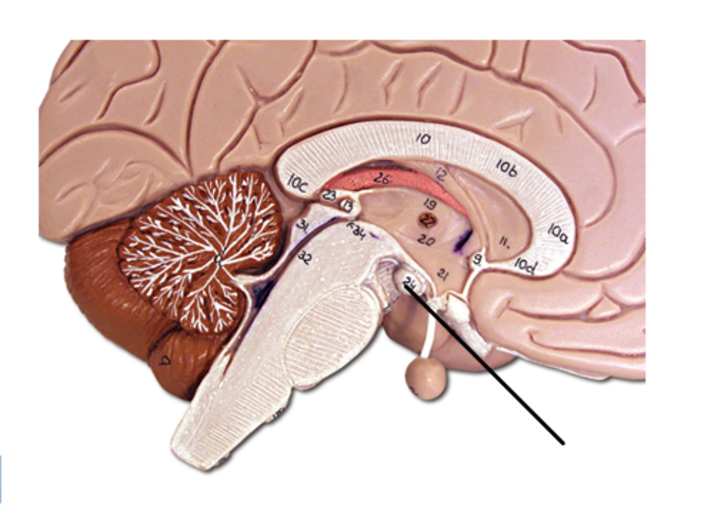
Pineal gland
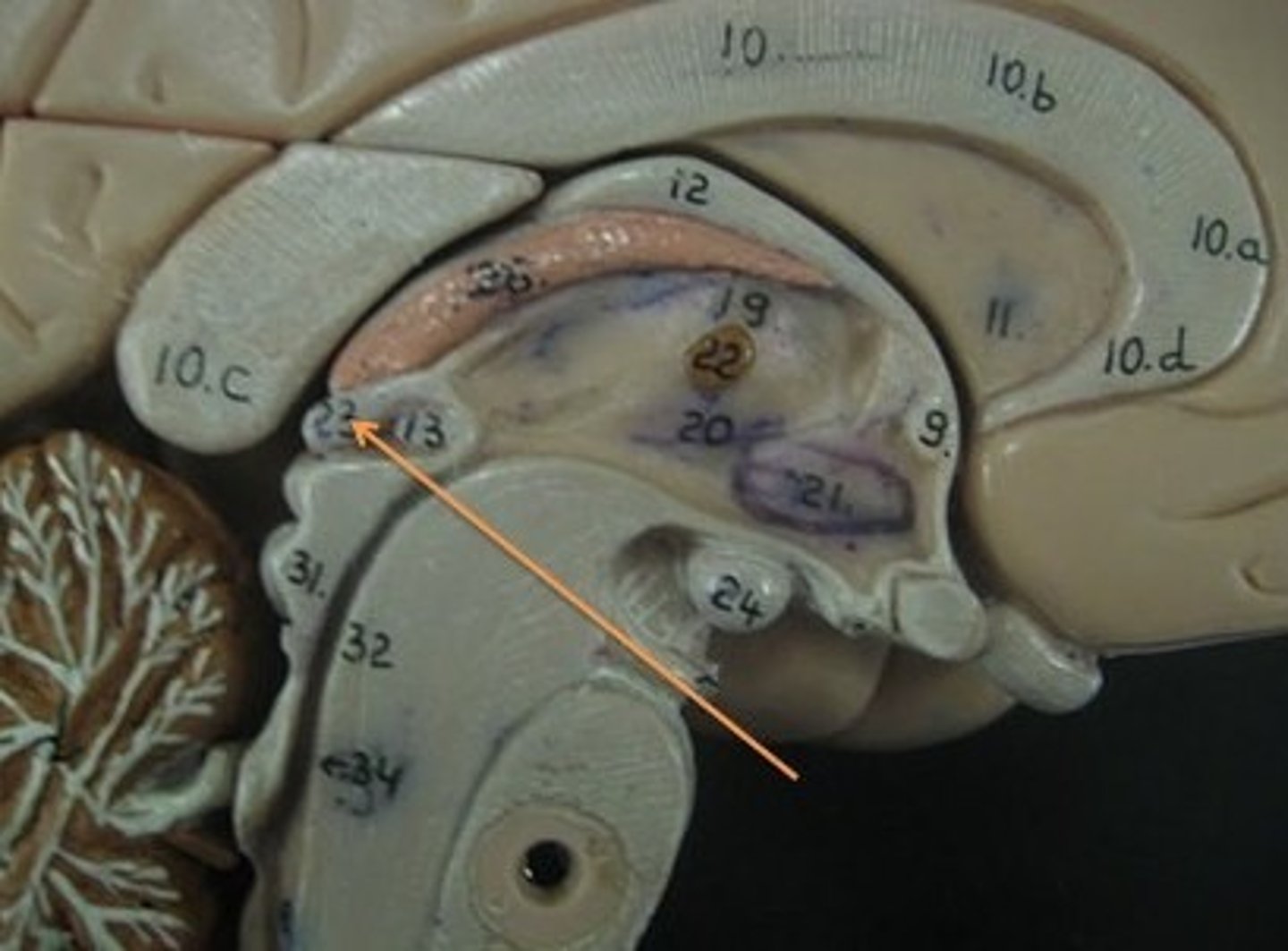
Thalamus
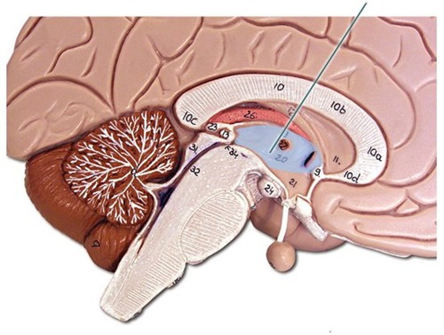
Hypothalamus
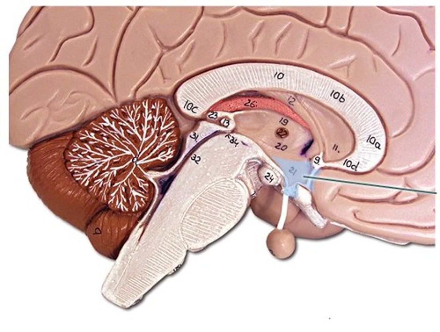
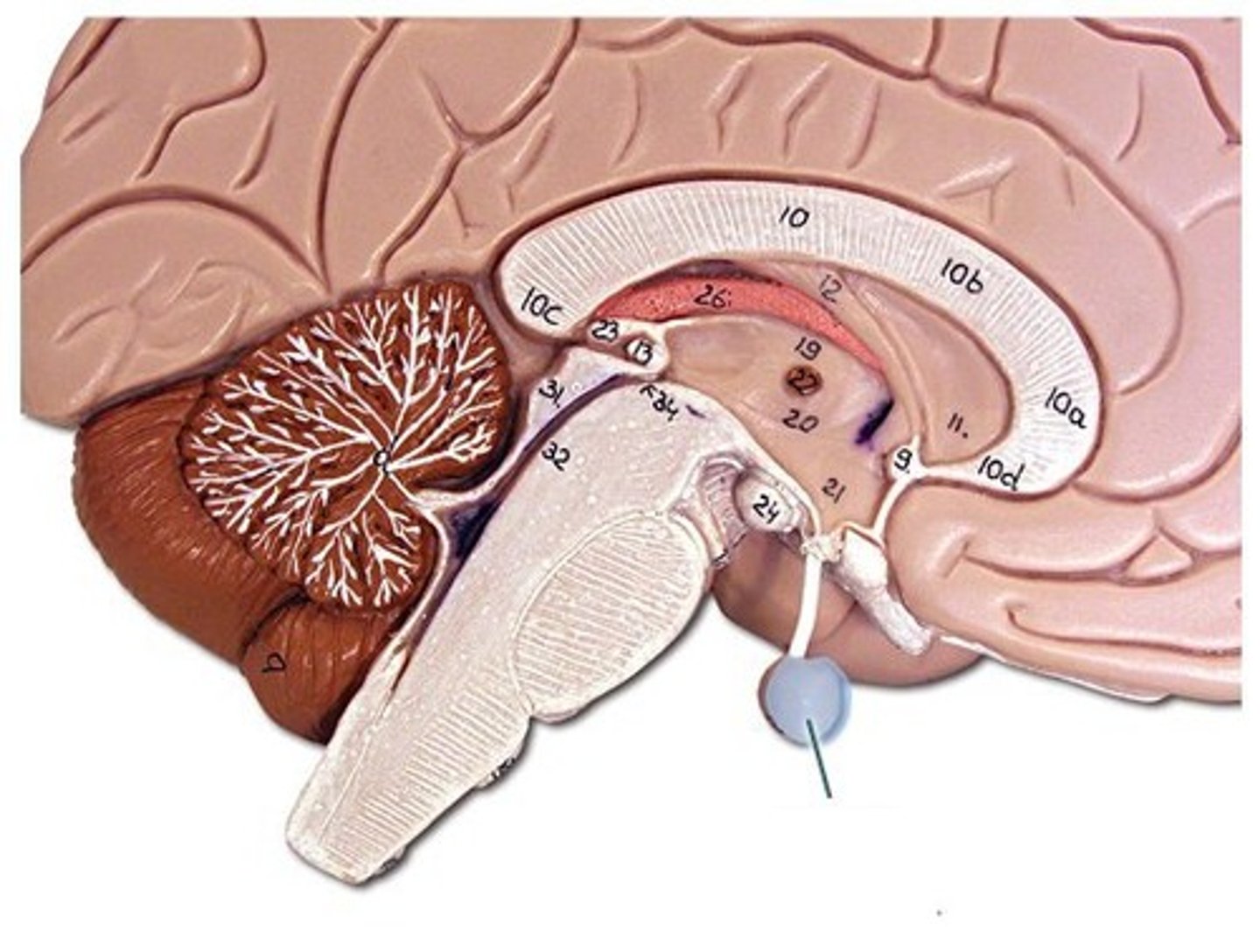
Pituitary gland
Looks like a large pea and is attached to the end of the infundibulum. The hypothalamus control it.
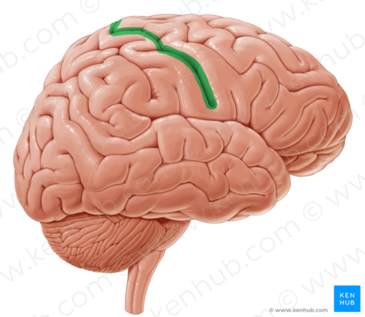
Central sulcus (furrow)
Shallow groove separating frontal lobe from parietal lobe.
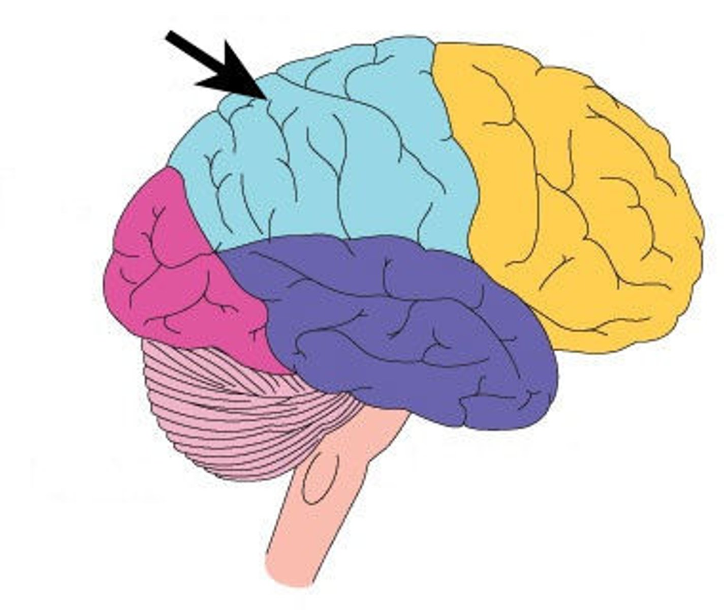
Parietal lobe
Occipital lobe
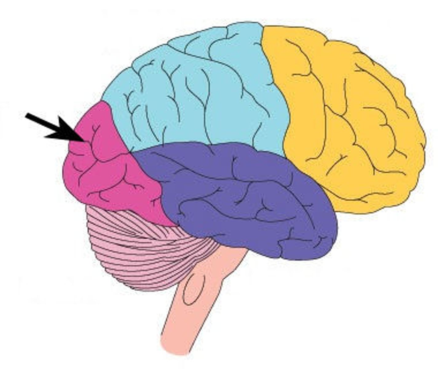
Frontal lobe
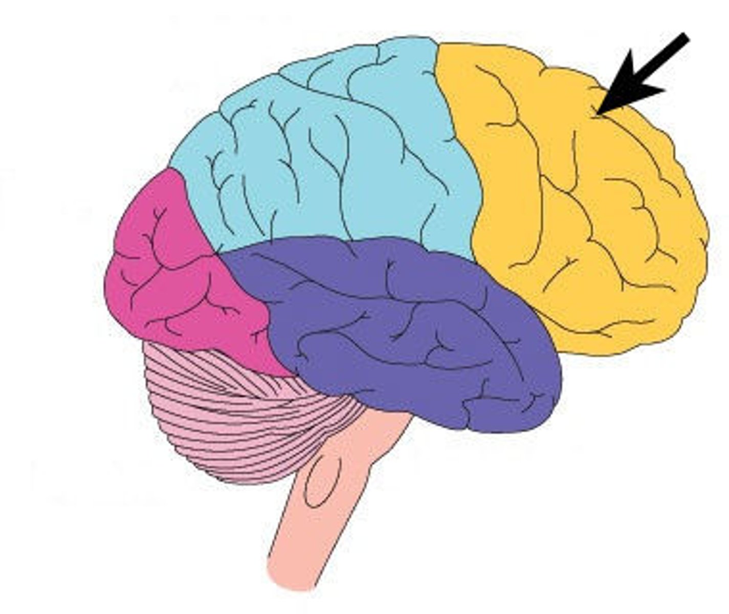
Insula
An inner lobe that lies deep to the lateral cerebral fissure and is not visible from the exterior.
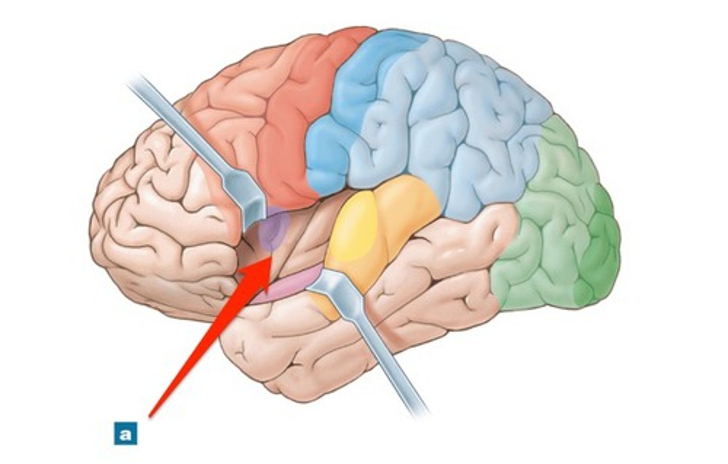
Temporal lobe
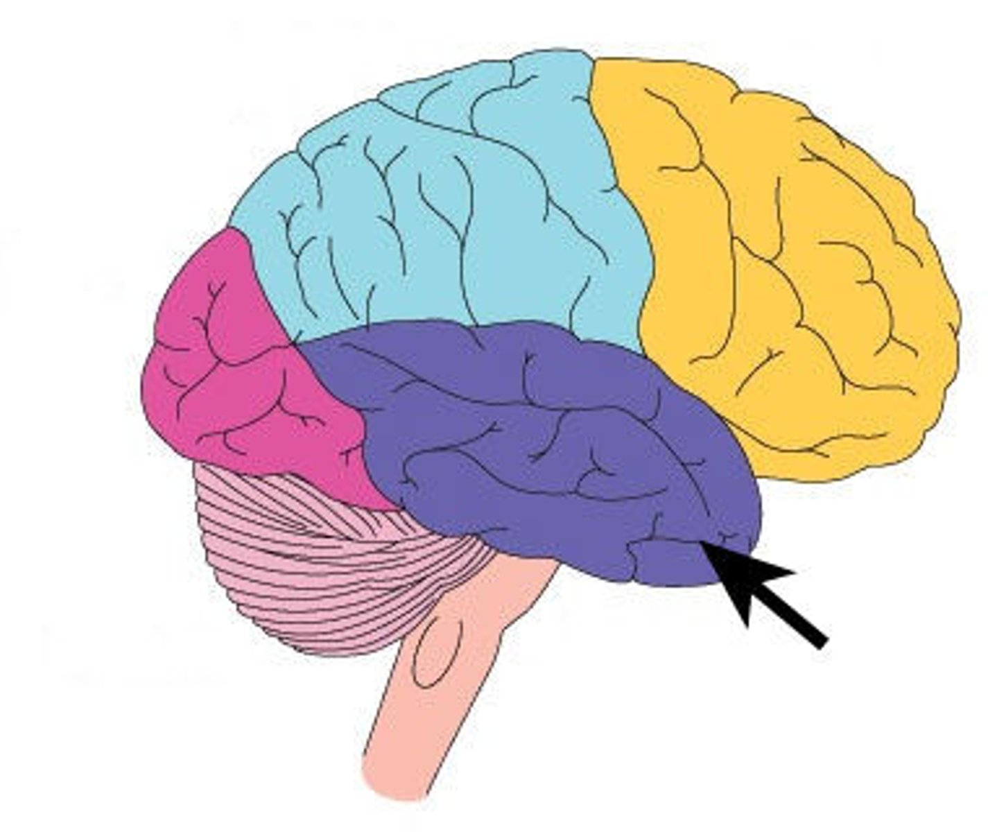
Central sulcus
Lateral ventricle
Choroid plexus
Third ventricle
Cerebral aqueduct
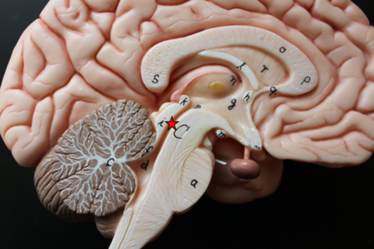
Fourth ventricle