Auditory Anatomy/Phisiology
1/51
There's no tags or description
Looks like no tags are added yet.
Name | Mastery | Learn | Test | Matching | Spaced |
|---|
No study sessions yet.
52 Terms
The Peripheral Auditory system includes:
outer ear
middle ear
inner ear
Sound localization
auditory system's ability to pinpoint the location of a sound source
• intensity and phase (time) difference
The Central Auditory system includes:
Auditory Brainstem (AB)
Auditory forebrain (AF)
The Auditory Forebrain includes:
Medial geniculate body
auditory cortex
Auditory brainstem includes:
cochlear nucleus
superior olivary complex
inferior colliculus
Medial geniculate body (MGB)
Processes and relays specific detailed
auditory information to the auditory cortex
AF
Auditory Cortex consists of:
primary auditory cortex (AI) and
secondary auditory cortex (AII).
Neurons in the Auditory cortex detect
Complex sound features: speech, pitch, and rhythm
Frequency Modulations of AC responds to
Changes in pitch over time (rising or falling tones)
Temporal Modulation of AC responds to
Changes in timing or rhythm of sound.
Superior olivary complex (SOC)
Receive bilateral inputs
• Localize sound
AB
What does binaural hearing in the auditory cortex allow?
Combines input from both ears to localize sound.
Cochlear nuclei (CN)
the first stop for auditory nerve fibers after they leave the cochlea.
AVCN, PVCN and DCN
AB
Nuclei of lateral lemniscus (NLL)
Helps with processing timing and temporal patterns
AB
Inferior colliculus (IC)
Combine the analysis of complex sound and the direction in space simultaneously
located in midbrain
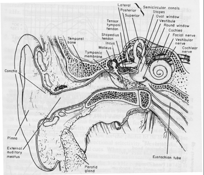
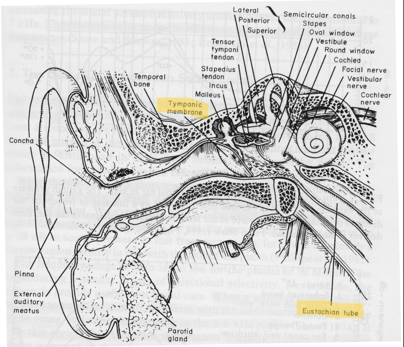
Function of the Outer Ear
Funnels sound waves to the ear drum using localization
provides intensity and phase difference
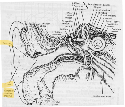
Pinna
External ear canal
Eustachian Tube
Connects middle ear to the nasopharynx
Equalizes air pressure between the middle ear and the atmosphere.
Sound pressure gain
amplification peaked around 2.5 kHZ
primary contribution from concha and outer ear canal (act as a resonator)
degrees is the angle faced
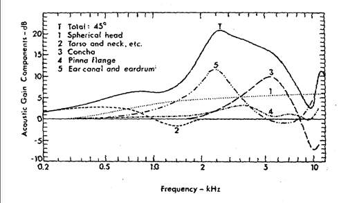
Pinna
Sound localization is in the midplane of the head
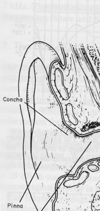
External Ear canal
The pathway for sound waves
tube shaped
protects ear canal by trapping dust
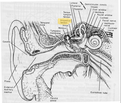
Middle Ear Includes
Tympanic Membrane
Tympanic Muscles
Auditory Ossicles
Eustachian Tube
Function of the Middle Ear
Transduction of sound: transferring sound energy from air (low impedance) to the fluid of the cochlea (high impedance).
The stapes pushes in and out on the oval window (membrane covered opening) creating a pressure wave in the fluid-filled cochlea
Acoustic Impedance
determines how much sound is reflected and transmitted at the boundary between two mediums
Problem with Sound Transmission
A large amount of acoustic energy will be reflected because the difference of acoustic impedance between the two sound media.
Solution for Sound Transmission
solutions: Increase pressure/force at the oval window,- Impedance Mismatch problem (makes sure sound enters instead of reflecting off the oval W)
Acoustic Reflexes in the Middle Ear
After receiving intense sounds, the two middle ear muscles are contracted to lower the sound transmission in the middle ear, providing a protection to the inner ear
Tensor Tympani
Contracts and increases tension on the tympanic membrane with intense sounds
in the middle ear
a tympanic muscle
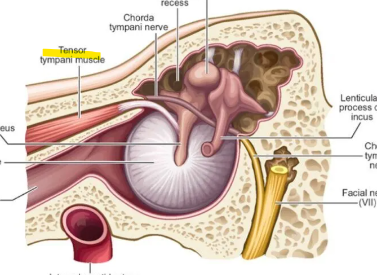
Stapedius
Contracts and works with the tensor tympani during high intensity sound to limit the motion of the ossicles and protect the inner ear
in the middle ear
a tympanic muscle
Inner Ear Includes
Vestibular Apparatus
Cochlea
Vestibular Apparatus
responsible for balance and spatial orientation, not hearing
Cochlea
coiled shape around modiolus
three scalae: scala vestibuli, Scala media (cochlear duct), Organ of Corti, Scala tympani
fluid is produced by vibration of stapes
Basilar Membrane
uses movement of fluid to vibrate up and down
Separates scala media from scala tympani
housed in cochlea
Reissners Membrane
Separates scala vestibuli and scala media.
Helps keep the two fluids (perilymph and endolymph) apart so their chemical balance stays correct
Scala Vestibuli
first chamber that recieves the vibration
filled with perilymph fluid, and the wave starts traveling through it
Scala Media (cochlear duct)
“middle” chamber
organ of corti
important for working with basilar membrane
Organ of Corti
contains hair cells (15,000) that detect vibration
convert sound vibrations into electrical signals that are transmitted to the brain for interpretation
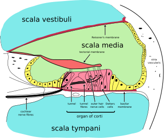
Scala Tympani
“bottom“ chamber: filled with perilymph
After the wave travels through the cochlea, it exits through this chamber and releases pressure at the round window
Helicotrema
joint opening at the apex of the cochlea for scala vestibuli and tympani.
allows fluid to move between them so low-frequency (bass) sounds can travel all the way to the top of the cochlea.
Supporting Cells
Structurally and metabolitically support the outer and inner hair cells
Frequency Selectivity
why we can hear a range of noise
BM tuned to a specific pitch
shown on ftc
Where each auditory nerve fiber is most sensitive to a particular sound frequency
nonlinearity
to prevent distortion and damage, hair cells adjust loud and soft sounds to either amplify or dampen
Auditory Nerve (AN)
A direct synaptic connection between the hair cells of the cochlea and the cochlear nucleus
Type I and Type II spiral ganglion cells
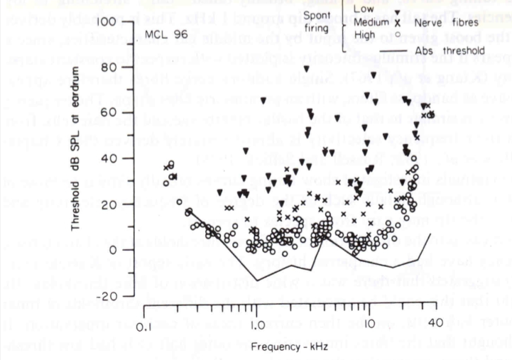
Spontaneous Firing Rate (AN)
baseline electrical activity of neurons, which occurs without external stimulation
Low, Medium, High
Higher spontaneous rate = lower threshold (more sensitive to quiet sounds)
Type 1
90 - 95% of spiral ganglion cells, connected to IHC
(20 fibers to one IHC in human)
responsible for transmitting the majority of auditory information to the brainstem
Type 2
5 – 10% of spiral ganglion cells, connected to
OHC, one to many
(one fiber to 10 OHC in human).
form widespread connections with outer hair cells
Frequency Threshold Curve (FTC)
x-axis: sound frequency (Hz)
y-axis: sound intensity (dB SPL)
It shows how much sound level is needed for that nerve fiber to respond to each frequency.
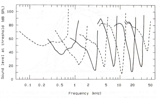
Tonotopic organization
the spatial arrangement of the basilar membrane where different regions respond to specific sound frequencies
High freq: Base of cochlea (near stapes)
Low freq: Along basilar membrane (apex of cochlea)
Intensity resolution
The ability to detect different sound levels
Threshold
The lowest sound level that causes a nerve fiber to start responding
for intensity resolution
Saturation
The highest sound level where the neuron’s firing rate stops increasing
for intensity resolution
Dynamic Range
range of sound intensities between threshold and saturation
20-50 dB: fiber can accurately represent sound intensity changes
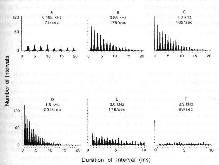
Phase locking
Neurons fire at a consistent phase of the sound wave
Frequency limit: up to about 4 – 5 kHz
Temporal Pattern: Spike timing matches the wave’s period