LIVER AND GB!!
 Call Kai
 Learn
Learn Practice Test
 Spaced Repetition
 Match
Match Flashcards
 Knowt Play
1/53

There's no tags or description
Looks like no tags are added yet.
Name | Mastery | Learn | Test | Matching | Spaced | Call with Kai | Chat |
|---|
No analytics yet
Send a link to your students to track their progress
54 Terms
the left lobe of liver is divided into medial and lateral segments by
left hepatic vein and ligamentum of teres
the left lobe of liver is seperated from caudate lobe by
ligamentum of venosum
what lies within falciform ligament and is previous fetal umbilical vein
ligamentum teres
alk phos elevation is associated with
obstructive jaundice
ALT elevation is associated with
cirrhosis, hepatitis, biliary obstruction
AST elevtion assocaited with
cirrhosis, hepatitis, mononucleosis
what are common causes of liver abscess
ascending cholangitis (m/c)
recent travel abroad
infection
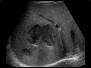
what is this
abscess
candidiasis is normally found with
immune-supressed patients
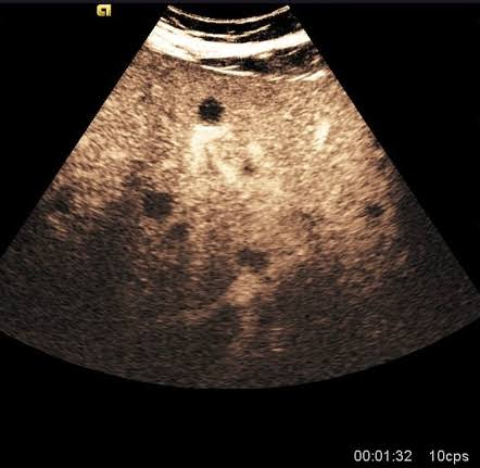
uniformly hypo lesions
wheel within wheel
candidiasis
what can cause echinococcal cyst
recent travel to underdeveloped countries
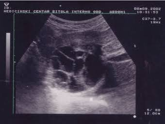
honeycomb appearance, daughter cysts, water lilly sign
echinoccocal cyst
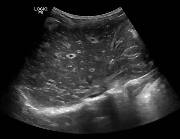
normal-hypo liver parenchyma, prominent portal veins, hepatomegaly, splenomegaly
hepatitis
what is schistosomiasis caused by
parasite entering the skin and travelling to lung then liver
what is the main clinical findings of schistosomiasis
rash, fever, diarrhea, lymphadenopathy, RUQ pain
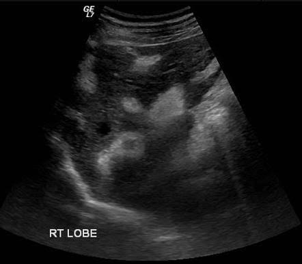
increase echogenicity of portal walls, thick gb wall
schistosomiasis
what is liver adenoma caused by mainly
long history of oral contraceptive use
type 1 glycogen storage disease
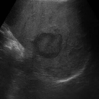
slightly hypo mass with a hypoechoic halo
adenoma
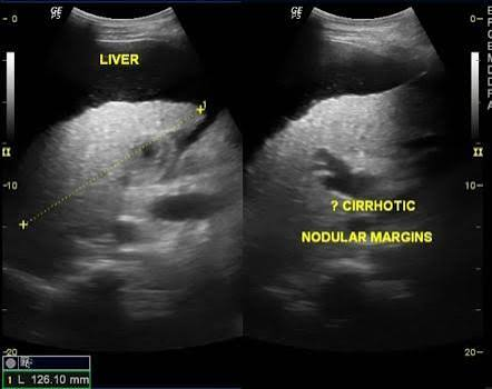
diffuse parenchymal echogenicity, irregular nodular contour, no portal wall distinguish, enlarged caudate lobe, splenomegaly, ascites
cirrhosis
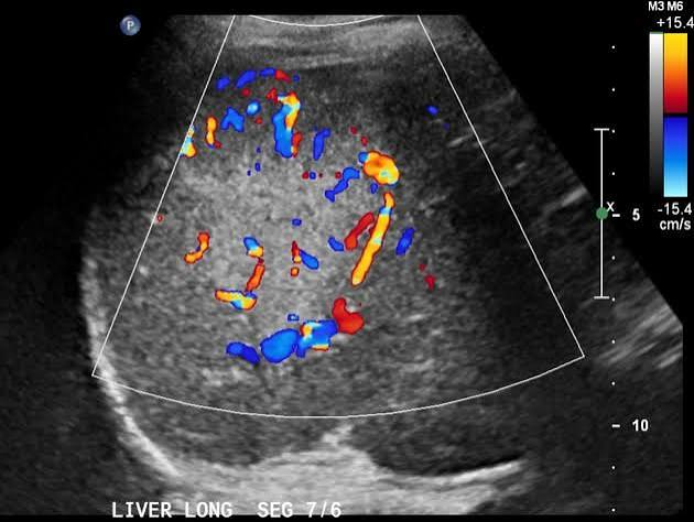
hyperechoic/isoechoic, spoke wheel pattern with a central scar
FNH
glycogen storage disease type 1
von Gierke disease
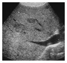
diffuse increase echogenicity, solid liver masses (adenomas)
glycogen storage disease
m/c malignant tumour in children
liver hepatoblastoma
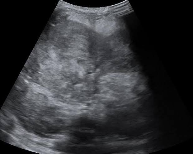
heterogenous, hyperechoic, cystic, internal spetations
heaptoblastoma
clinical of HCC
palp mass, abdo pain, weight loss, jaundice
elevated ALT, AST, alk phos
previous cirrhosis, chronic hep b
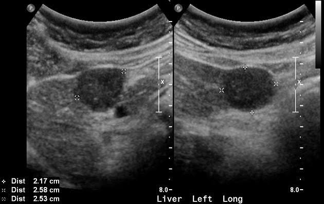
HCC
variable echogenicity, hypo halo
what are common patterns for metastases
bulls eye/ target, hyperehchoic, cystsic, complex, diffuse
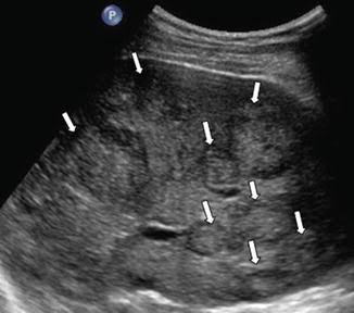
weight loss, jaundice, increase AST, ALT, bilirubin
mild increase alk phos
mets
budd chiari
thrombus within hepatic veins
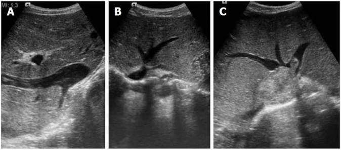
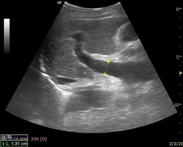
sonographic findings for portal hypertension
MPV exceeding 13mm, splenomegaly, ascites, collaterals
RI exceeding 0.8 in hepatic arteries
changes in portal venous flow
hepatofugal, pulsatile, decrease in velocity
TIPS
shunt placed behind portal vein and hepatic vein
m/c right portal vein and right hepatic vein
m/c cause of cirrhosis
alcohol abuse
HCC has a ____ in serum albumin
decrease
hepatic abscess is most likely to develop in which condition
asceinding cholangitis
what separates the left lobe from right lobe
middle hepatic vein and main lobar fissure
pt presents with RUQ pain, fever, leukocytosis. Pt discloses recent travel abroad. Complex mass is seen in rt lobe of liver. this is most likely
hepatic abscess
pts with hep b have an increase risk for developing
cirrhosis and adenoma
daughter cysts are associated with
echinococcal cyst
normal blod flow pattern in MPV is
phasic, hepatopedal
what is related to oral contrceptives/hormone levels
adenoma and FNH
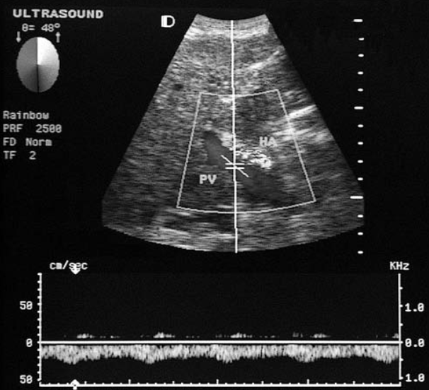
pt presents with hx of hep c and abdominal distention. This finding in the porta heptatits is most commonly associated with
portal hypertension
heptofugal flow
what liver pathology is associated with immune suppresed patients
candidiasis
metastatic lesions involving liver m/c originate from
colon
what is cirrhosis
severe insult to the liver cells leading to subsequent necrosis
paraumbilical vein courses from umbilicus to the
left portal vein
hepatic veins demonstrate ____ flow
multiphasic
decrease in prothrombin time can be associated with
acute cholecystitis
caroli disease
segmental, saccular, beaded appearance of intrahepatic ducts
multiple cystic structures in liver that communicate with biliary tree
what is klatskin tumour
small echogenic mass near hepatic hilum, dilatation of intrahepatic ducts, normal extrahepatic ducts
choledochal cyst
cystic mass medial to the gallbladder and lateral to head of pancreas
mirizzi syndrome
immobile calc in cystic duct or neck of GB
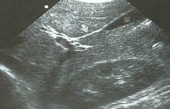
main lobar fissure
gravity dependent low level echoes within bile duct
hemobilia
dilatation of the intrahepatic ducts with normal extraheptatic ducts is characteristic of
klatskin tumour