Lab 19: The Heart Anatomy
1/52
There's no tags or description
Looks like no tags are added yet.
Name | Mastery | Learn | Test | Matching | Spaced | Call with Kai |
|---|
No analytics yet
Send a link to your students to track their progress
53 Terms

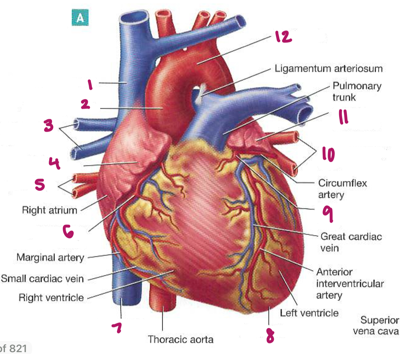
Label figure 1.
aorta (arch)

Label figure 2.
superior vena cava

Label figure 3.
right pulmonary artery

Label figure 4.
right pulmonary veins

Label figure 5.
right atrium

Label figure 6.
pulmonary trunk/artery

Label figure 7.
tricuspid valve
Label figure 8.
right ventricle

Label figure 9.
inferior vena cava

Label figure 10.
left pulmonary artery

Label figure 11.
left pulmonary veins

Label figure 12.
left atrium

Label figure 13.
bicuspid (mitral) valve

Label figure 14.
aortic valve

Label figure 15.
left ventricle

Label figure 16.
descending aorta
What is the name of the blood vessel that carries blood into the right atrium? Is the blood oxygenated or deoxygenated?
superior vena cava and inferior
deoxygenated
What is the name of the blood vessel that carries blood away from the right ventricle? Is the blood oxygenated or deoxygenated?
pulmonary artery
deoxygenated
What is the name of the blood vessel that carries blood to the left atrium? Is the blood oxygenated or deoxygenated?
pulmonary veins
oxygenated
What is the name of the blood vessel that carries blood away from the left ventricle? Is the blood oxygenated or deoxygenated?
aorta
oxygenated
What does the systemic circuit do?
left side that pumps blood whole body
BP + pulse
What does the pulmonary circuit do?
right side that pumps blood (CO2) to lungs
Start:
inferior/superior vena cava → _____1_____atrium →
______2_______valve → right ______3______ → ______4_____valve → ____5_____trunk/artery → deoxygenated (__6__gas) blood enters the ______7_____, where gas exchange occurs through the process of _____8_____. Oxygenated (___9___gas) blood leaves the lungs going back to the heart via the → pulmonary _____10_____ → enters the ______11_____ atrium → ______12_______valve → left _____13_____ → _____14______ valve → ____15_____ → end of body
right
tricuspid
ventricle
pulmonary
pulmonary
CO2
lungs
diffusion
O2
veins
left
bicuspid/mitral
ventricle
aortic
aorta

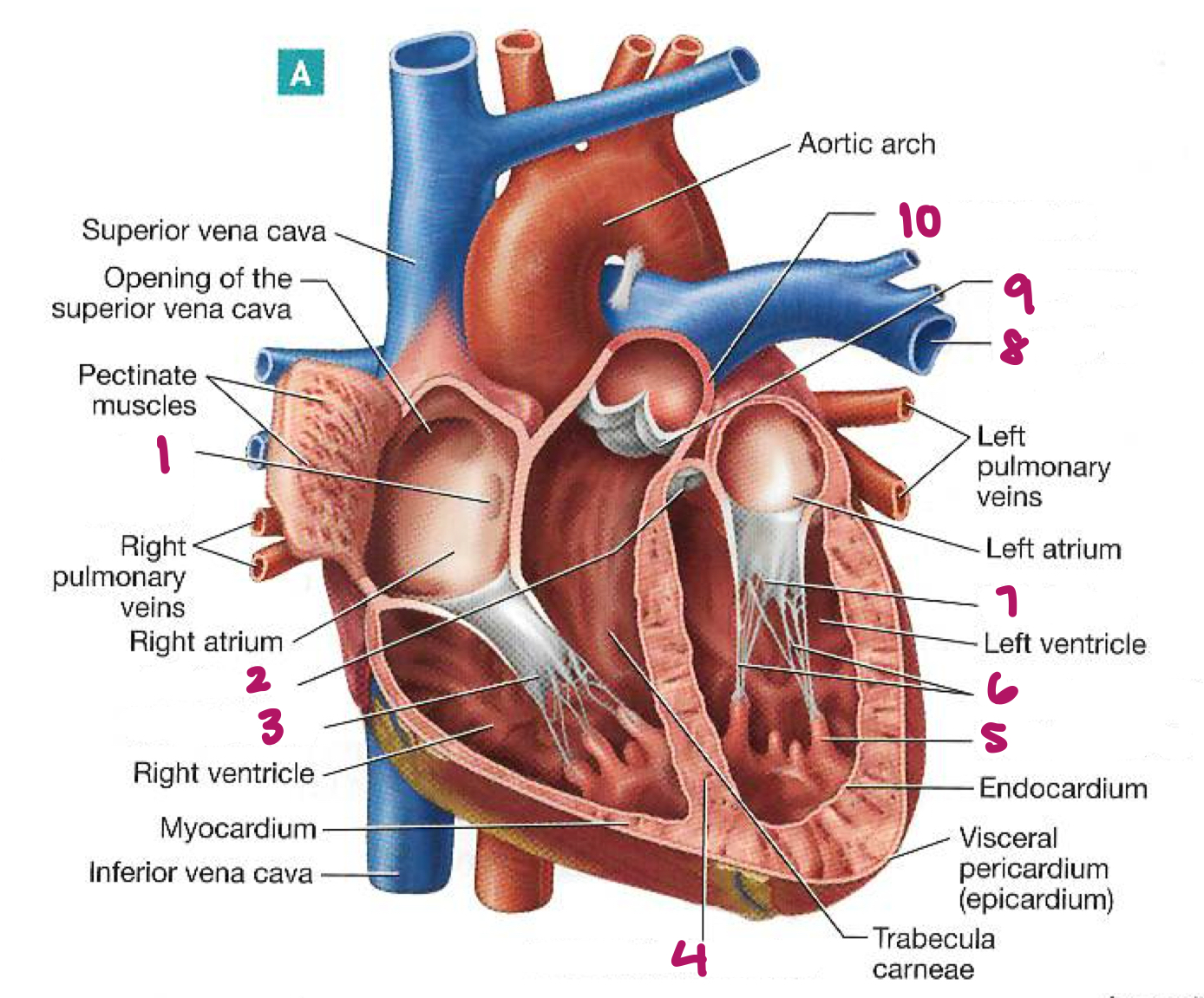
Label figure 1.
superior vena cava

Label figure 2.
ascending aorta

Label figure 3.
branches of right pulmonary artery
Label figure 4.
auricle of the right atrium

Label figure 5.
right pulmonary veins

Label figure 6.
right coronary artery

Label figure 7.
inferior vena cava

Label figure 8.
apex of heart

Label figure 9.
left coronary artery

Label figure 10.
left pulmonary veins

Label figure 11.
auricle of left atrium

Label figure 12.
aortic arch
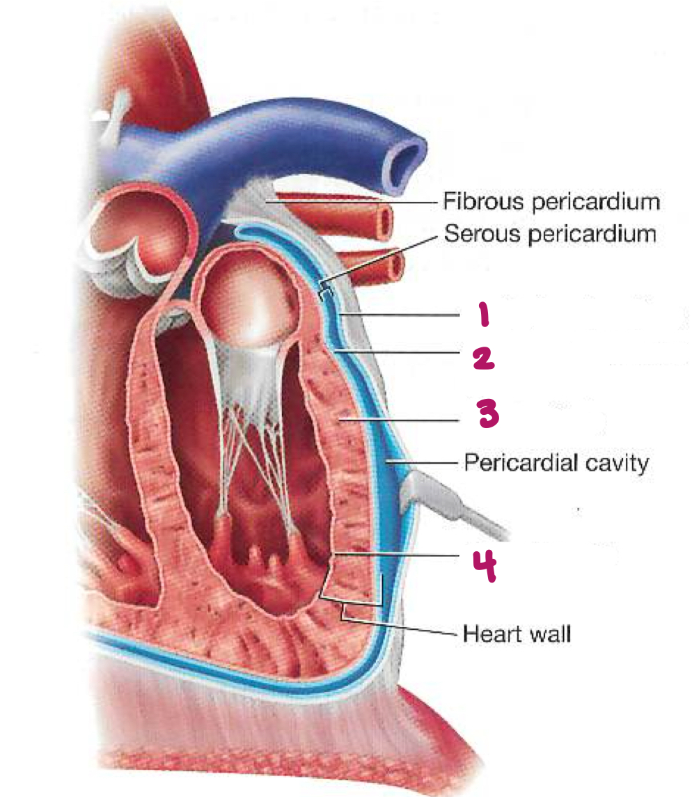
Label figure 1.
Parietal pericardium

Label figure 2.
visceral pericardium (epicardium)

Label figure 3.
myocardium

Label figure 4.
endocardium
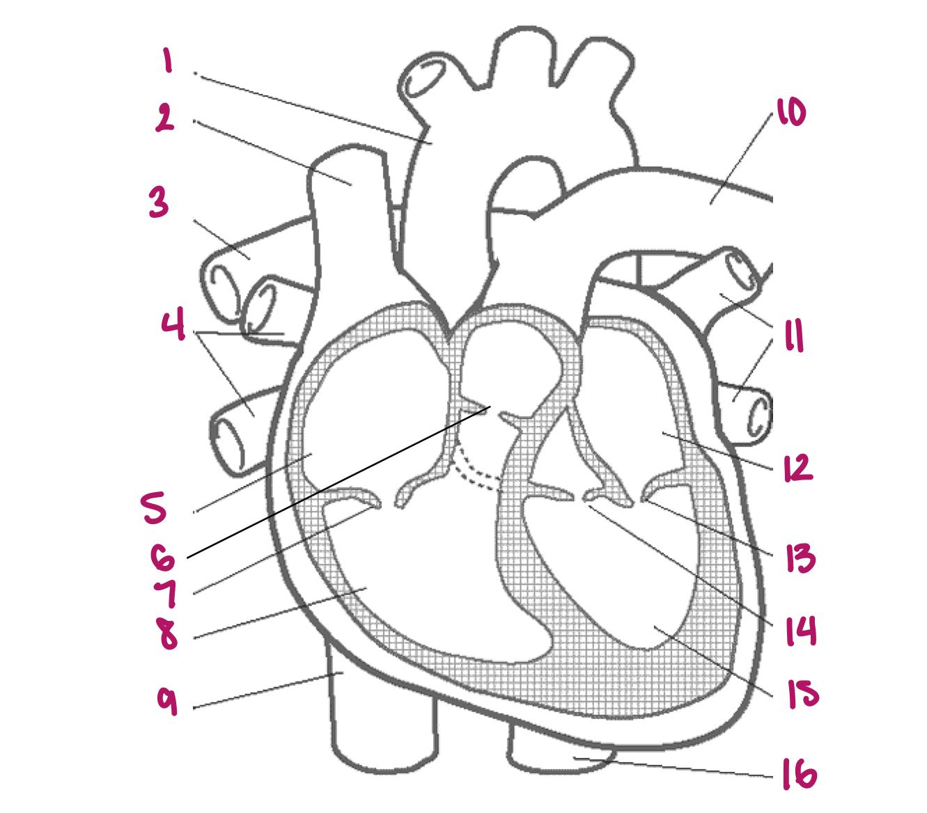
Label figure 1.
fossa ovalis in the interatrial septum

Label figure 2.
aortic valve

Label figure 3.
tricuspid

Label figure 4.
interventricular septum

Label figure 5.
papillary muscle

Label figure 6.
chordae tendineae

Label figure 7.
bicuspid (mitral) valve

Label figure 8.
left pulmonary artery

Label figure 9.
pulmonary valve

Label figure 10.
pulmonary trunk
ignore (type that for ans)
ignore

Label figure 1.
sinoatrial (SA) node
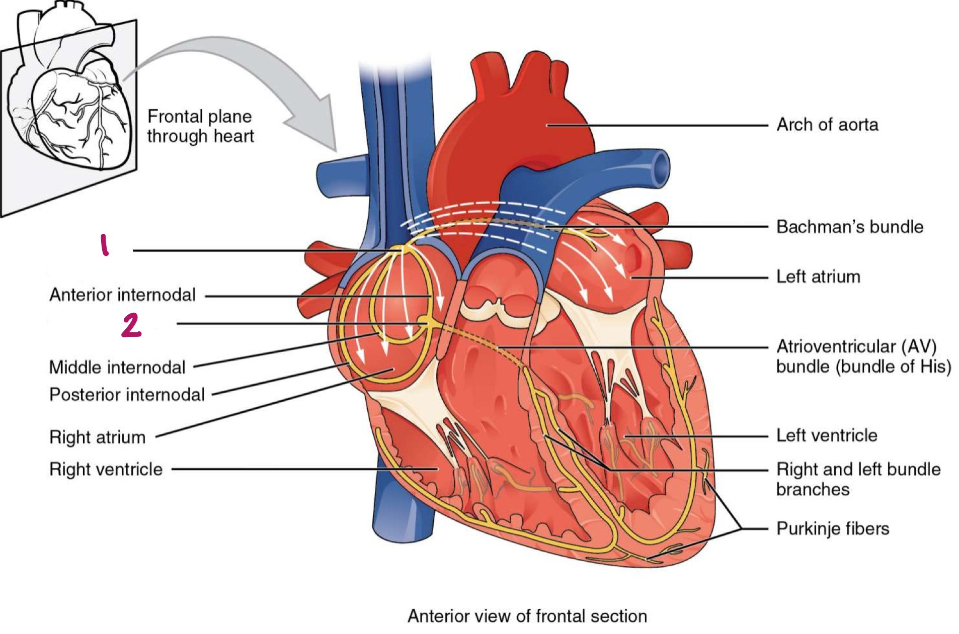
Label figure 2.
atrioventricular (AV) node
What is the top of the heart called?
base