Neural Cognition (Chapter 2) *axons not included
1/77
There's no tags or description
Looks like no tags are added yet.
Name | Mastery | Learn | Test | Matching | Spaced | Call with Kai |
|---|
No analytics yet
Send a link to your students to track their progress
78 Terms
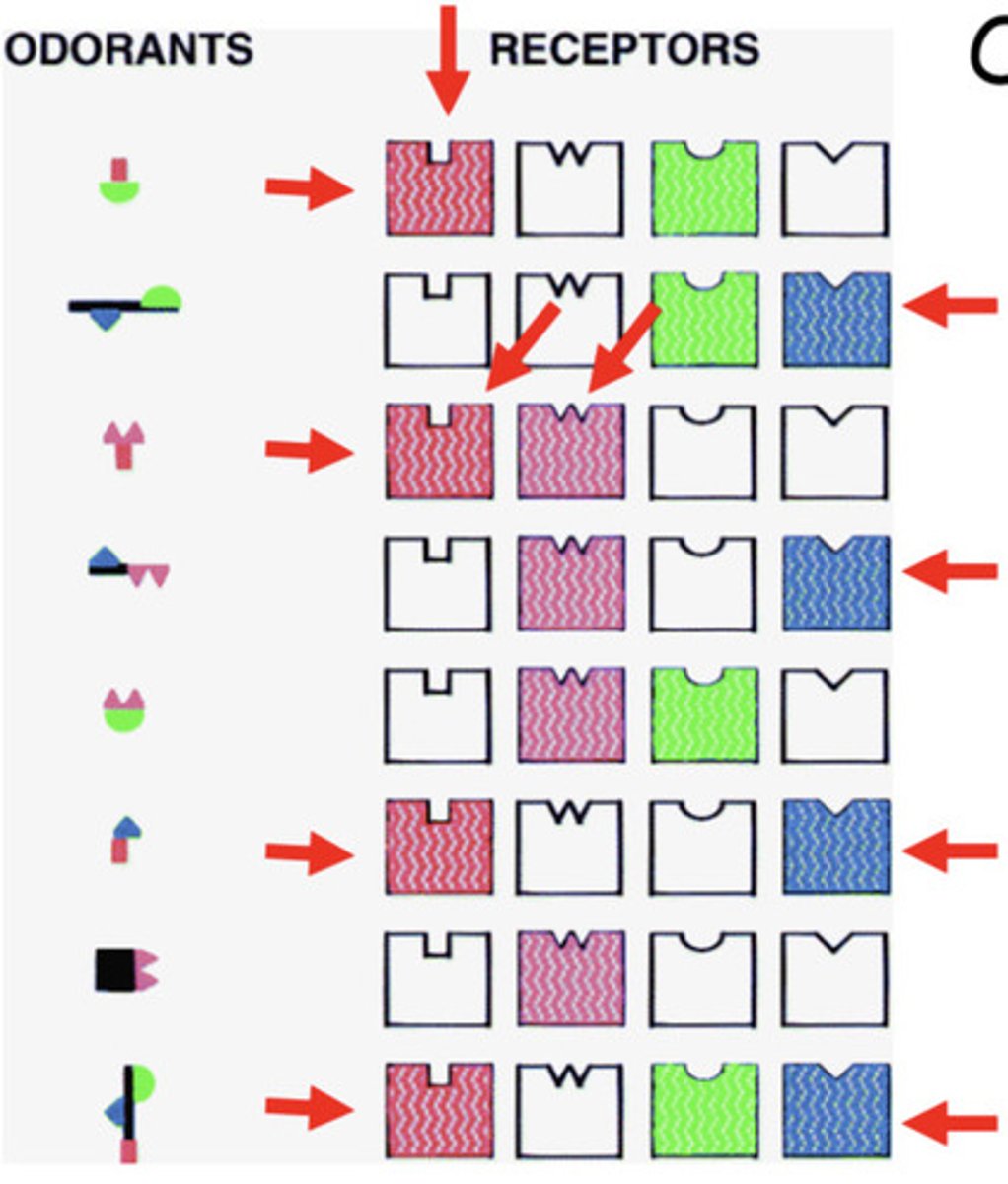
How is information represented by neurons?
Specific neurons can, in some cases, represent specific stimuli.
Pattern Coding
Coding of information in sensory systems based on the temporal pattern of action potentials.
-E.g., turning the page in a book, swiping your phone screen, realizing you are late
Cognitive Appraisal determines:
WHO IS IT?
Emotional Appraisal determines:
WHY DO I CARE?
Fregoli Syndome
Patients hold the delusional belief that different strangers are actually the same known person, in different disguises.
-This leads to an inappropriate emotional arousal for
faces.

Capgras Syndrome
Patients can recognize loved ones, but patients think that they are actually impostors.
-This leads to an intellectual identification without a familiarity response.
Capgras syndrome has been linked to abnormalities in the:
Amygdala and Prefrontal Cortex
Damage to Amygdala can cause:
Lack of emotional response
-People with Capgras syndrome won’t experience the warm sense offeeling safe and secure when looking at a loved one’s familiarface.
Damage to Prefrontal Cortex
Especially active when doing tasks that require planning or careful analysis.
-Can impair analytical reasoning.
Illogical thoughts (e.g., that someone who looks exactly like a loved one is not that loved one, or that strangers are actually loved ones) are not dismissed as they normally would be. 11
Amygdala is also involved with:
-Feeling of familiarity
-Memory for emotional events
-Emotional decision Making
-Threat Detection
Demonstrates how our intellectual understanding of the world is intertwined with our basic emotional reactions to it!
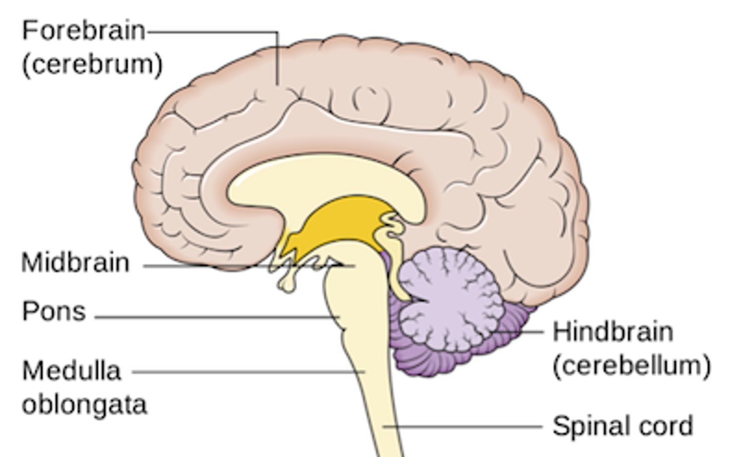
Brain Structures:
Hindbrain, Midbrain, Forebrain
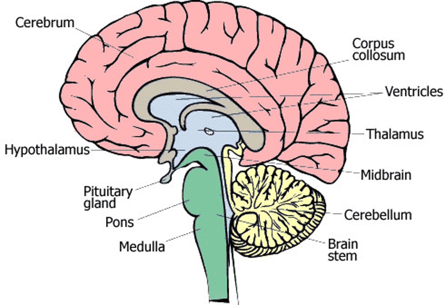
Hindbrain
Includes structures that are critical for key life functions:
-Cerebellum
-Medulla
-Pons
"CMP"
Cerebellum
Coordinates movements and balance (Largest region)
Also involved in other sensory and cognitive roles – damage to this area can cause problems with spatial reasoning, discriminating sounds, and integrating input from various sensory systems.
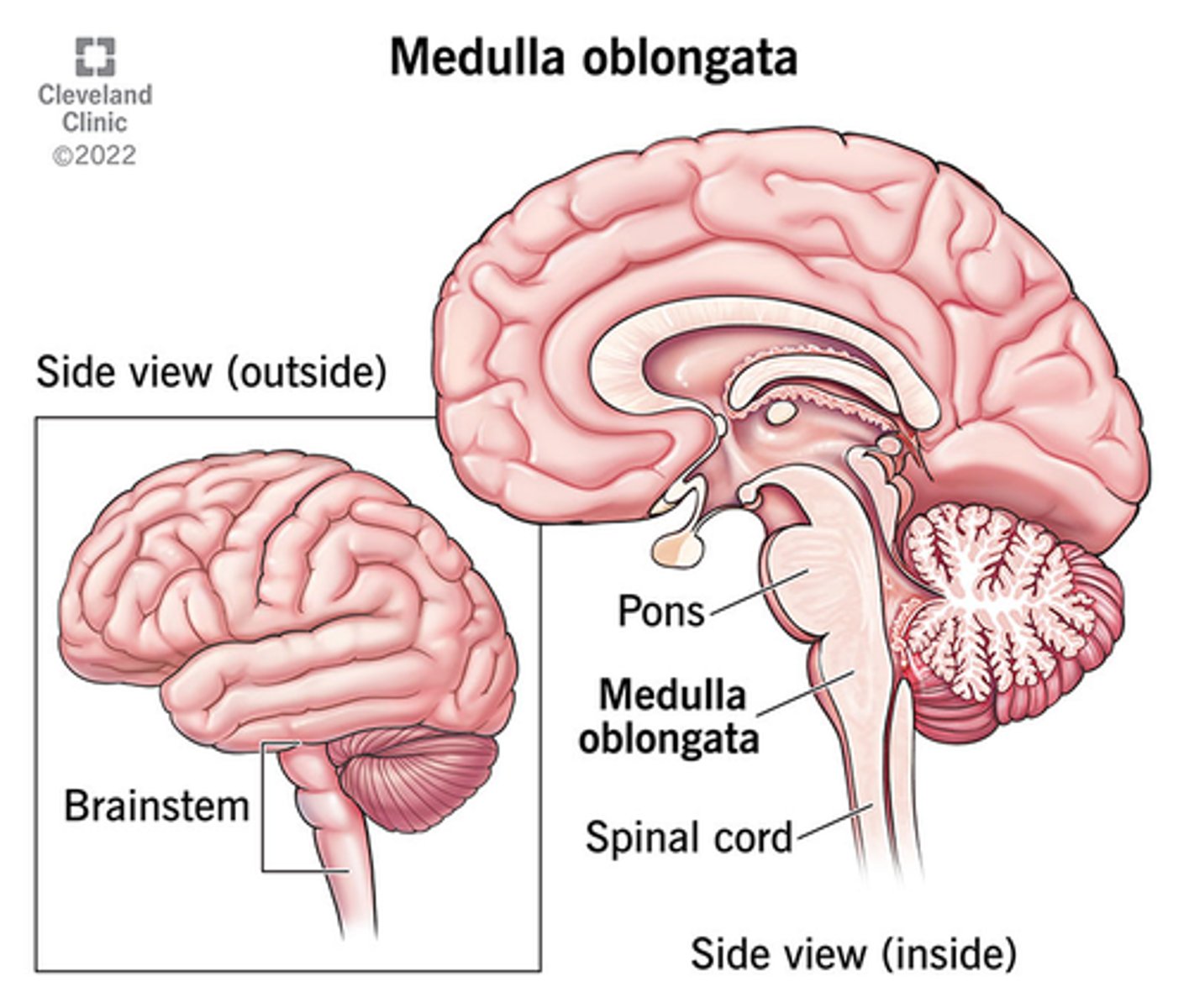
Medulla
Controls vital functions such as breathing and heart rate
Pons
Aspects of sleep/wake cycles, sensory functions.
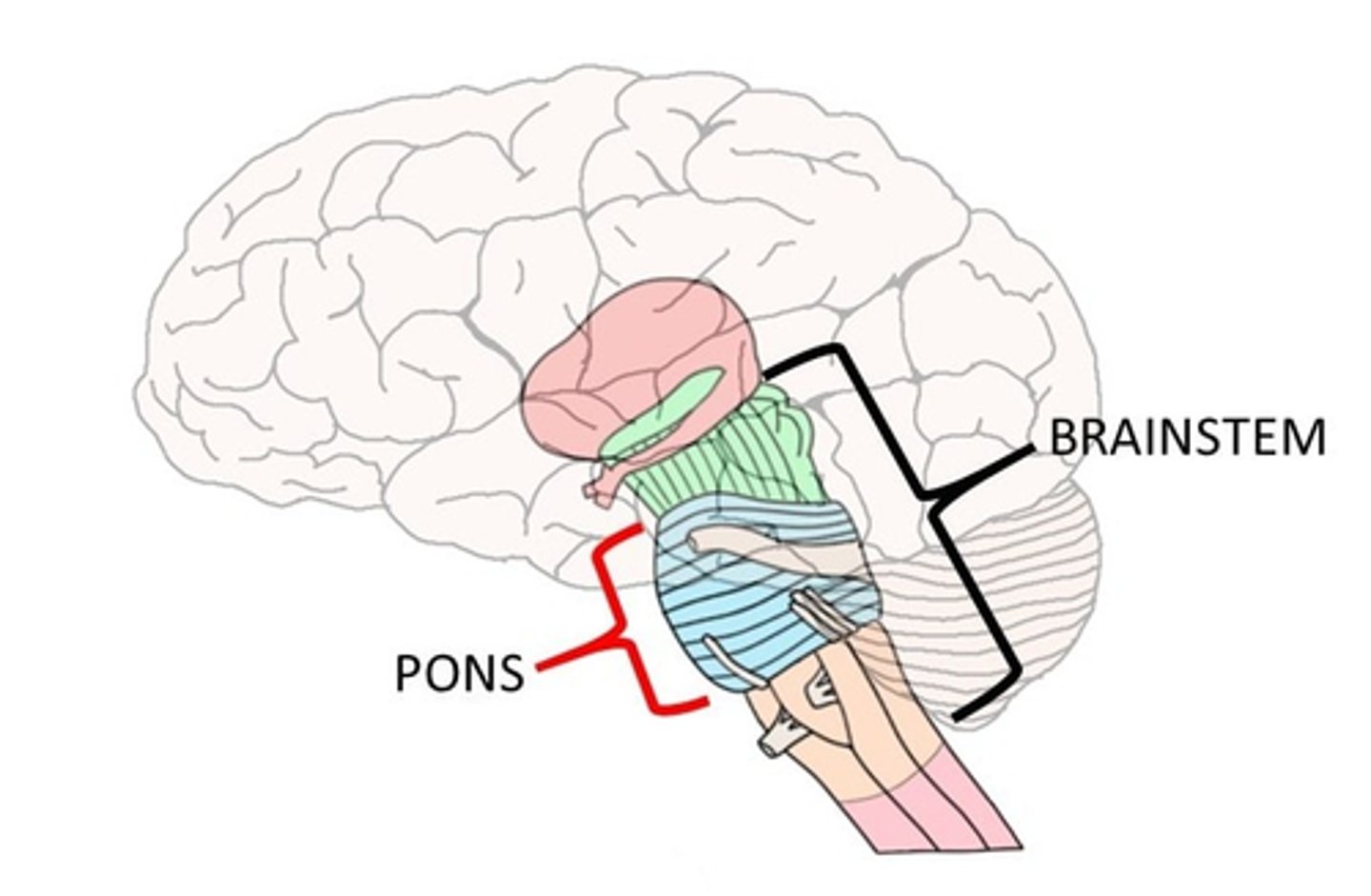
Pons is the ______________ between the cerebellum and the rest of the brain.
"Bridge"
-Links the brain to the spinal cord

Midbrain
Coordinating precise eye & eyelid movements
Relaying auditory information from ears to forebrain
Regulating pain experiences
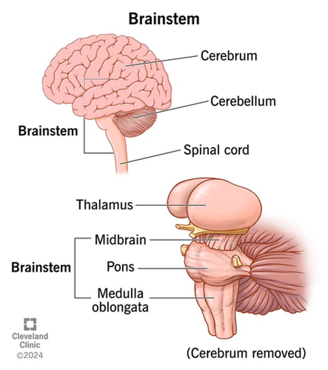
Forebrain
The largest and most complicated region of the brain, includes the thalamus, hypothalamus, limbic system, and cerebrum.
-Surrounds the midbrain and most of the hindbrain
The Forebrain includes:
-Subcortical structures
-Cortex
-Four lobes
What is the outer surface of the forebrain?
Cortex
-(3mm thick; approx. 80% of the brain)

Fissures/Sulci
Deep groves separating areas of the brain.
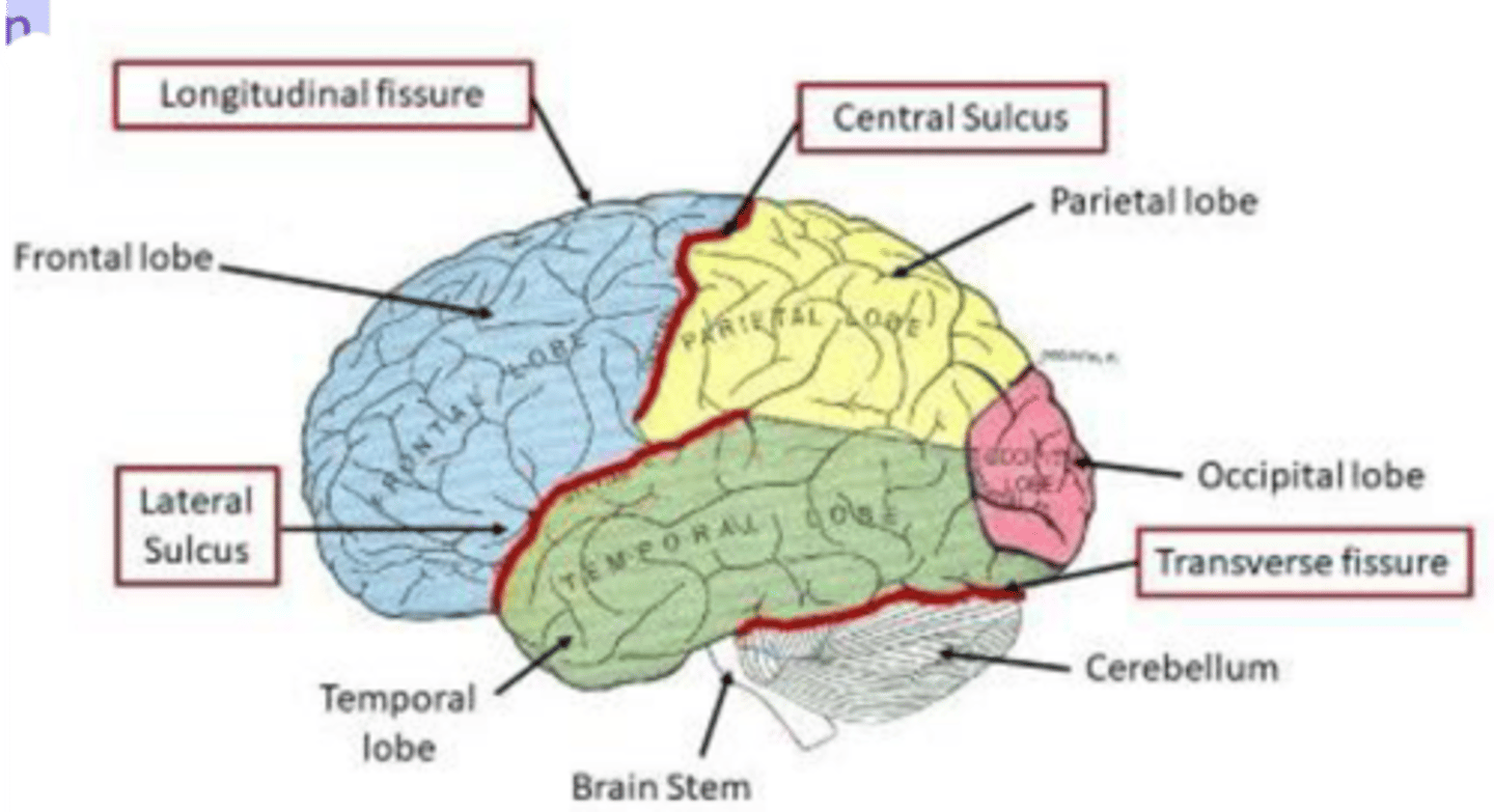
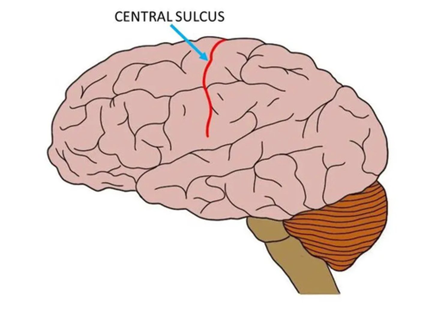
The two cerebral hemisphere is divided by:
Central Sulcus
-Sometimes called the: cerebral fissure/sulcus fissure of Rolando
The subcortical parts of theforebrain include:
-Thalamus
-Hypothalamus
-Limbic System
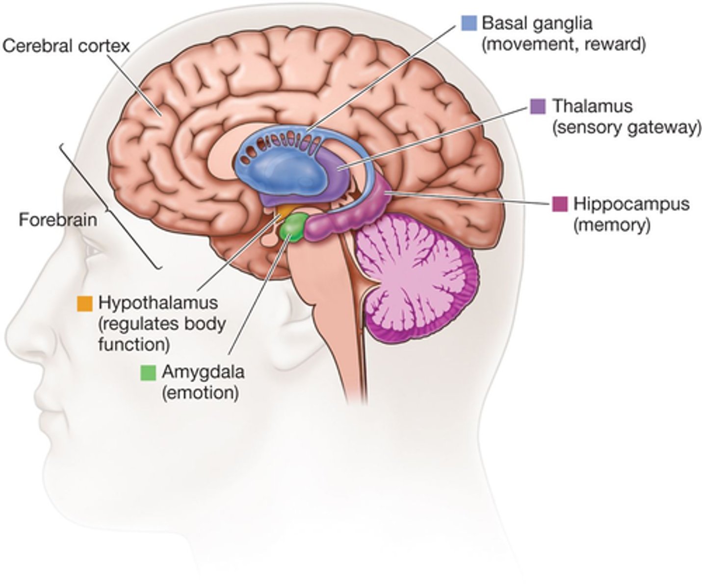
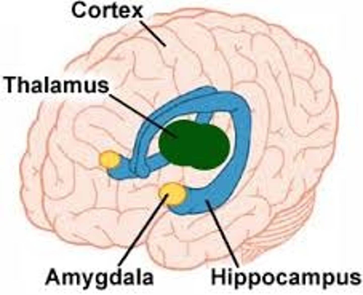
Thalamus
Sensory Relay Station

Hypothalamus
Controls behaviors that serve specific biological needs
(e.g., eating)

Limbic system
Involved in our behavioral and emotional responses, especially when it comes to behaviors we need for survival
-Feeding
-Regulating brain activity during sleep
-Reproduction and caring for our young,
-Fight or flight responses.20
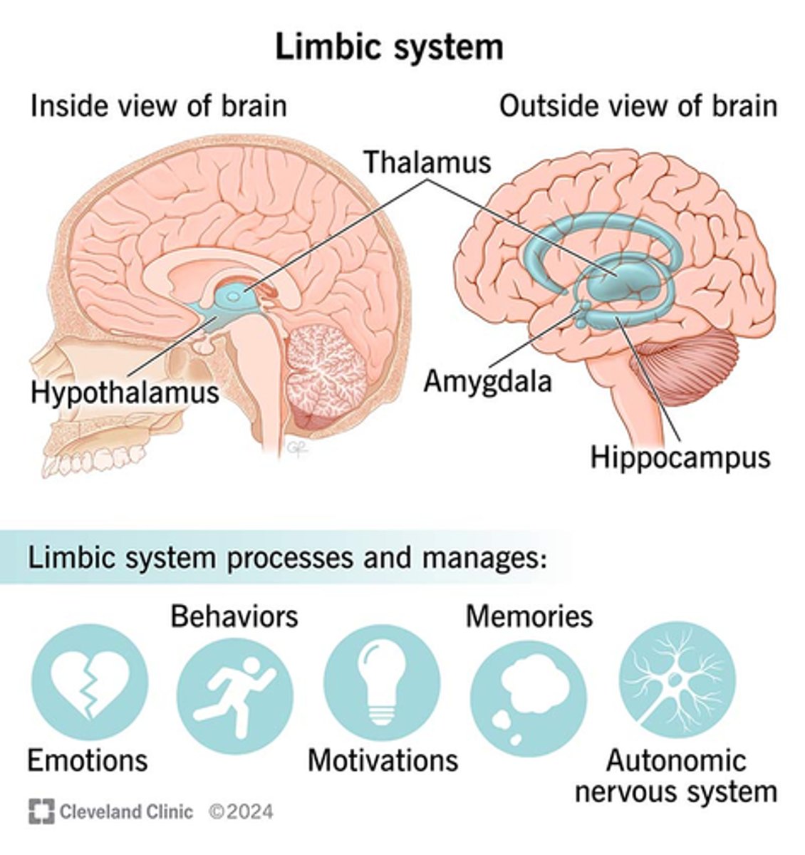
Limbic system is located:
Underneath the cortex in the temporal lobe

The Limbic System includes:
-Amygdala,
-Hippocampus
-Thalamus
-Hypothalamus
-Basal ganglia
-Cingulate gyrus

Amygdala
Emotional Center

Hippocampus
Learning and Memory
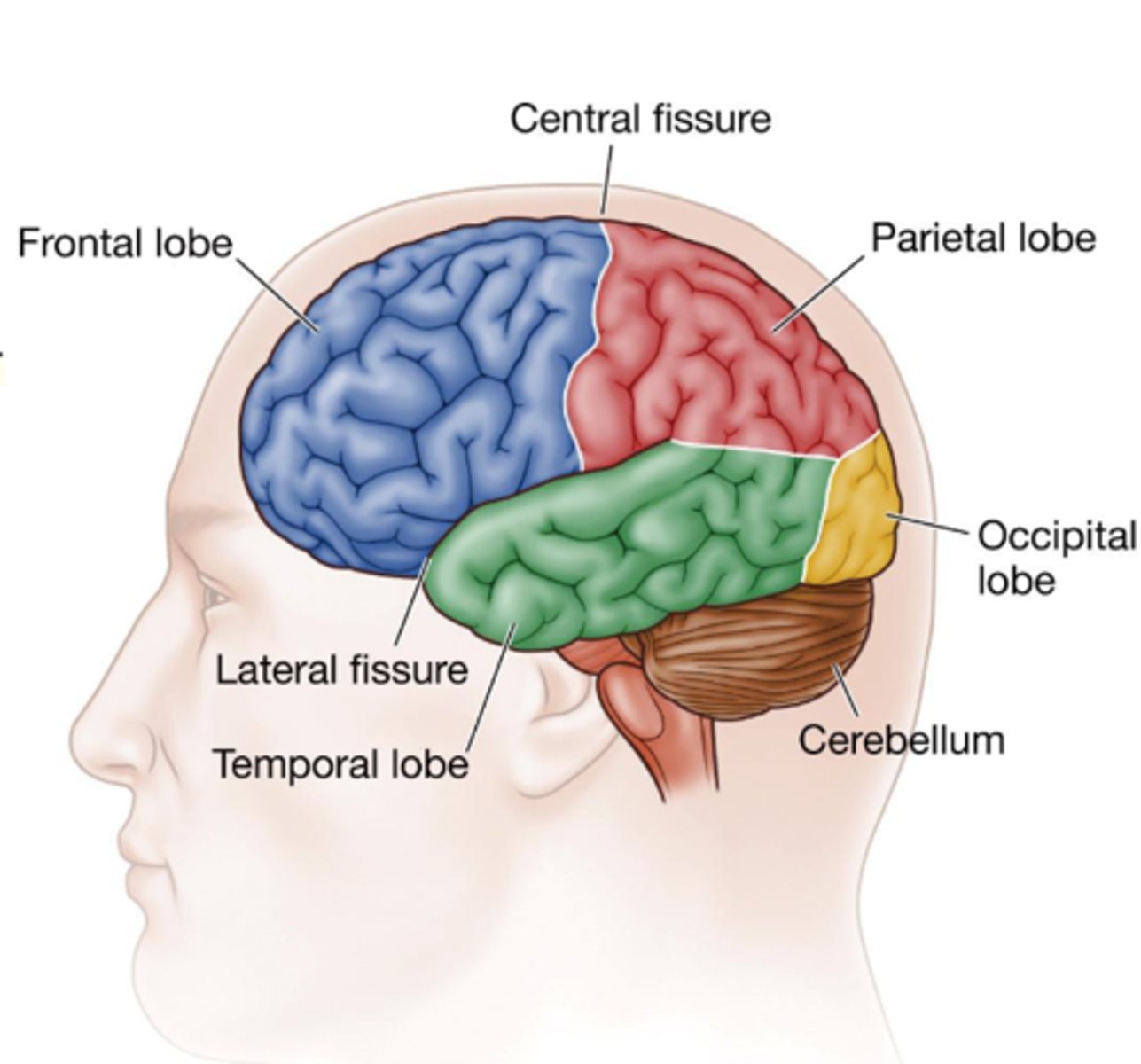
Cerebral Cortex
The largest portion of the human brain
-Thin layer of tissue covering the cerebrum (i.e., forebrain)
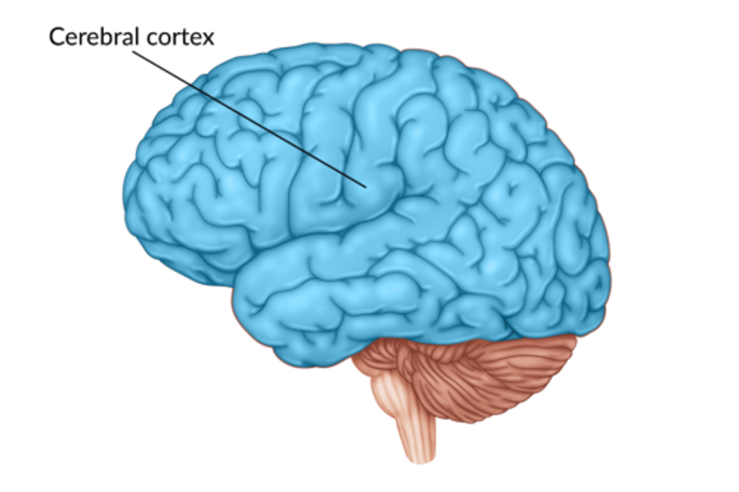
Regions of the Cortex:
-Motor Areas
-Sensory Areas
-Association Areas

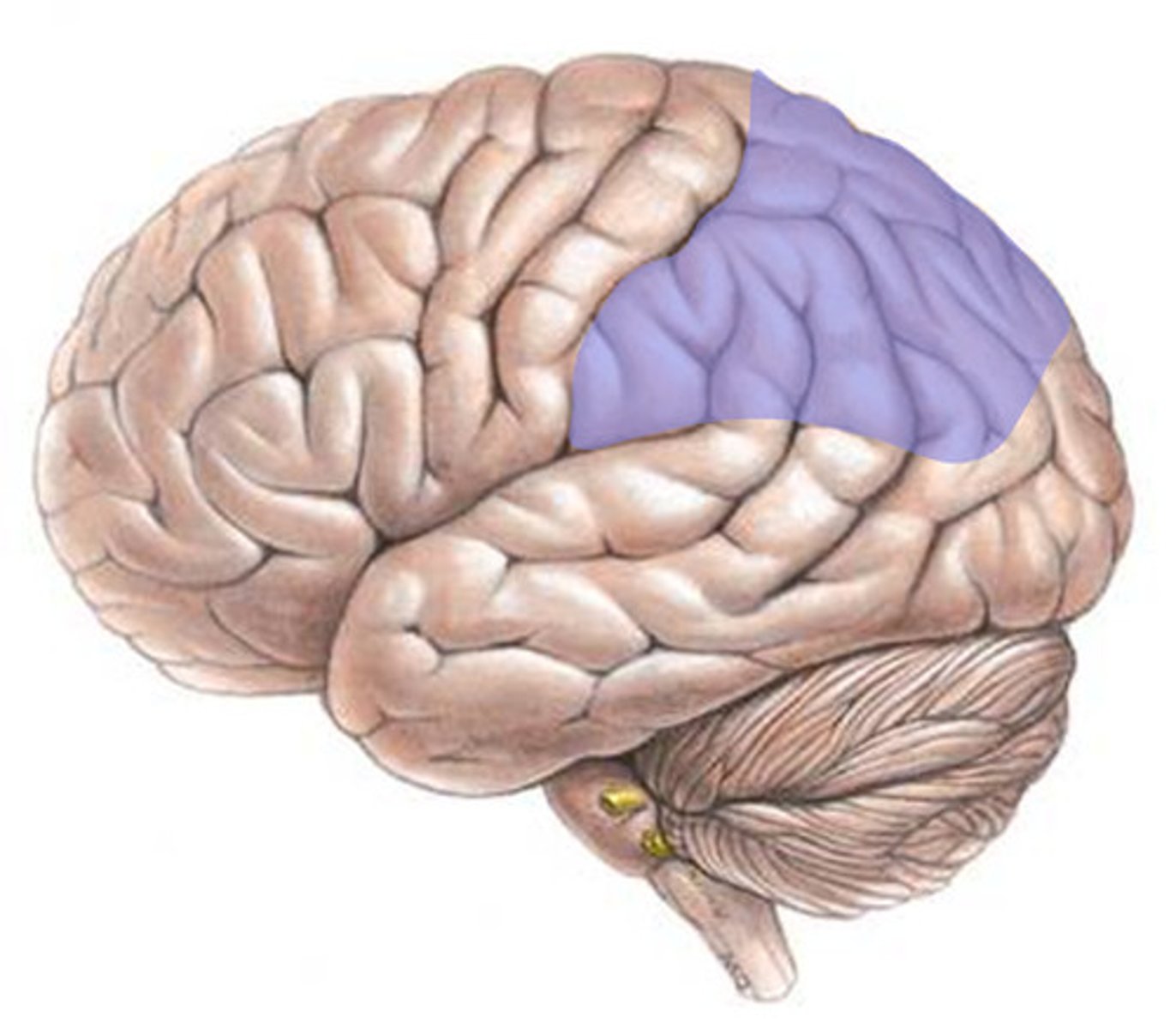
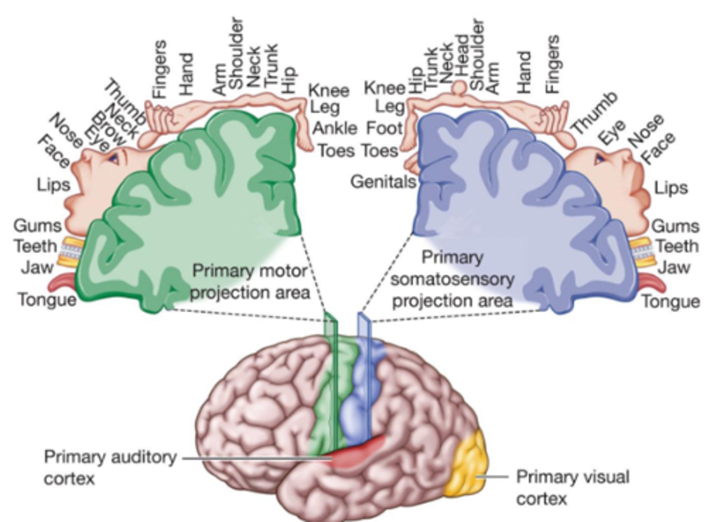
Primary Motor projection areas
Departure points in the motor cortex (frontal lobe) for signals that control muscle movement

Contralateral Control
Stimulation to the left hemisphere leads to movement on the right side of the body, and vice versa
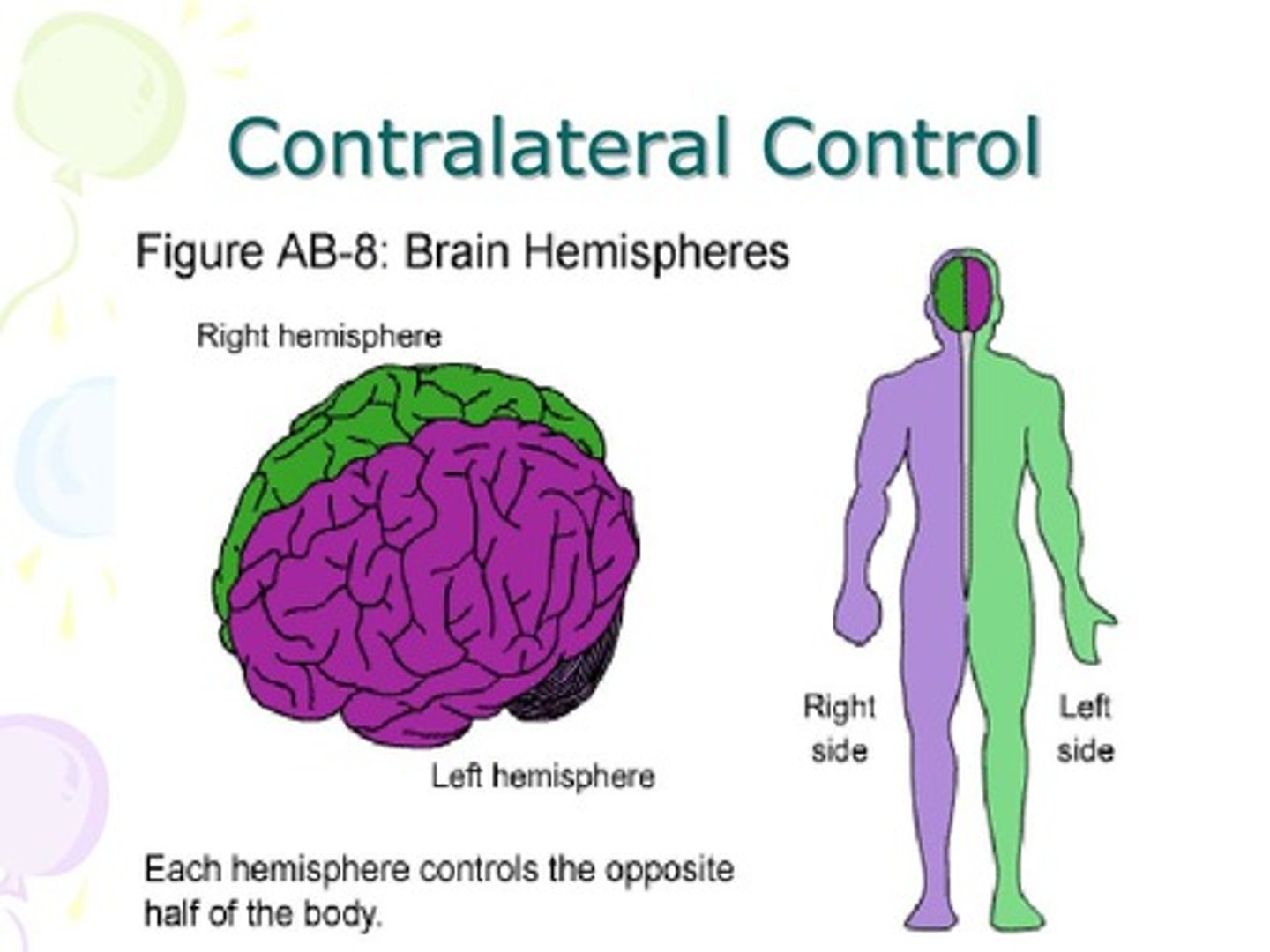
More ____________________ reflects greater motor precision, and greater sensitivity
Cortical Coverage
Primary Somatosensory Projection area:
Information arriving from the skin senses (touch, temperature) is projected to this region in the parietal lobe
-Contralateral organization
-Cortical space assigned based on acuity

Each part of the body's surface is represented by its region on the:
Association cortex.

Association cortex
Regions of the cerebral cortex that integrate simpler functions to perform more complex functions
PSP: Areas of the body that are near to each other are typically represented by _______________
Nearby areas
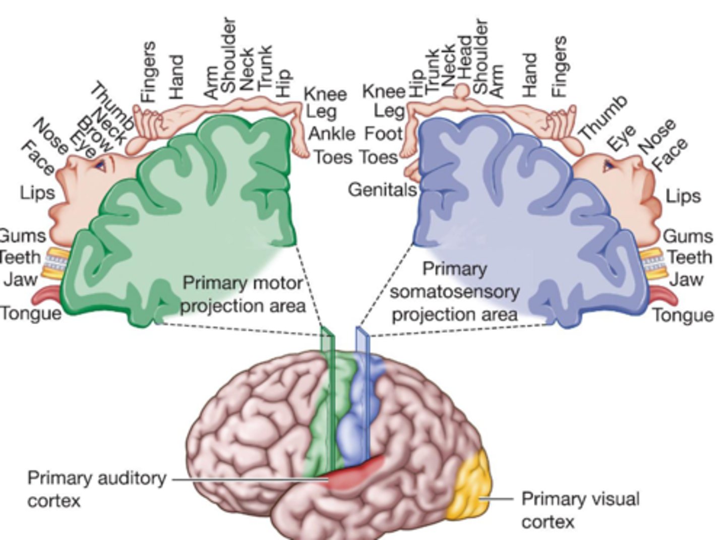
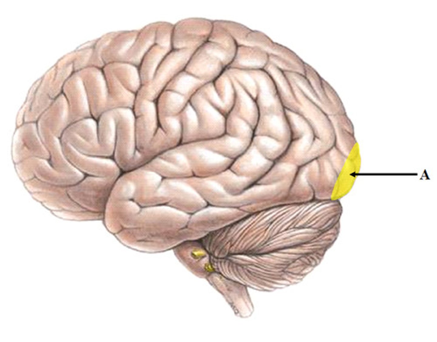
Primary Visual Cortex (Occipital lobe):
Visual sensation
–Each region of visual space has its cortical representation, and adjacent areas of visual space are usually represented by adjacent brain sites

Primary Auditory Cortex (Temporal lobe):
Auditory sensations
-Different sound frequencies have their cortical sites, and adjacent brain sites are responsive to adjacent frequencies.
Which of these Cortex are almost 100% Contralateral?
Primary Visual Cortex (occipital lobe)
Contralateral to physical space (right hemisphere receives information from the left visual field sites)
What is not as clear a contralateral distinction?
Primary Auditory Cortex (temporal lobe)
-But, 60% of the nerve fibers from each ear send information to the brain's opposite side.
PRIMARY PROJECTION AREAS
The first to receive information from another system (typically external environment) before relaying it for integration.

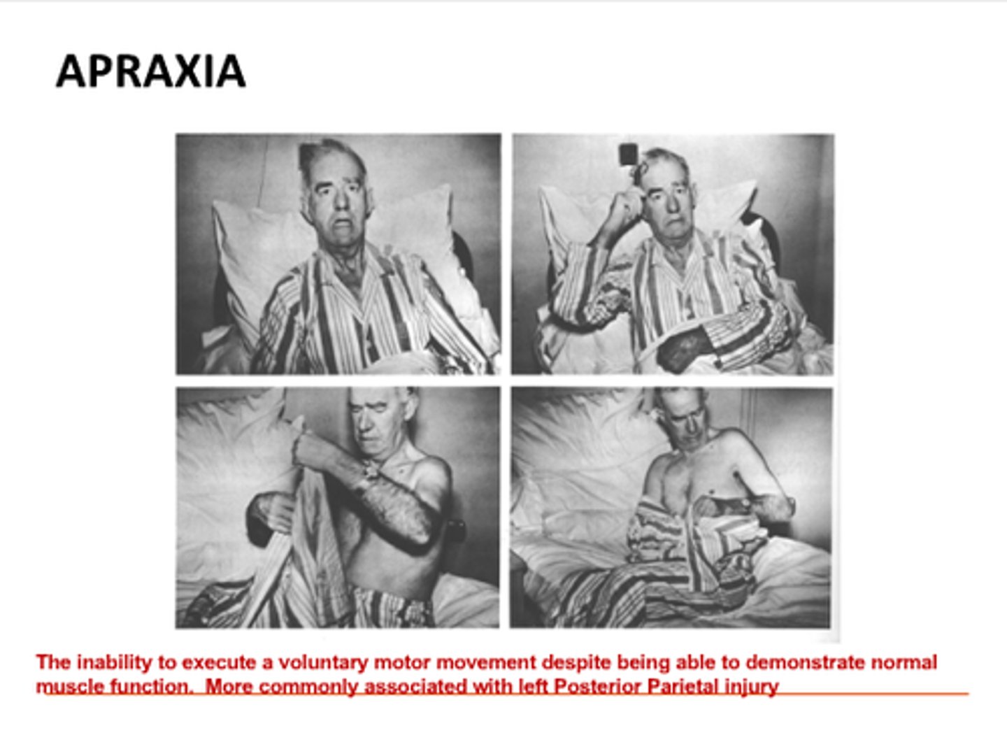
Apraxia
Problems with the initiation or organization of voluntary action
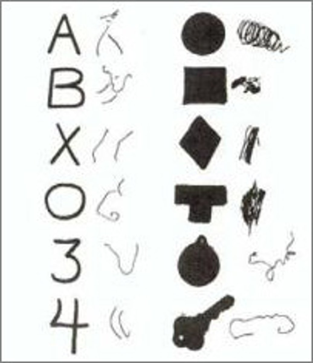
Agnosia
Problems identifying familiar objects; typically affect one modality only. (e.g., visual agnosia, auditory agnosia)
E.g., Lesions in the occipital or auditory cortex
Unilateral Neglect Syndrome
Problems in which half the visual world is ignored.

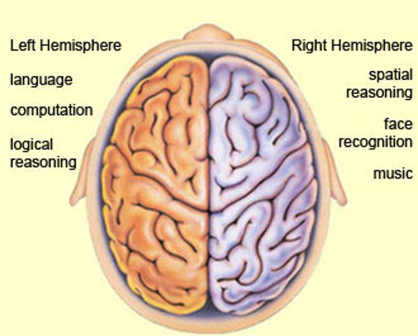
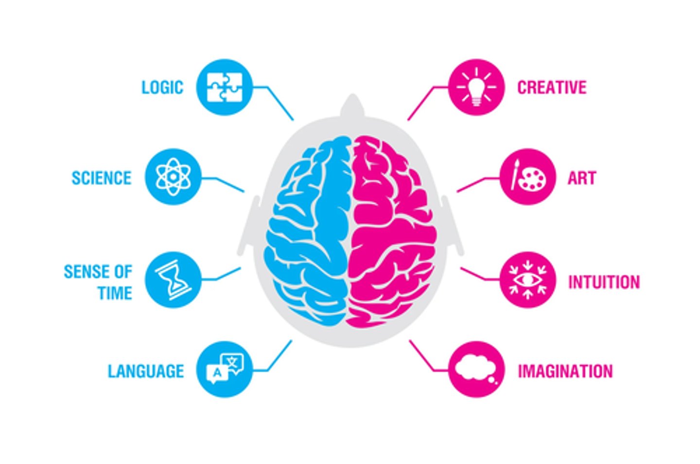
Lateralization
A term that refers to the brain being divided into roughly symmetrical left and right hemispheres
-Virtually all parts of the brain come in pairs, but there are some differences in functions between the left- and right-side structures
The the two hemispheres are connected by:
Commissures
(thick bundles of fibers that carry information between the two hemispheres)
What is the largest commissure?
Corpus Callosum
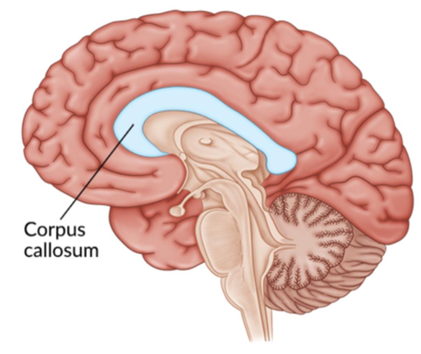
Split-brain patients:
Severed corpus callosum;
-Was a last-resort treatment for severe epilepsy
-Severely limits communication between the hemispheres
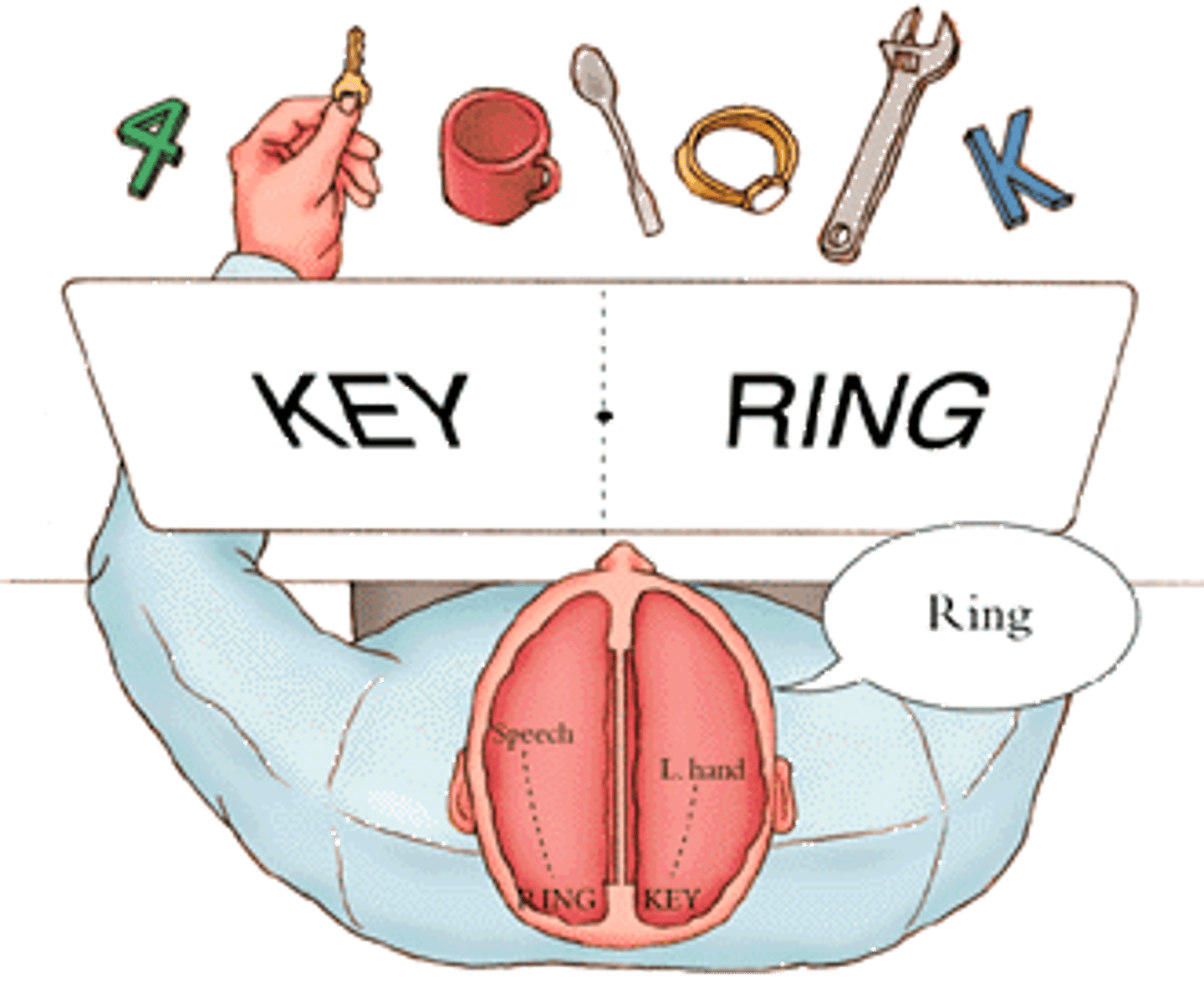
Evidence for some hemispheric specializations of functions:
Left hemisphere is responsible for language capacities.
Right hemisphere is crucial for tasks involving spatial judgment.
Neuropsychology
The study of the brain’s structures and their relation to brain function
Clinical Neuropsychology
Seeks to understand the functioning of intact, undamaged brains by examining cases involving brain damage

Lesions
A specific area of damage, will have specific consequences depending on the location.
E.g., Damage to left frontal lobe results in language disruption, but we do not see this effect if the right frontal lobe is damaged.
Several types of neuroimaging techniques...slightly different but all produce:
Precise, 3D pictures of a living brain
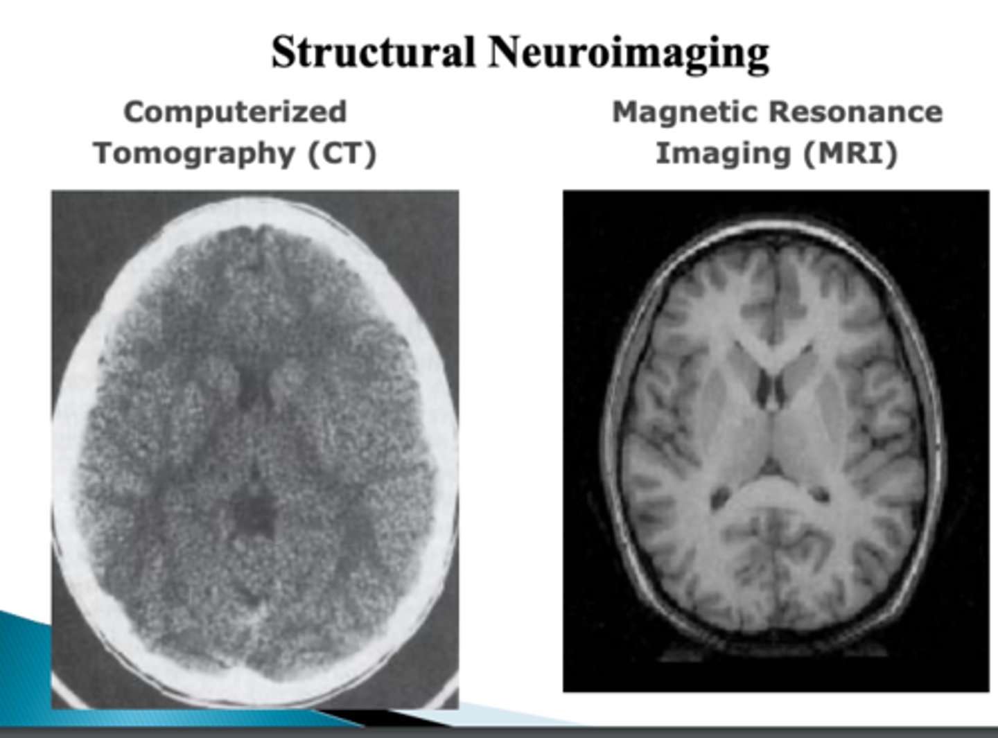
Structural Neuroimaging Technique:
Provide a detailed portrait of the shapes, sizes, and positions of the brain’s components:
Functional neuroimaging:
Tells us about activity levels throughout the brain.
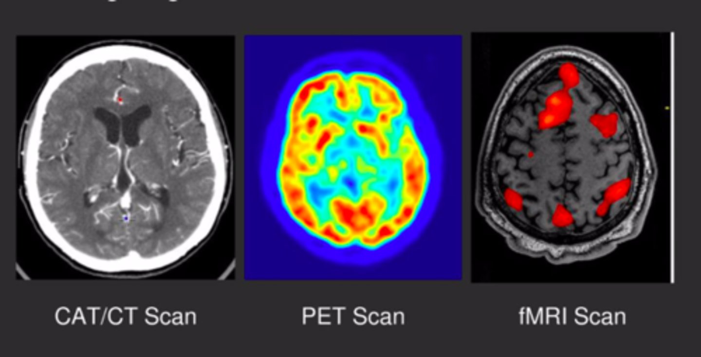
Computerized axial tomography (CT) scans
3D X-ray pictures of the brain only.
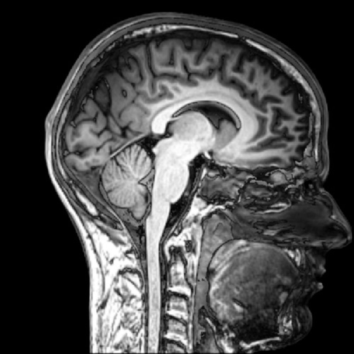
Magnetic resonance imaging (MRI) scans
Relies on the magnetic properties of the atoms that make up the brain tissue
–Very detailed structural details

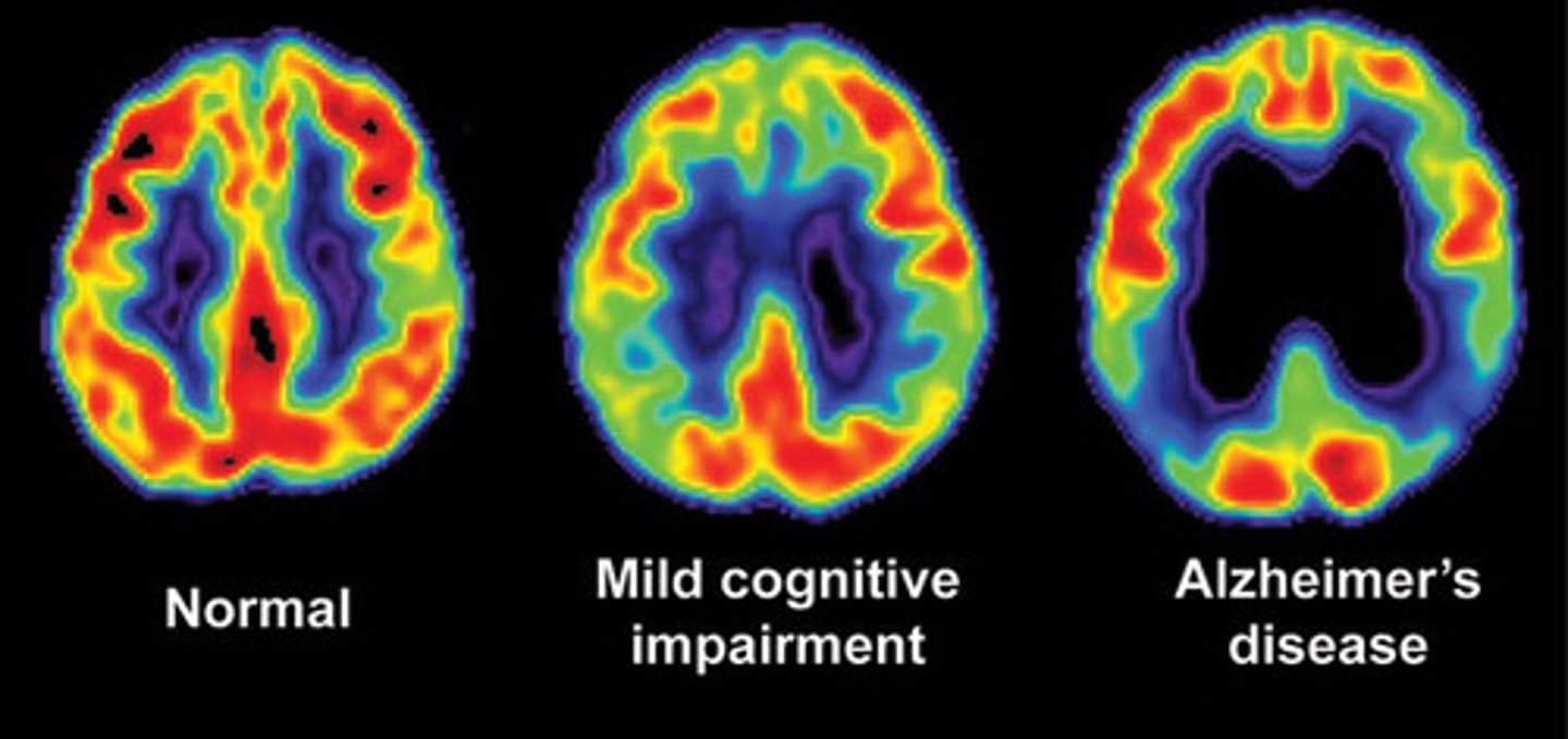
Positron emission tomography (PET) scans
A tracer, like glucose tagged with low-dose radioactivity, is used in scans to track tissue activity based on glucose usage, providing insights into brain activity levels.
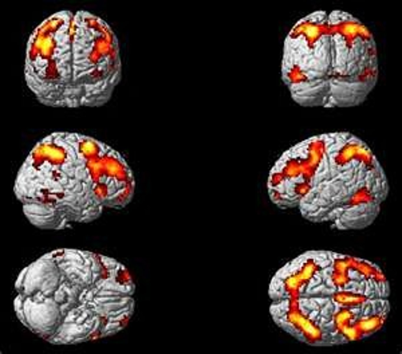
Functional magnetic resonance imaging (fMRI) scans
Measure the oxygen content in blood flowing through each region of the brain.
-Provides an index of the level of neural activity in that region
fMRI: Parahippocampal Place (PPA)
Activated after looking at a face, to a house or a place.
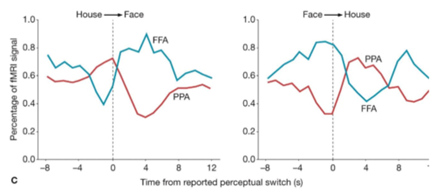
fMRI: Fusiform Face Area (FFA)
Activates during facial recognition

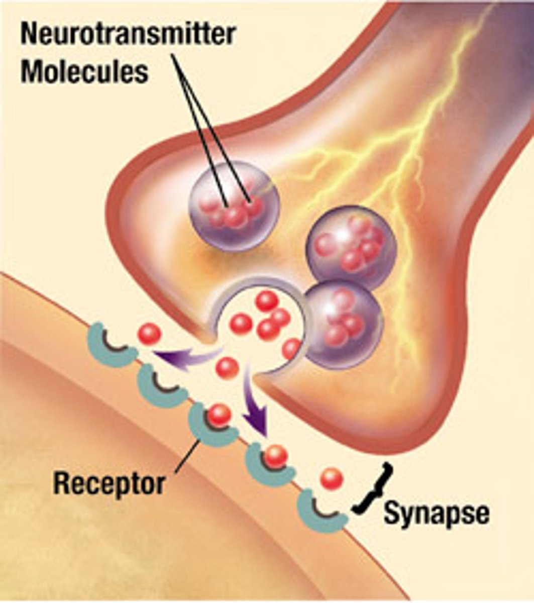
Communication between neurons is:
Chemical
Neurons communicate with one another via neurotransmitters.
-Once a neuron is activated, it releases the transmitter, and this chemical can then activate (or de-activate) other, adjacent neurons.
Communication within a neuron is:
Electrical
"Input" end receives neurotransmitters; "output" end releases
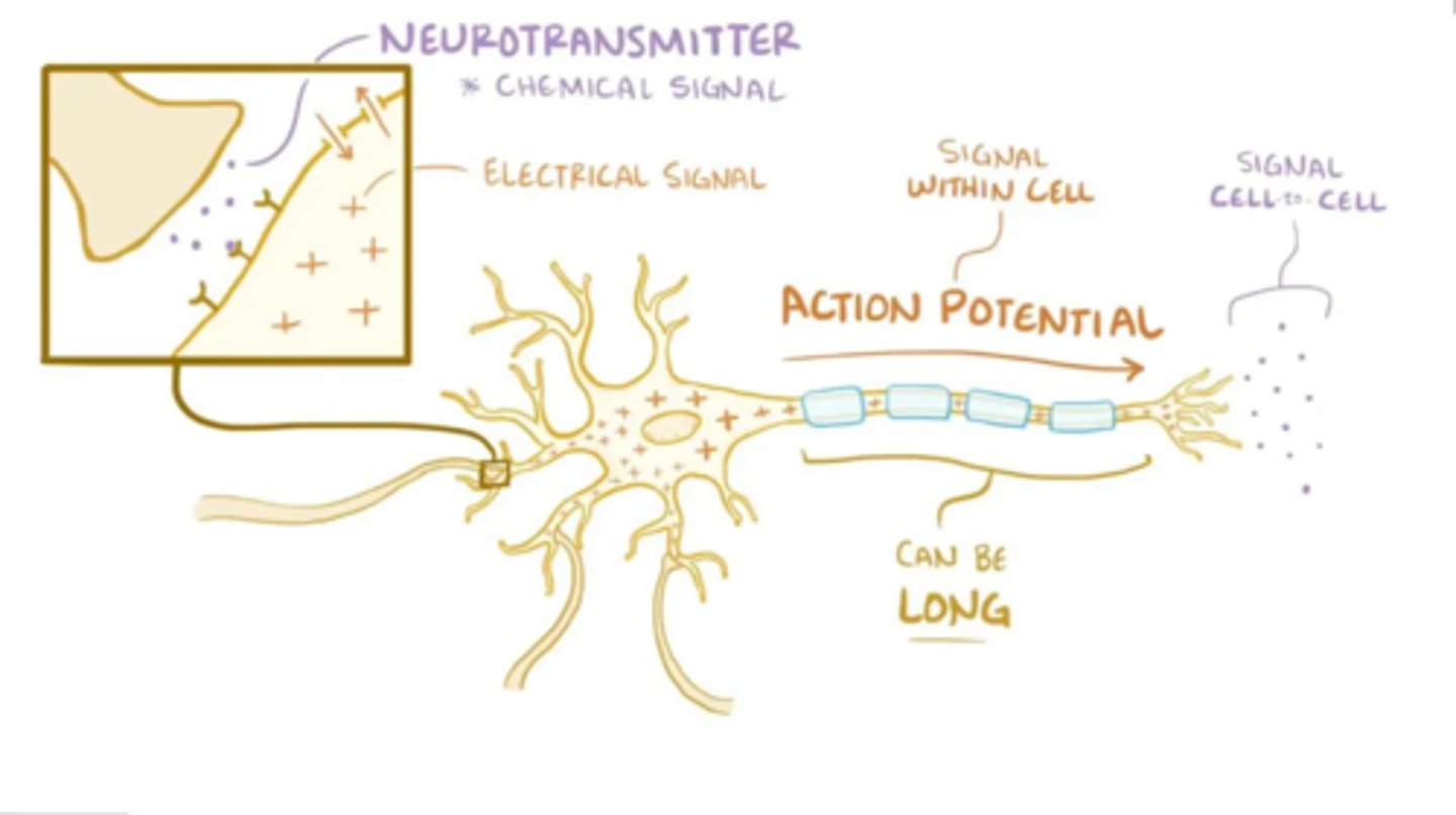
Electrochemical Process
Any conversion between chemical energy and electrical energy
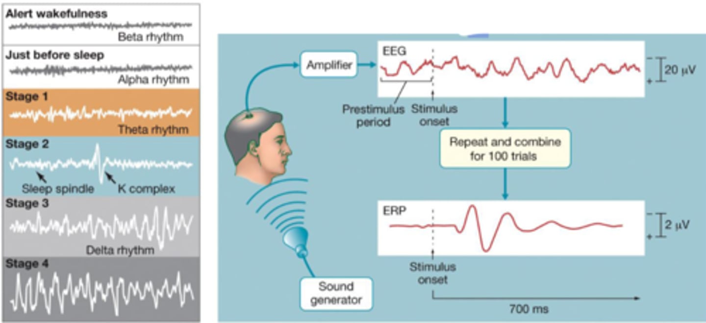
Electroencephalogram (EEG)
Recording of the electrical communication within neurons.
-The current generated by all of the activated neurons is strong enough to be detected by electrodes placed on the surface of the scalp.
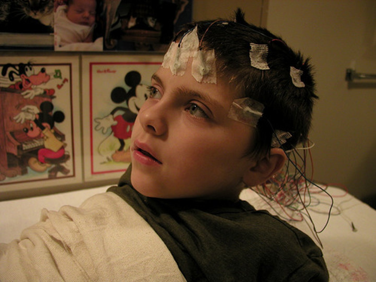
EEG is used to study:
Broad rhythms (e.g., sleep stages
Event-related potentials (ERPs) – changes in electrical activity to a specific input.
EEG is a _____________ (Electrical, Chemical) Process!
Electrical
Brain function can also be studied through techniques that manipulate the functions:
Chemical effects on neurotransmitters
Electrical stimulation
Gene manipulation (almost always with mice and rats)
EEG Strength and Weaknesses:
Knows when, but not where
Strength: Temporally locating neural activity
Weakness: spatially locating neural activity
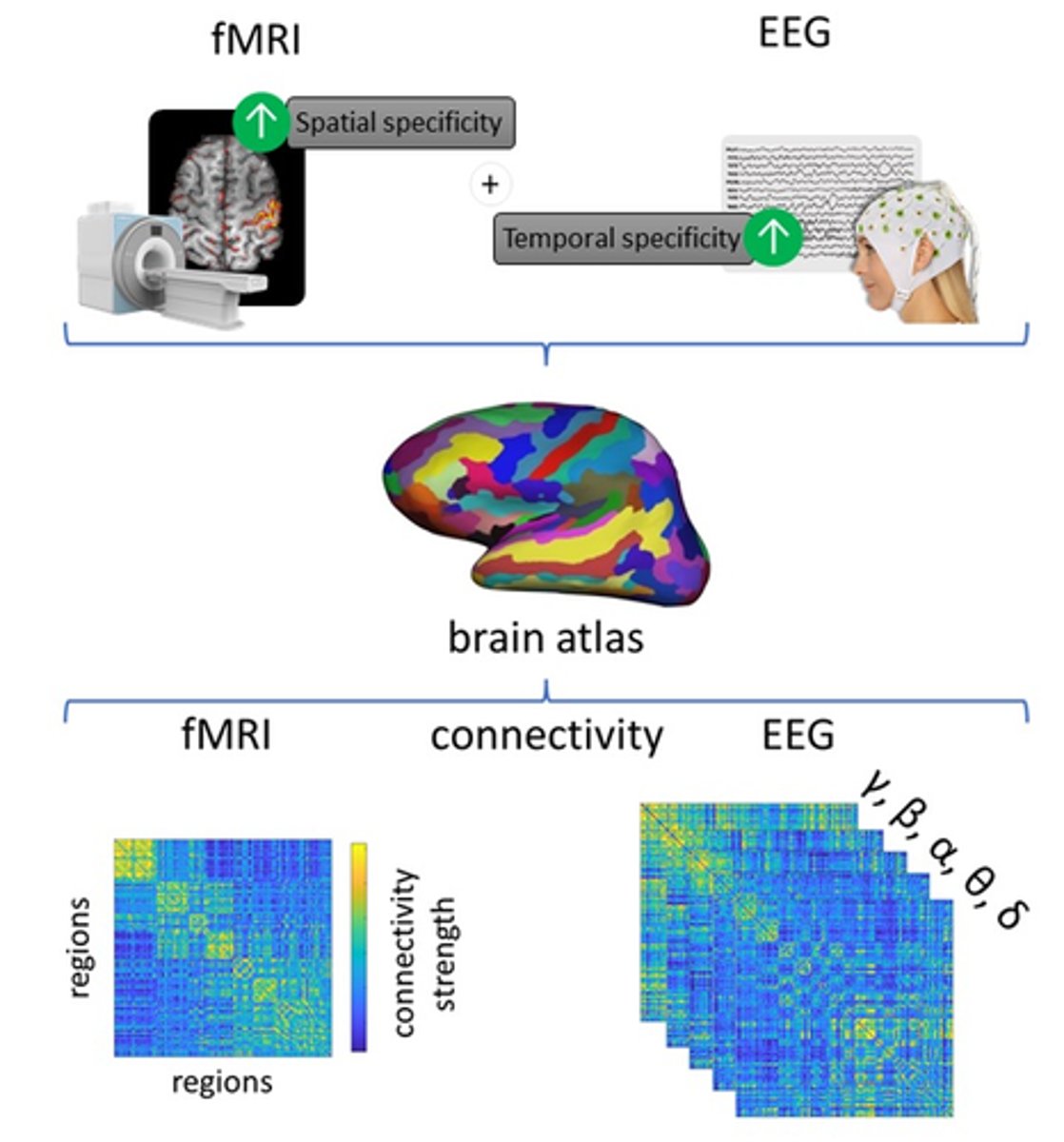
fMRI Strength and Weaknesses:
Knows where, but not when
Strength: spatially locating neural activity (where?)
Weakness: temporally locating neural activity (when?)

MRI scans
Detect brain structures, not activity
Most neuroimaging techniques used to study brain activity and structures provide only, what kind of data?
Correlational
The identified brain region may not be necessary; it may be only correlated with the task
e.g., a speedometer is correlated with the movement of a car
However, if damage to a brain site disrupts a particular function, it is an indication that the site does play some role in _________ that function.
Supporting
So we can have some casual explanations!
Transcranial Magnetic Stimulation (TMS)
The use of strong magnets to briefly interrupt normal brain activity as a way to study brain regions.
-Magnetic pulses activate neurons
-Produces temporary lesions
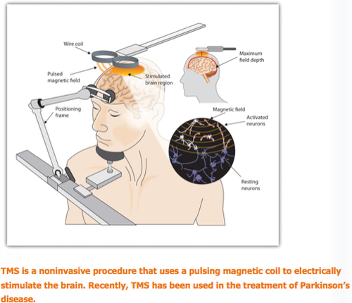
TMS can provide us some:
Casual Data
Since we are manipulating our variables.