Conventional Radiography & Imaging Principles
 Call Kai
 Learn
Learn Practice Test
 Spaced Repetition
 Match
Match Flashcards
 Knowt Play
1/26

There's no tags or description
Looks like no tags are added yet.
Name | Mastery | Learn | Test | Matching | Spaced | Call with Kai | Chat |
|---|
No analytics yet
Send a link to your students to track their progress
27 Terms
What is the purpose of medical imaging?
To visualize internal body structures and recognize abnormalities such as congenital anomalies, tumors, and fractures.
What does the introduction of contrast media allow us to study?
Luminal or vascular organs and potential or actual spaces, including:
Digestive tract
Blood vessels
Kidneys
Synovial cavities
Subarachnoid space
What are the five common diagnostic imaging techniques?
Conventional radiography (X-ray)
Computed tomography (CT)
Ultrasonography (US)
Magnetic resonance imaging (MRI)
Positron emission tomography (PET)
What is conventional radiography?
An imaging technique in which a highly penetrating beam of X-rays passes through the body, producing an image based on differences in tissue density.
How does conventional radiography produce different shades on an image?
Different tissues absorb different amounts of X-rays depending on their density. Dense tissues absorb more X-rays, while less dense tissues allow more X-rays to reach the detector.
Why does compact bone appear white on an X-ray?
Because it is very dense and absorbs many X-rays (radiopaque), so few X-rays reach the detector.
Why does air appear black on an X-ray?
Because it absorbs very few X-rays (radiolucent), allowing most X-rays to reach the detector.
Define radiopaque.
A substance that absorbs many X-rays and appears white or light on a radiograph
Define radiolucent.
A substance that absorbs few X-rays and appears dark or black on a radiograph.
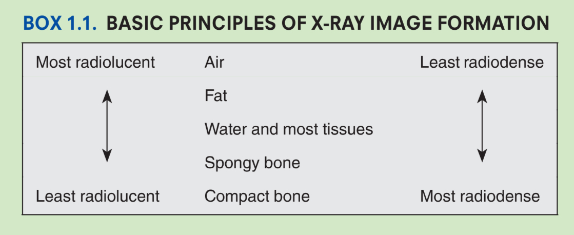
Arrange the following from most radiolucent → most radiopaque:
Water/soft tissue, Fat, Air, Compact bone, Spongy bone
Air
↓
Fat
↓
Water & soft tissue
↓
Spongy bone
↓
Compact bone
Which tissue is more radiopaque: compact bone or spongy bone?
Compact bone.
Which tissue absorbs more X-rays: compact bone or soft tissue?
Compact bone.
Why is the body part placed close to the detector during radiography?
To maximize image sharpness and minimize magnification
Why are radiographs considered two-dimensional representations?
Because they compress a three-dimensional structure into a two-dimensional image, causing structures to overlap.
Why are at least two radiographic projections usually obtained?
Because a single radiograph cannot accurately localize abnormalities due to superimposition of structures.
What does PA stand for?
Posteroanterior projection.
The X-ray beam passes posterior → anterior.
In a PA projection, where are the X-ray tube and detector?
X-ray tube = posterior
Detector = anterior
What does AP stand for?
Anteroposterior projection
The X-ray beam passes anterior → posterior.
In an AP projection, where are the X-ray tube and detector?
X-ray tube = anterior
Detector = posterior
How are PA and AP radiographs viewed?
As if you are facing the patient.
The patient’s right side appears on your left.
What do the radiopaque letters “R” and “L” indicate on lateral radiographs?
The side of the patient that was closest to the detector.
Why are contrast media used in radiography?
To visualize structures that are not visible on plain radiographs.
Give two examples of commonly used radiopaque contrast media.
Iodine compounds
Barium
Name four structures commonly examined using contrast media. (+an extra one)
Digestive tract
Blood vessels
Kidneys
Synovial cavities
(Also: subarachnoid space)
Why can abnormalities be missed if only one radiographic projection is taken?
Because overlapping anatomical structures in a 2D image can hide or obscure pathology. Multiple projections improve localization and detection.
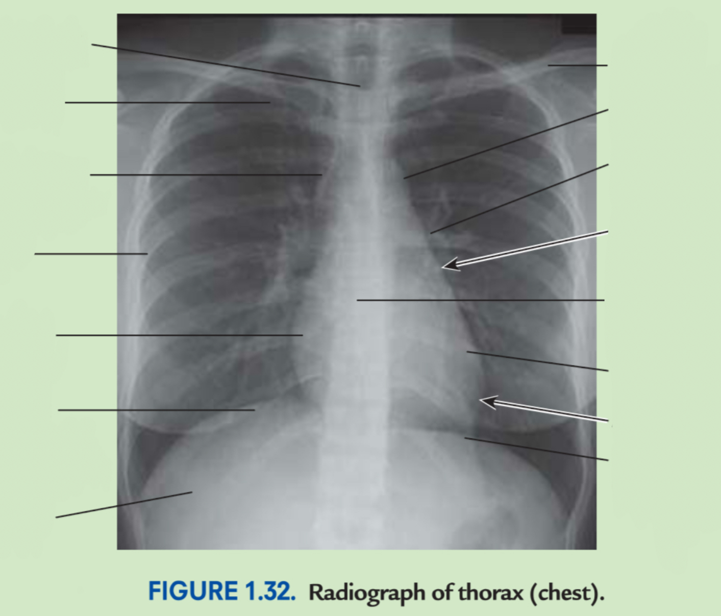
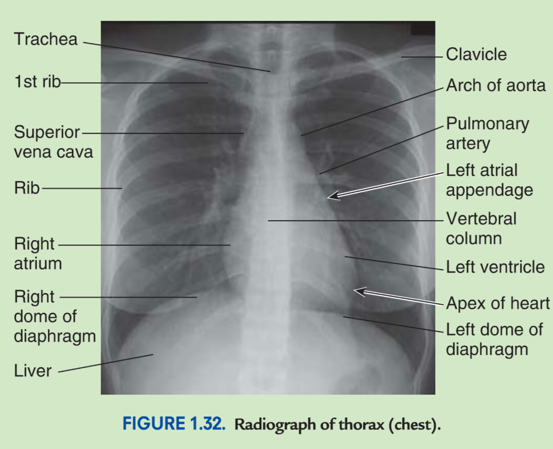
Label chest X-ray
Memorise
Good