Aorta (lil more info)
 Call Kai
 Learn
Learn Practice Test
 Spaced Repetition
 Match
Match Flashcards
 Knowt Play
1/74

There's no tags or description
Looks like no tags are added yet.
Name | Mastery | Learn | Test | Matching | Spaced | Call with Kai | Chat |
|---|
No analytics yet
Send a link to your students to track their progress
75 Terms
Blood vessel anatomy
Tunica intima - inner layer endothelial cells
Tunica media - Middle layer, muscle thicker and more organized layer in arteries
Tunica Adventitia/Externa - outer later, epithelial cells, connective tissue
First branches of ascending aorta are
Coronary arteries
Aortic arch starts at level of ___________ and extends distal to left subclavian artery
Innominate artery
Bovine arch
Most common anatomic variant of the arch anatomy
Two branch arch formation: the innominate and left CCA origin are combine and the left subclavian artery is the second branch
Although “bovine” indicates a cow, this arch formation is NOT related to the arch formation in cattle
Aorta is ________ to IVC until umbilicus
Posterior
__________ resistance in the upper abdominal aorta due to low resistance branches
Lower
__________ resistance in the distal aorta due to high resistance branches
Higher
Common iliac arteries
Originates at the aortic bifurcation at L3 or L4, umbilicus level
Supply blood to the legs pelvis
Bifurcate into the internal and external iliac arteries
High resistance flow
Common iliac arteries are anterior to the common iliac veins
Eval to arterial pathology in long and trans
Internal iliac artery
Hypogastric artery
Travels medially and supplies the pelvic organs, NOT GONADS
Tri or Bi, lower resistance than the common iliac
External iliac artery
Travels laterally and passes under the inguinal ligament to become the common femoral artery
Courses along medial side of the psoas muscle
Supplies the extremities with blood
Tri, high resistance
USA abdominal indications
Abdominal pain
Pulsatile mass
AAA on plain film
F/U AAA
Trauma
Decreased pedal pulses
The distal aorta can usually be palpated by pressing on the ___________ at the level of the iliac crest
Abdomen
US in aorta
Best anatomic landmark for abdominal structures
Long and trans
Coronal can show Aorta and IVC
AP and trans measurements should be obtained
Gradual distal tapering and becomes more anterior distally
Common iliac artery ends at distal bifurcation into external and internal iliac arteries
Normal Aorta measurements:
Proximal: 2.0-2.6cm
Mid: 1.6-2.4cm
Distal: 1.1-2cm
Iliacs: 0.6-1.4cm
Doppler of aorta
Velocity: 70-100cm/s
Biphasic above renal arteries due to low resistance branches
Triphasic below renal arteries
Coarctation
Congenital defect causing narrowing of the aorta
Occurs with other heart defects and is component of Shone complex
Occurs distal to the origin of left subclavian
Causes lower extremity ischemia
Decreased bilateral pedal pulses
Systemic HTN usually present
Elevated pressures is activated causing increased systemic pressure
Takayasu arteritis
Presents as hypertrophic areas of inflamed tissue usually found in aorta and its branches
Commonly affects subclavian and carotid arteries
Can also affect renal arteries
Most commonly seen in young females
Aortic ectasia
Lack of tapering of the Ao as it travels distally, size remains constant from proximal to distal portions
Can be precursor to aneurysm formation
AAA is most commonly caused by
Degenerative disease
Symptoms of AAA
Pulsatile mass, low back pain, abdominal bruit
AAA most commonly forms in __________ segment
Infrarenal
>60% of pts with a pop aneurysm also have a
AAA
Iliac artery considered aneurysmal when diameter exceeds _____ or when diameter increased by 50% as compared to adjacent segment
1.5cm
AAA above the renal arteries -
Surgical intervention asap
AAA below renal arteries -
Most common; surgical intervention at a diameter >5.5cm
Fusiform AAA
Vessel wall stretches in a circumferential manner; all three wall layers intact
Saccular AAA
Formed with a stalk connecting dilated wall portion to main vessel; usually associated with wall dissection
Berry AAA
Tiny out pouching, usually found in cerebrum/circle of Willis; usually congenital
Mycotic AAA
Due to infection in the artery and the arterial wall; syphilis most common
Vasculitis AAA
Due to inflammatory process of vasa vasorum and tunica adventitia
Aneurysm diameter change of >4mm on a yearly follow up exam, the next follow up should be at __________
3 months
Ruptured AAA can cause
Hypotension, severe abdominal pain, shock, death
Complications of AAA
Rupture is MOST COMMON and most critical
Decreased flow extremities
Blue Toe syndrome with thrombus accumulation and embolization to the toes
Affects renal circulation and systemic BP if proximal to renal artery
Aneurysmal measurement ____
>3cm
Possible ________ accumulation causes increased echogenicity in AAA
Thrombus
Blue toe syndrome
Thromboembolic disease
Embolic material lodges in distal artery
Causes acute ischemia distal to location of embolism and blue color changes in distal tissues
Causes of Blue toe syndrome
Thrombus in prox aneurysm
Arteritis
Ulcerated atherosclerotic lesions
Some angioplasty procedures
Thrombus in a vein
Cardiac arrythmias
EVAR
Used to improve and streamline flow through a stenosis or AAA
Easier and better for PT
Kissing stents used for stenosis at the origin of the common iliac arteries
Intravascular ultrasound (IVUS)
Can be used to guide the graft during placement
Physician uses an US catheter to visualize the lumen prior to the procedure and eval best location for endoluminal graft placement
Advantages of IVUS over Angiography during angioplasty
No need to cut the aorta to insert the graft
No radiation
No contrast
Can provide 2D and 3D images of vessel
Limitations of IVUS
Catheter size limits vessels that can be evaluates
Introduction of the catheter can lead to arterial spasm
Significant atherosclerosis can blur the image
Images suffer from ringdown artifact caused by dead space
Cost of disposable catheter; cannot be heated/sterilized b/c the elements will lose their US properties
Intraoperative Duplex US
Used to assess the flow changes during and after procedure
Surgeon places transducer directly on open wound or exposed vessel; requires sterile technique
Sonographer may be asked to assess distal flow at the ankles during and after procedure
Ankle pressures should demonstrate a mild increase after the graft is placed
Complications of EVAR
Stenosis, thrombosis, endoleak
Endoleak type 1
Leak at site of attachment
1a - leak at prox attachment site
1b - leak at distal attachment site
Flow pattern resembles the flow pattern within the graft
Most dangerous type
Type 2 endoleak
Will see retrograde flow into aneurysm sac and increases AAA diameter
IMA, internal iliac, lumbar arteries and accessory arteries are potential sources for leak
Bidirectional flow pattern, Doppler waveform reflects end source pathology
MOST COMMON
Type 3 endoleak
Caused by tear in the graft or separation of the main graft from the iliac limbs
Type 4 endoleak
Transgraft leak through porous material of the graft
Type 5 endoleak
Endotension - no leak but there is continued expansion of the aneurysmal sac greater than 5cm
Post surgical EVAR US
Performed 1 month post-op, then at 3, 6, 9, 12 and 18 months followed by annual exams
Measure outer wall to outer diameter of AAA to compare to prior exam
>0.5cm increase in diameter from last exam = possible leak
Use color and PW
Document proximal attachment site and confirm it has not migrated
Eval velocities prior and distal to EVAR
True endoleak will be reproducible
Potential signs of an unstable AAA sac
Echolucent areas within the sac
Changes in aneurysm shape and/or sac size
Increased pulastility of sac
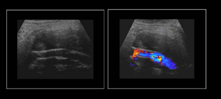
Normal Endovascular stent
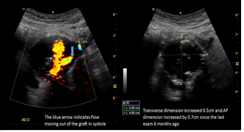
Endograft leak
Dissection
Intimal layers tear and allows flow between intima and media layers into a blind pocket
Causes weakened vessel wall, increased risk of rupture
Most commonly in ascending aorta
Second most commonly is just distal to subclavian
Significant AAA can lead to dissection (>5cm)
CTA is preferred for diagnosis
Dissection is most commonly found in
Ascending aorta
USA dissection
Linear echogenicity seen in lumen of the vessel, separating it into two channels
Color flow demonstrates two lumens, both with turbulence
Bidirectional flow
Stanford Classification:
Type A: dissections that involve the ascending aorta and arch
Type B: Dissections that involve the aorta from the left subclavian artery to iliac bifurcation
DeBakey Classification:
Type 1: Dissection that involves the ascending AO and Descending
Type 2: Dissections that involve only the ascending AO
Type 3: Dissections that involve descending AO only
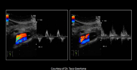
Aortic dissection
Aortic stenosis is most commonly caused by
Atherosclerotic changes
Aortic stenosis
Resistance will increase proximal to stenosis
Distal stenosis = tardus parvus
If the stenosis is superior to the renal artery, renal ischemia will activate renin-angiotensin system causing systemic HTN
Effects will be similar to coarctation
Leriche syndrome is also known as
Aortoiliac occlusive disease
Leriche syndrome
Due to occlusion of the abdominal aorta just above the site of its bifurcation
Causes BILATERAL symptoms
Cause of erectile dysfunction
Low resistance waveforms and post-stenotic changes in legs
Leriche syndrome symptoms
Fatigue of both lower limbs, intermittent bilateral claudication with ischemic pain, absent or diminished femoral pulses and pallor or coldness of both lower extremities
Retroperitoneal fibrosis is also known as
Ormond disease
Retroperitoneal fibrosis most commonly occurs at the level of the
Bifurcation
Retroperitoneal fibrosis
Idiopathic overgrowth of fibrous tissue around atherosclerotic aorta
Can be ruptured to drugs, infection, malignancy or cancer therapy
May lead to ureteral obstruction causing hydro, IVC compression causing bilateral edema, scrotal swelling from compression of gonadal vein
Retroperitoneal fibrosis USA
Soft tissue mass surrounding great vessels
Hypoechoic
Smooth borders
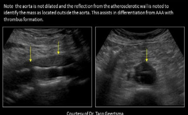
Retroperitoneal fibrosis
If the aortic arch only has two branches instead of 3, what is this variant called
Bovine arch
The external iliac arteries become the common femoral arteries when:
They cross under the inguinal ligament
AP measurements of AO are most accurate in what plane
Long
What causes elevated brachial pressures and diminished ankle pressures bilaterally?
Coarctation
Most common complication of AAA
Rupture
Ying-Yang sign is seen on Doppler related to ?
Aneurysm
Kissing stents are used to correct:
Iliac artery stenosis
Which of the following arteries are involved in a Type 2 EVAR endoleak?
Lumbar and inferior mesenteric
Hallmark sign for endoleak
Increased in aortic sac size