Human Anatomy Review
 Call Kai
 Learn
Learn Practice Test
 Spaced Repetition
 Match
Match Flashcards
 Knowt Play
1/41

Earn XP
Description and Tags
Brentwood Ambulance Post 325
Name | Mastery | Learn | Test | Matching | Spaced | Call with Kai | Chat |
|---|
No analytics yet
Send a link to your students to track their progress
42 Terms
Terminology
Anatomy:
Physiology:
Pathophysiology:
Anatomy: The study of the structure of the body and its parts (what the body is made of).
Physiology: The study of how the body works (how the organs and body system functions).
Pathophysiology: They study of how diseases, injuries, or disorders change the normal function of the body.
Anatomical Position
Facing forward; palms facing outward
Planes of the body:
Coronal (Frontal) : Divides the body front / back
Sagittal (Lateral): divides the body : left / right
Mid-sagittal (midline): Divides the body into equal left and right halves
Transverse (axial): divides the body top / bottom
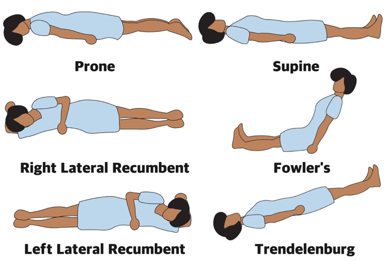
Positional terms:
Prone : Facing down
Supine: Facing up
Shock Position or Trendelenburg: Head down, legs up , increases BP ; Increases HR
Fowlers Position: Sitting up 45º or 90º
Recover Position: Right or left Recumbent
Directional Terms:
Cranial/ Superior: Top; Closer to head
Caudal / Inferior: Bottom; away from head
Ventral/ Anterior: Front
Dorsal/ Posterior: Back
Lateral: Away from midline
Medial: Close to midline
Proximal: Close to
Distal: Away from / far away
The importance of Anatomical Position
The position patients are found help fit into pieces of a bigger puzzle
You can determine in the most basic sense:
How it possibly happened
what the patient may have injured
Common conditions that may improve or determine the patients condition
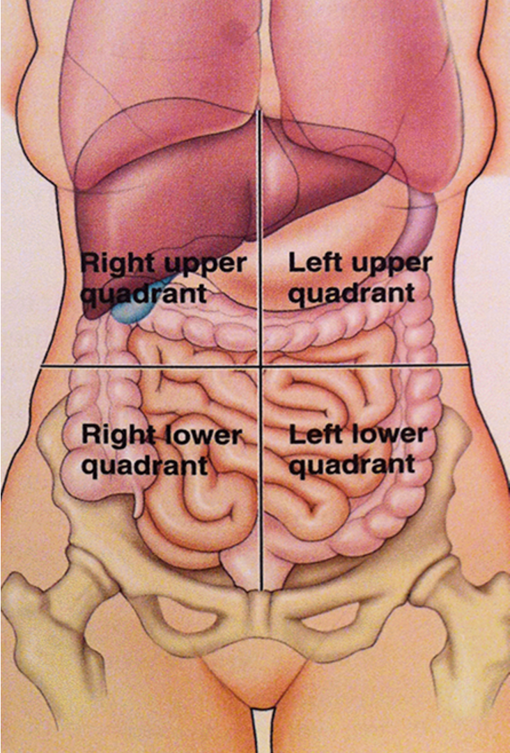
The 4 Quadrants:
Right Upper Quadrant: Liver
Right Lower Quadrant
Left Upper Quadrant : Stomach
Left Lower Quadrant
Anatomical Terms
Right and left: YOUR PATIENT’S RIGHT AND LEFT!!!
Mid-Clavicular: Line that runs down the middle of the clavicle (the nipple of the breast)
Bilateral: Both Sides
Dorsal: Back side, or tip (dorsal fin of fish)
Ventral: opposite of dorsal, front side
Superficial VS. Deep
Superficial: Near or on the skin; scrapes and minor cuts
Deep: Farther Away from the surface; Gun shots or stabs
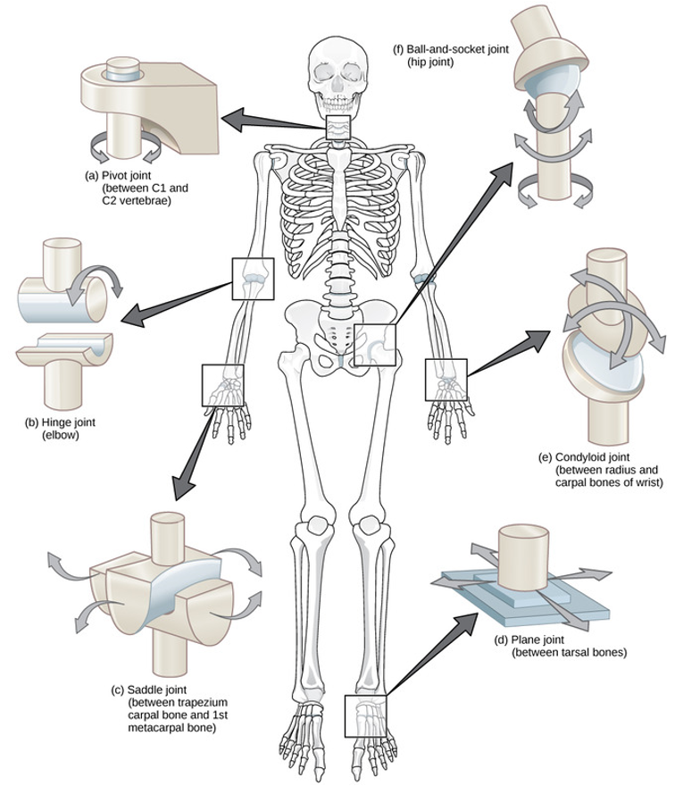
Joints
Occurs wherever bones come in contact
Consist of the ends of the bonds and the connecting and supporting tissues
Two types of joints:
Ball and Socket Joint
Allows rotation and bending
Hinge Joint
Motion restricted to flexion and extension
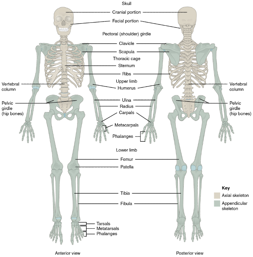
Skeletal System:
Forms the body’s internal framework
Made of 206 bones and cartilage
Gives the body its shape
Two Divisions:
Axial Skeleton
Skull
Spine (Vertebral Column)
Ribs (Thoracic Cage)
Sternum (Breast Bone)
Function: Protects the brain, Spinal Cord, Heart, and Lungs
Appendicular Skeleton:
Arms
Legs
Shoulders Clavicles & Scapulae
Pelvis
Function: Allows Movement
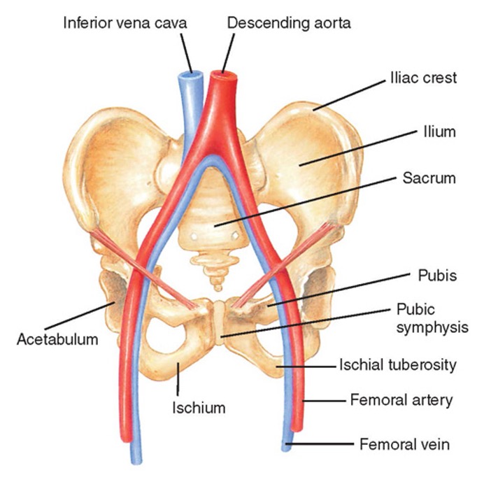
Pelvis
Connects the legs to the trunk
Supports body weight
protects pelvic organs
The pelvic girdle consists of:
two coxae (hip bones)
sacrum
coccyx
each pelvic bone is formed by fusion of the ilium, ischium, and pubis ( by the sacrum)
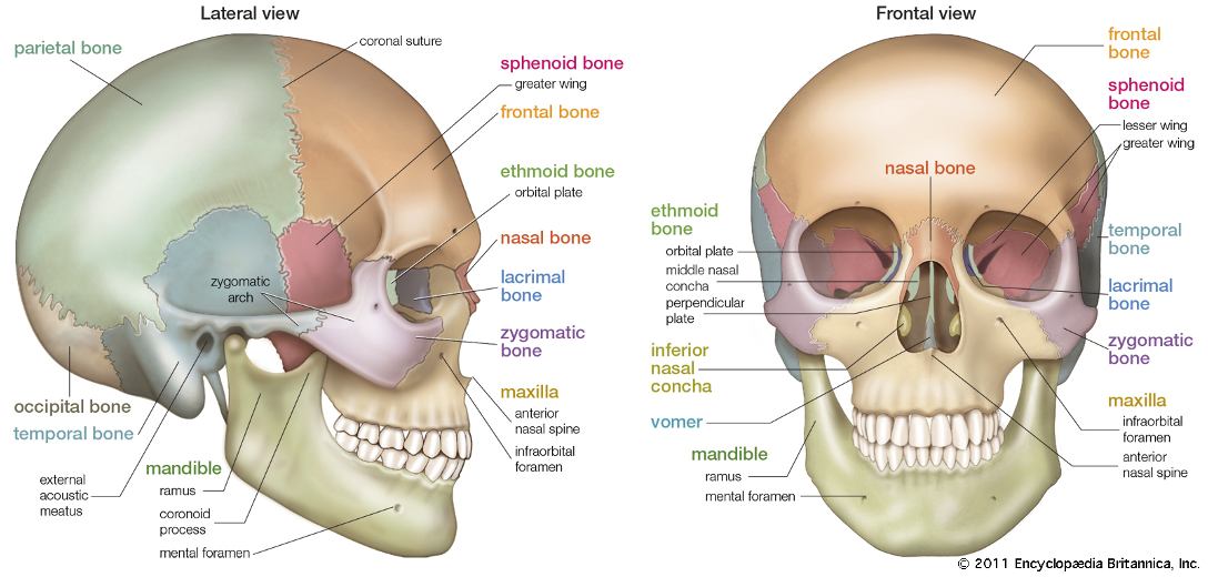
The Cranium
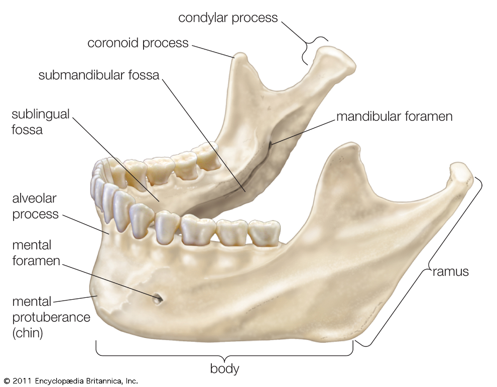
Structure of the Mandible
Functions of the Skeletal System
Support
Protection
Movement
Injury Term: Fracture = Broken Bone
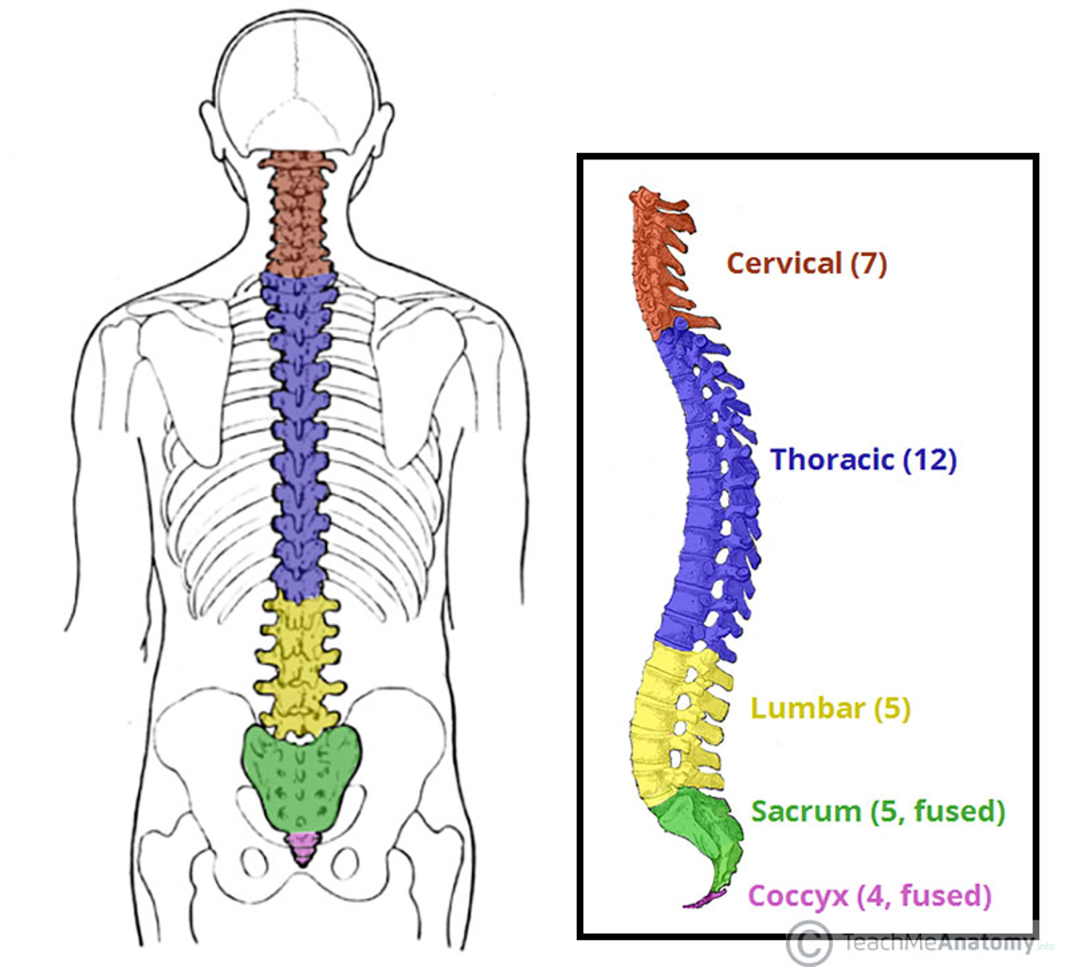
The Spinal Column
Coffee at 7 —> Cervical
Tea at 12 —> Thoracic
Licorice at 5 —> Lumbar
Soda at 5 —> Sacrum
Cookies at 4 —> Coccyx
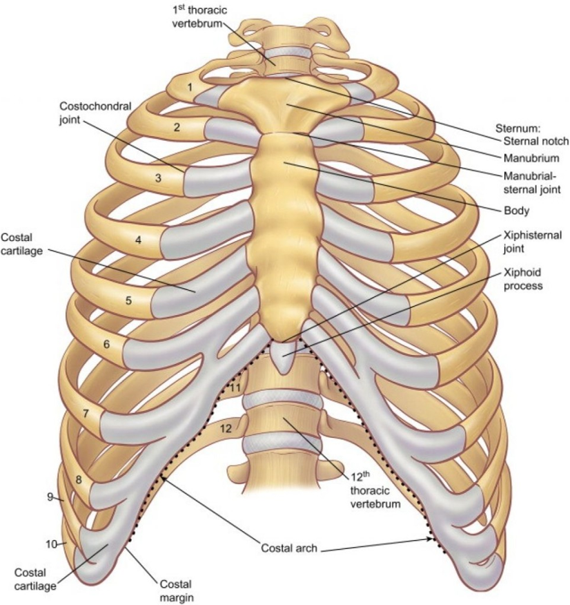
The Thorax & Upper Extremities
Formed by 12 thoracic vertebrae and pairs of ribs.
Thoracic cavity contains:
Heart
Lungs
Esophagus
* Chest Injuries can cause multiple internal injuries.
Upper Extremities:
Shoulder
Clavicle
Scapula
Humerus
Radius
Ulna
Carpals
Metacarpals
Phalanges
Lower Extremities:
Pelvis
Femur
Patella
Tibia
Fibula
Metatarsals
Phalanges
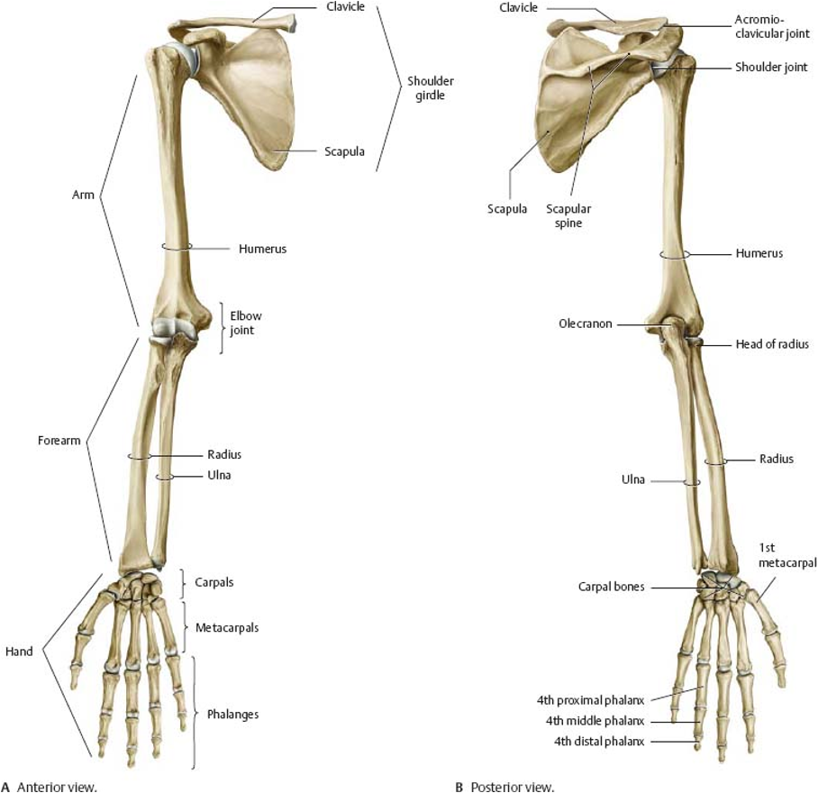
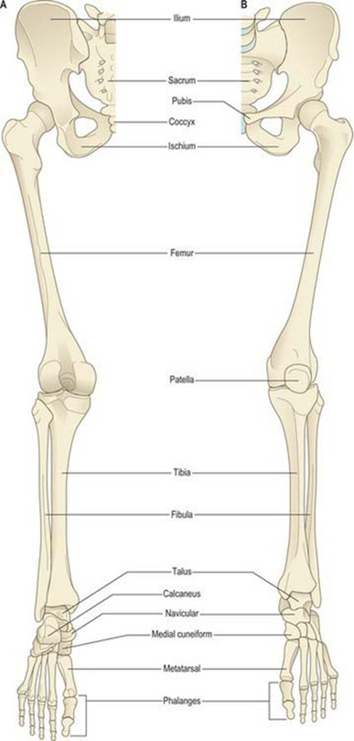
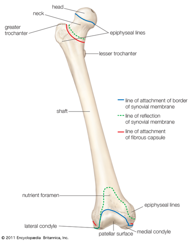
Femur
Femur (thigh) is the longest and strongest bone in the body
The neck of the femur connects the head and shaft at a 125º angle.
Can withstand 1,200-2,500 Ibs of compression force.
Femur fractures are life-threatening because they can cause severe internal bleeding
EMT care: stabilize the leg and provide immediate treatment/ transport
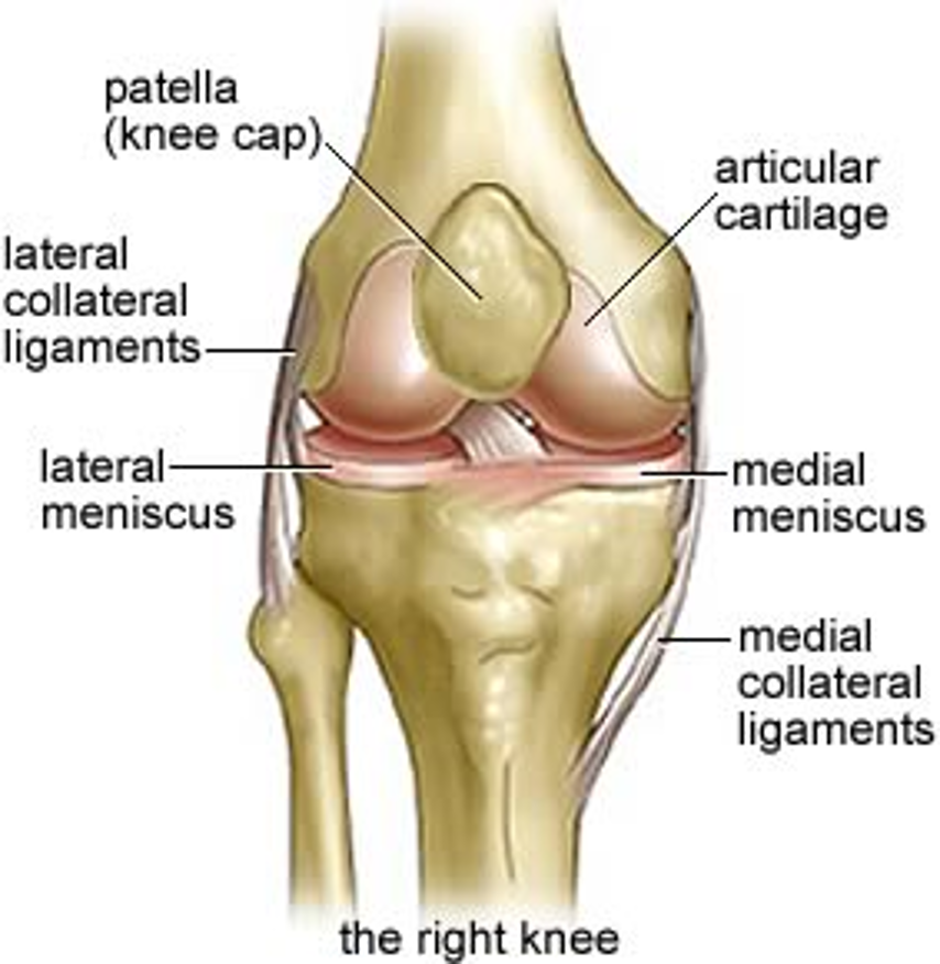
Knee
The Knee is a complex structure and one of the most stressed joints in the body.
It is the largest joint in the body
It is vital for movement and vulnerable to injury
The knee is a hinge joint responsible for weight-bearing and movement
It Consists of:
Bones
Meniscus
Ligaments
Tendons
Functions of the Knee:
Supports the body on an upright position without muscles working
Helps lower and raise the body
Provides Stability
Acts as a shock absorber
Allows twisting of the leg
Makes walking more efficient
Helps propel the body forward
EMT importance:
Knee injuries are common because the joint bears body weight
Assess for pain, swelling, deformity, and limited movement
Immobilize the injury if a fracture or serious ligament injury is suspected
Musculoskeletal System:
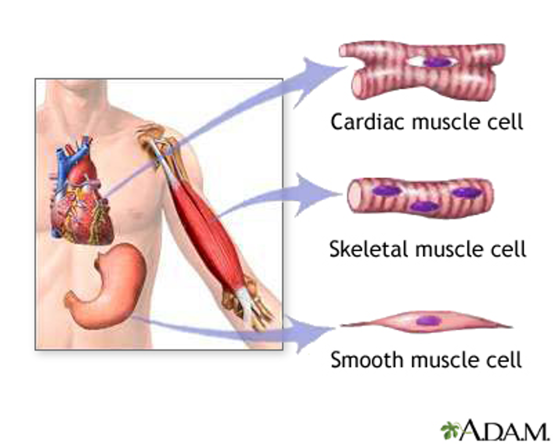
3 types of Muscles:
Skeletal (Striated)- Voluntary; moves the body
Smooth- Involuntary; found in organs and blood vessels
Cardiac - Involuntary; found only in the heart
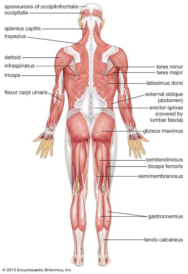
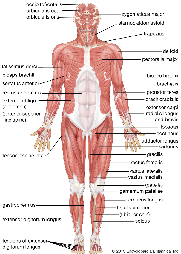
Skeletal muscles work with the skeletal system
They are under voluntary control
Function:
Enable movement
Maintain Balance
Maintain Posture
Muscles also help keep the body warm
when you get cold, the body shivers involuntarily to produce heat
shivering helps maintain homeostasis (the body’s normal internal balance).
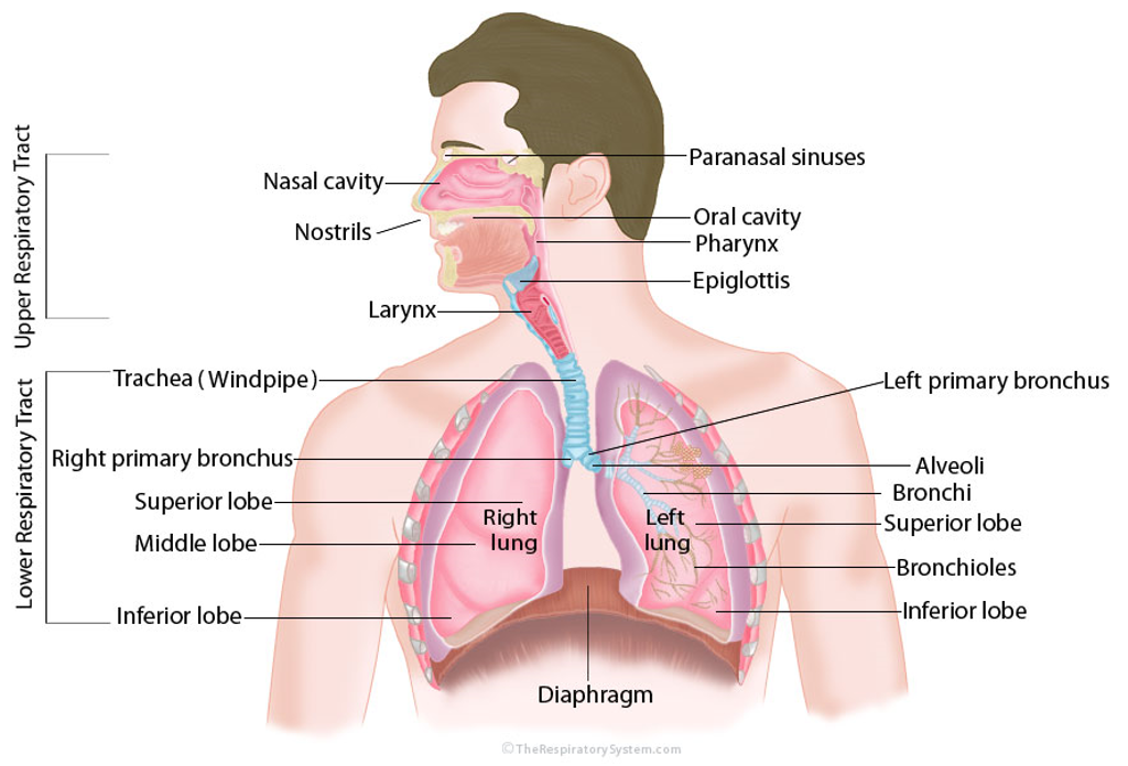
Respiratory System
The respiratory system consists of all the structures involved in breathing (respirations).
It includes:
Nose
Mouth
Thorat (Pharynx)
Larynx (Voice Box)
Trachea (Windpipe)
Bronchi
Bronchioles
Lungs
Diaphragm
Chest Wall
Muscles
Accessory Muscles for Breathing
Functions:
Oxygen (O2) passes from the lungs into the blood
Carbon dioxide (CO2) is removed from the blood and exhaled
The diaphragm is the primary muscle used for breathing
Upper and Lower Respiratory
Upper Airway:
Nose and Paranasal Sinuses
Oral Cavity
Pharynx
Larynx
Lower Airway:
Trachea
Lt and Rt Principal Bronchi
Secondary (Lobar) Bronchi
Segmental (Tertiary) bronchi
Terminal Bronchiole
Respiratory Bronchiole
Alveolar Duct and Sacs
Airway Adjuncts:
EMTs can use airway adjuncts to help maintain an open airway
Used for patients who are unresponsive or in cardiac arrest
Types:
Oropharyngeal Airway (OPA) - used in unresponsive patients without a gag reflex
Nasopharyngeal Airway (NPA) - Used in patients with a gag reflex or who are semi-conscious (unless facial/ skull trauma is suspected)
Supraglottic Airway (i-gel) - Advanced airway device used during cardiac arrest or when a patient cannot maintain their airway.
Breathing
Inhalation (Active):
Diaphragm and Intercostal muscles contract
Thoracic Cavity increases in size
Diaphragm moves downward
Ribs Move upward and outward
Air flows into the lungs
Exhalation (Passive):
Diaphragm and intercostal muscles relax
Thoracic cavity decreases in size
Diaphragm moves upward
Ribs move downward and inward
Air flows out of the lungs
Muscles of Breathing:
Diaphragm is the primary muscles of breathing
Also involved are:
Neck (Cervical) Muscles: help during difficult breathing
Intercostal Muscles: move the ribs during breathing
Abdominal muscles: help with forceful exhalation (coughing, sneezing)
Pectoral (Chest) muscles: Assist with breathing when a person is in respiratory distress.
Respiratory System Physiology:
Alveolar-Capillary Exchange:
Occurs in the alveoli (air sacs of the lungs)
Oxygen-rich air enters the alveoli during inhalation (inspiration)
Oxygen-poor blood flows through the surrounding capillaries
Oxygen (O2) moves from the alveoli —> capillaries (blood)
Carbon dioxide (CO2) moves from capillaries —> alveoli
CO2 is then exhaled from the lungs
Capillary - Cellular Exchange:
Occurs between the capillaries and body cells
Capillaries deliver oxygen (O2) to the cells
Cells give carbon dioxide (CO2) to the Capillaries
The blood carries CO2 back to the lungs to be exhaled
Chemical Control:
The brain stem controls breathing
It monitors carbon dioxide (CO2) levels in the blood and spinal fluid
The primary reason for breathing is to remove excess carbon dioxide (CO2)
Hypoxic Drive is a backup breathing drive that is triggered by low oxygen (O2) levels (Seen in some patients with chronic lung disease, such as (COPD)
Infant and Child Airway Considerations
Mouth and nose are smaller and more easily obstructed
Pharynx: The tongue takes up more space than in adults, making airway blockage more likely
Trachea (windpipe): Narrower and more easily blocked
Softer and More Flexible
Diaphragm: Infants and Children depend more on the diaphragm for breathing because the chest wall is softer
Cardiovascular System
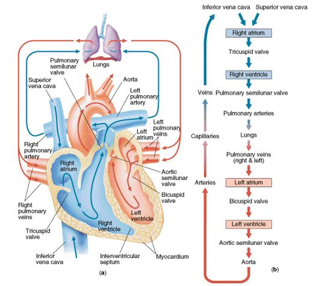
Heart: pumps blood throughout the body
Right Atrium: Receives oxygen-poor blood from the body and pumps it to the right ventricle
Left Atrium: Receives Oxygen-rich blood from the lungs and pumps it to the left ventricle
Right Ventricle: Pumps oxygen-poor blood to the lungs
Left Ventricle: Pumps Oxygen-Rich blood to the body
Valves: Prevent back flow and keep blood flowing in one direction
Arteries:
Coronary Arteries: Supply the heart muscle with blood
Aorta:
Largest artery in the body
Begins at the left ventricle of the body
travels through the chest (thoracic cavity) and abdomen (abdominal cavity)
Splits near the navel into the iliac arteries
Pulmonary Artery:
Starts at the right ventricle
Carries Oxygen-poor blood to the lungs
Carotid Arteries:
Located in the neck
Supply blood to the head and brain
Pulse: Felt on either side of the neck
Femoral Artery:
Located in the thigh/groin
Supplies the lower extremities
Pulse: Felt in the groin
Radial Artery:
Located in the forearm / wrist
Pulse: Felt on the side of the wrist
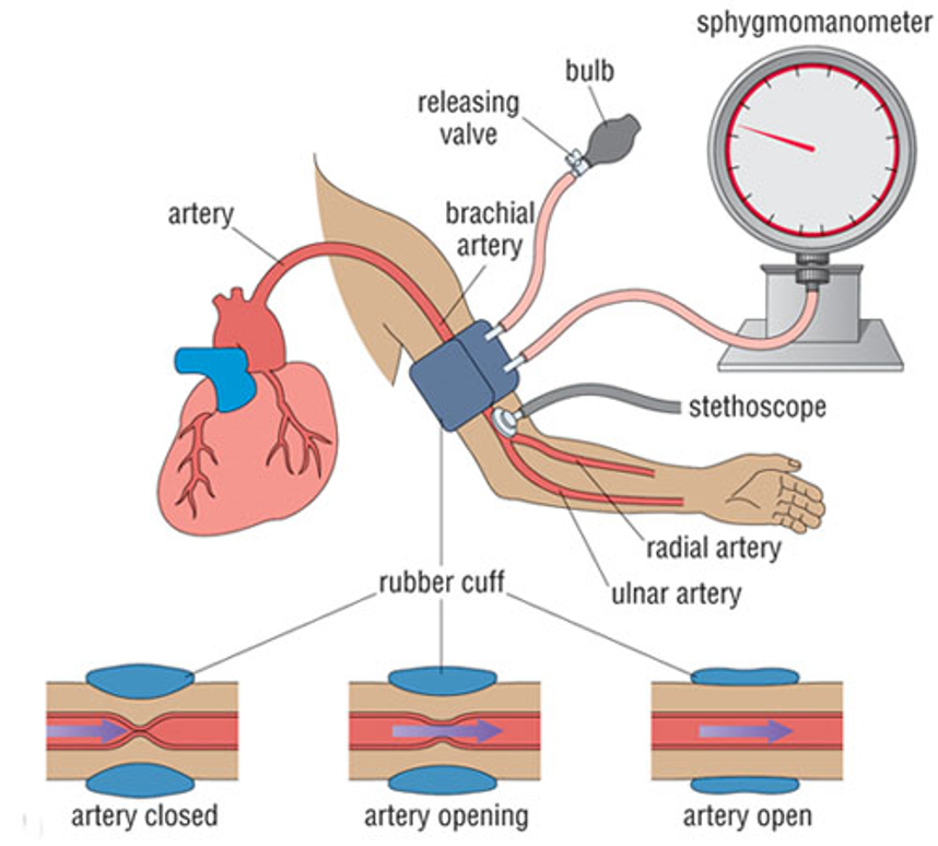
Brachial Artery:
Located in upper arm
Pulse: Felt inside the arm between the shoulder and elbow
used to measure blood pressure
Posterior Tibial Artery:
Located behind the medial Malleolus (inside ankle bone)
Pulse: Felt behind the inside ankle
Dorsalis Pedis Artery:
Located on the top (anterior surface) of the foot
Pulse: Felt on the top of the foot
Arterioles:
Smallest branches of arteries
Carry blood from arteries to capillaries
Capillaries:
Tiny blood vessels that connect arterioles to venules
Found throughout the entire body
site where oxygen, nutrients, carbon dioxide, and waste products are exchanged between then blood and body cells.
Venules
The smallest veins
collect blood from the capillaries
Merge together to form larger veins, which return blood to the heart
Veins
Carry Blood back to the heart
Usually carry oxygen-poor (deoxygenated) blood, expect for the pulmonary veins
Pulmonary Veins
Carry oxygen-rich blood from the lungs to the left atrium if the heart
Exception: They are veins that carry oxygen-rich blood
Venae Cavae:
Superior Vena Cava : Returns Oxygen-Poor blood from the head, neck, arms, and chest to the right atrium
Inferior Vena Cava: Returns Oxygen-Poor Blood from the abdomen, pelvis, and legs to the right atrium
Heart Circulation
Blood Pressure: 120/80 mmHg
Systolic Pressure:
The pressure exerted against the walls of the arteries when the left ventricle contracts
Represents the highest pressure in the arteries
The top # in a blood pressure reading
Diastolic Pressure:
The pressure exerted against walls of the arteries when the left ventricle is at rest (relaxes)
Represents the lowest pressure in the arteries
The bottom # in a blood pressure reading
Organs: The Digestive System: Anatomy
Abdomen:
The Second major body cavity
Contains the major organs involved in digestion and excretion
Divided into 4 quadrants:
Right Upper Quadrants (RUQ)
Left Upper Quadrants (LUQ)
Right Lower Quadrants (RLQ)
Left Lower Quadrant (LLQ)
Mouth:
Includes:
Lips
Cheeks
Gums
Teeth
Tongue
Begins the process of digestion
Salivary Glands:
Produce Saliva to help break down food
Oropharynx:
Passageway that connects the mouth to the esophagus
Esophagus:
Tube that moves food from the mouth to the stomach
Stomach:
Breaks down food using acid and enzymes
Pancreas:
Produces enzymes that help digest food
helps regulate blood sugar
Liver:
Produces bile
Helps process nutrients and Remove toxins
Bile Ducts:
Carry bile from the liver to the Small intestine
Small Intestine:
Main site where nutrients are absorbed into the bloodstream
Large Intestine:
Absorbs water and forms waste (feces)
Appendix:
Small pouch attached to the large intestine
Rectum:
Stores feces before elimination
GI Pathway (Food Route):
Mouth —> Oropharynx —> Esophagus —> Stomach —> Small Intestine —> Large Intestine —> Rectum —> Outside the Body
The Integumentary System (Skin):
Skin:
the largest single organ in the body
made up of 2 layers:
Epidermis: the outer (superfical) layer
Dermis: the inner (deeper) layer
Subcutaneous Tissue (Hypodermis)
located beneath the skin
contains fat (adipose tissue)
Functions:
insulates the body
stores energy
cushions and protects underlying tissues
Three major functions of the skin:
Protects the body from the environment (injury, bacteria, chemicals)
Regulates body temperature (through sweating and blood vessel changes
Transmits information from the environment to the brain (touch, pain, pressure, temperature)