Looks like no one added any tags here yet for you.
Anterior
Toward the front
Posterior
Toward the back
Dorsal
Toward the top of the head/back of the body
Vernal
Toward the bottom of the skull/front of the body
Lateral
Toward the side of the body/away from the middle
Medial
Toward the middle of the body/away from the side
Ipsilateral
On the same side of the body
Contralateral
On the opposite side of the body
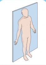
Coronal (frontal) section
Slice of brain parallel to the forehead
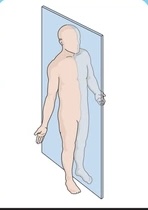
Sagittarius (lateral) section
Slice of brain perpendicular to the ground and parallel to the temporal lobes
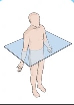
Transverse (axial) section
Slice of brain parallel to the ground
Central Nervous System (CNS)
Brain + Spinal cord
Peripheral nervous system (PNS)
Sensory and motor neurons that connect central nervous system to the rest of the body
Autonomic nervous system
Part of peripheral nervous system that regulates involuntary functions
Sympathetic Nervous system
Part of autonomic nervous system that controls arousal, energy expenditure, increases heart rate, blood sugar, and causes goosebumps
Involves adrenal medulla (secretes epinephrine and norepinephrine)
Fight or flight response
Parasympathetic Nervous System
Part of Autonomic nervous system that controls relaxation, energy storage, increases salivation, gastric, and intestinal motility
Activated by yoga and meditation
Rest and digest response
Forebrain, Midbrain, Hindbrain
The three divisions of the human brain
Cerebral Cortex
The outermost layer of gray matter w/ 26 billion neurons and is 3mm thick
Part of the forebrain
Best distinguishes humans from other animals
Convolutions/wiggles increase surface area for information storage
Consists of mainly glia and cell bodies that give it grayish brown color
Neocortex
Part of cerebral cortex that is phylogenetically the newest part of the cortex composed of 4 lobes
Limbic cortex
Older part of cerebral cortex involved in emotion and memory
Frontal lobe
Lobe involved in speaking, muscle movement, planning, judgement, and emotional control
Its size is directly related to size of person’s social networks
Primary motor cortex
Posterior part of frontal lobe that controls voluntary movements
Prefrontal cortex
anterior part of frontal lobe involved in planning, judgement, and decision-making
Parietal Lobe
Lobe that contains primary somatosensory cortex
Somatosensory cortex
Anterior Part of parietal lobe that maps the body’s surface
Occipital Lobe
Contains primary visual cortex
Visual cortex
Posterior part of occipital lobe that receives and processes visual information
Temporal lobe
Contains primary auditory cortex
Auditory cortex
Superior part of temporal lobe that receives auditory information
Aphasia
Difficulty producing/comprehending speech caused by brain damage
Broca’s Aphasia (expressive aphasia)
Damage to left frontal lobe that causes difficulty in speech production
Meaningful but halting, labored, and ungrammatical
Function words (a, the, in, about) are omitted
Wernicke’s aphasia (fluent aphasia)
Damage to the left posterior superior temporal lobe that causes difficulty in speech comprehension/ability to understand speech & produce meaningful words
Patients do not recognize that they cannot produce meaningful speech or understand others
Corpus Callosum
Bundle of axons that interconnects the corresponding regions of the association cortex of the two hemispheres of the brain
Left cerebral hemisphere
Hemisphere that is analytical, logical, and processes language & math
Right cerebral hemisphere
Hemisphere that synthesizes information, holistic view, pattern recognition, and emotion perception
Limbic cortex
part of the phylogenetically ancient cortex and is a key component of the limbic system.
Specifically, it includes the insular cortex and cingulate cortex.
The limbic cortex integrates sensory, affective, and cognitive components of pain and processes information regarding the internal bodily state
Limbic system
Doughnut shaped system of neural structures at border of brainstem and cerebral hemispheres
Associated with emotions and memory
Amygdala
Part of the limbic system responsible for motion regulation, fear, aggression, shaped like two almonds
Hippocampus
Part of the limbic system that is tough nut shaped and is important in memory
Basal ganglia
Collection of subcortical nuclei important for controlling movement
Related to Parkinson’s disease and OCD
Parkinson’s disease
Degeneration of caudate nucleus’s and putamen in basal ganglia
Notes by tremors, rigidity, balance issues
Obsessive compulsive disorder (OCD)
Increased activity in the caudate nucleus and in the frontal area of the basal ganglia
Nucleus Accumbes
Reward center of the brain
Dopamine release here is linked to drug addiction
Thalamus
Inferior to basal ganglia
Relay station for sensory and motor signals
Directs all in-coming and out-going information to sensory receiving area in the cortex and sends replies to cerebellum and medulla
Hypothalamus
Smalls neural structure beneath the thalamus that control autonomic nervous system, pituitary glands, and basic survival behaviors like four Fs (feeding, fighting, fleeing, and mating)
Brain stem
Oldest part and central core of the brain, beginning where spinal cord swells as it enters the skull
Responsible for autonomic survival functions (like breathing and heartbeat)
Reticular formation
Network of neural tissue in central part of brainstem that regulates sleep, arousal, attention, and various vital reflexes
More active in introverts and extroverts
Pons
Bulge in brainstem part of reticular formation that is important in sleep, arousal, and sensory analysis/movement
Medulla Oblongata
Base of brainstem that controls vital functions like heart rate, breathing, and blood pressure
Cerebellum
“Little brain” at rear of brainstem that helps coordinate voluntary movement and balance, motor learning, and higher cognition like math
Default Mode Network (DMN)
Brain areas that are active when participants in resting state (When brain is not focused on a task)
Daydreaming, self-reflection, reminiscing
Posterior cingulate cortex, precuneus, medial frontal cortex, and temporal parietal junction
Correlation with its deterioration and Alzheimer’s From rising levels or amyloid beta and tan proteins
Human Connectome Project (Brain mapping)
Launched in 2009 to map functional brain connectivity
Uses diffusion spectrum imaging (MRI Technique) to track brain fiber connections
Marcel Just
Decoded thoughts using fMRI with team at Carnegie Mellon
Can distinguish between autistic individuals vs neurotypical controls, suicidal individuals, and can help patients communicate via imagining different scenarios
Neuroplasticity
Brain’s capacity to adapt to damage and modify brain/reorganize following damage (especially in children)
Hemispherectomy
Removal of a brain hemisphere
Young children can recover nearly full brain function
Constraint induced therapy
Used in stroke recovery where patients are forced to use their weaker link to rewire the brain