Lecture 2: Diagnosis Techniques for Studying numerical + structural alterations to the human genome
 Call Kai
 Learn
Learn Practice Test
 Spaced Repetition
 Match
Match Flashcards
 Knowt Play
1/43

There's no tags or description
Looks like no tags are added yet.
Name | Mastery | Learn | Test | Matching | Spaced | Call with Kai | Chat |
|---|
No analytics yet
Send a link to your students to track their progress
44 Terms
**What are the 2 categories of cytogenetic testing?
Constitutional: Genetic changes are present in majority of all cells in the body (generally present from conception)
Cancer: genetic changes confined to a specific population of abnormal tumor or blood cells
*****What are the Tissue sources for CONSTITUTIONAL cytogenetic testing in pre vs. postnatal situations
hint post (2) pre (4)
POST
Peripheral blood lymphocytes
Skin Fibroblasts
PRE
Amniotic fluid
Chorionic villi
Product of conception
Fetal tissue
*****What are the Tissue sources for CANCER cytogenetic testing
hint (2)
bone marrow aspirate
Solid tumor - tissue
Overview of the study of the genome:
cytogenetics vs. Molecular vs. Molecular cytogenetics/cytogenomics
History of the 3 techniques
Cytogenetics: the microscopic study of whole chromosomes, large structural changes, and chromosome number (low resolution)
Molecular genetics: is the study of DNA, genes, and nucleotide sequences at the molecular level (very high resolution)
Molecular cytogenetics and cytogenomics: bridge the gap between traditional microscopic chromosome analysis and high-throughput molecular DNA technology
3 techniques (in chronological order): Banding, FISH, Microarray

*****Which of the 3 Techniques (banding, FISH, Microarray) is used on Cytogenetics?
G Banding
***What is Colcemid in the process of chromosome preparation from cultured cells?
Inhibits the Mitotic spindle formations

***What is the Application of G-banding
what technique is used to view G banding
KARYOTYPING
Seen at the MICROSCOPE

Example of a Diagnosis we can see from a karyotype

What is a Phildelphia chromosome?
Tiny chr 22
What is the difference between the chr. from a blood vs. cancer sample visually in a Karyotype?
SIZE
Blood = BIGGER
Cancer = SMALLER
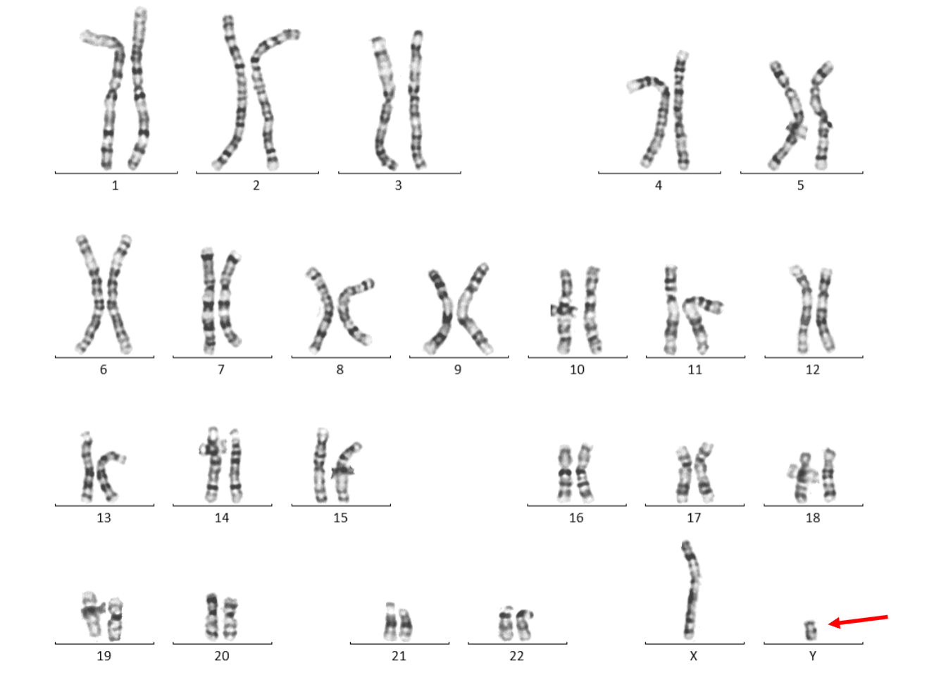
Male, 27 y
Clinical indication: INFERTILITY
What is the only thing we can tell from this karyotype?
ALL we can say is the Y is kinda short
*****Which of the 3 Techniques (banding, FISH, Microarray) is used in Molecular genetics??
FISH
Fluorescence in-situ Hybridization
Unlike karyotype it is a TARGETED test
***How does fish work/ what are the principles/ basic steps
Design DNA probes
Label the probes
Denature probes + Target DNA
Hybridize the single strand probs and DNA
Visualization (probes will bind to area of interest they are designed to bind to)

****When performing FISH the Target DNA can be obtained from what 2 stages of the cell cycle?
METAPHASE chromosomes (more condensed)
INTERPHASE nuclei (less condensed)
****What are the 4 most commonly used classes of FISH PROBES
postnatal/prenatal
Locus specific (Y)
Centromere specific (X)
Sub telomeric region (6)
Whole chromosome paints

*****FISH has a Targeted approach
What things does it target
What Abnormalities is this method typically used to DETECT?
Targets GENE/REGIONS of a chromosomes
Detects MICROdeletions/MICROduplications
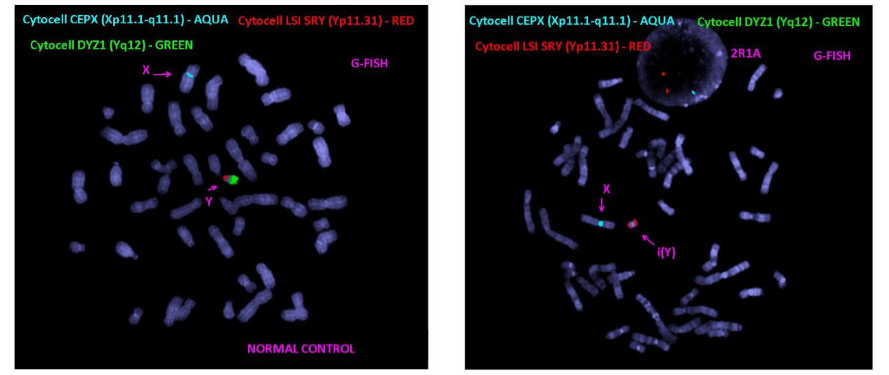
****Going back to the example of the 27yo male with fertility (SHORT Y chr. from karyotype) a FISH was used
What can be interpreted from the FISH results
Normal on left. Patient on Right
WHY did he have INFERTILITY
He has a ISOCHROMOSOME of Y
Mistake in centromere division that caused the loss of both the long arms on his Y chromosome
The deleted region contained lots of genes related to fertility
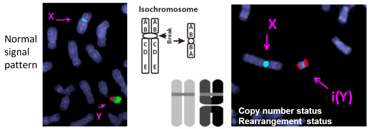
****what is an ISOCHROMOSOME?
An abnormal chromosome where the two arms are mirror images of each other
*****What is CBFB break apart rearrangement probe
HOW DOES IT WORK
FISH assays to detect structural changes, breaks, or rearrangements in the CBFB gene located on chromosome 16q22
How:
2 colored probes are used in the 16q22 region
each overlaps with a section of the CBFB gene
In NORMAL cases the 2 probes would be seen together when visualized
But in this case there was an inversion causing there to be a separation between the red and green probes

*****What is FISH Scoring
how does it work
WHAT DOES IT DETECT
QUANTITATIVE FISH test
count fluorescent signals of a specific probed gene under a microscope to determine if specific gene mutations, amplifications, deletions, or chromosomal abnormalities are present
Determines COPY NUMBER STATUS
can compare copy numbers between a control gene and a target gene


*****What can a MULTICOLOUR FISH detect from this Karyotype?
B-ALL = B cell acute lymphoblastic leukemia
REARRANGEMENT STATUS
paint each chromosome its own color = easily tell which pieces of which chromosome moved where

****RECAP: What is the APPLICATION (aka what can it detect) of each of the Common classes of FISH Probes?
Locus/ gene specific
Chromosome specific CENTROMERE probes
Chromosome specific PAINTING PROBES (whole chr + arm-specific)
Multi color WHOLE CHR. painting probes
Diff between Chromosome specific Painting probe vs. Multicolor whole chr. painting probe =
Chromosome specific = stains ONE specific chromosome (eg. chr 16)
Whole chr = stains ALL chromosomes in the genome

***There is a 23yo male with Developmental delays + seizures
which Genetic technique does the presence of developmental delays indicate for?
WHY?
MICROARRAY
Not cell by cell anymore but a more gene based technique
Microscopic and submicroscopic chromosomal imbalances, known as copy number variations (CNVs) or microdeletions/microduplications (such as 22q11.2 deletion syndrome), frequently underlie neurodevelopmental abnormalities
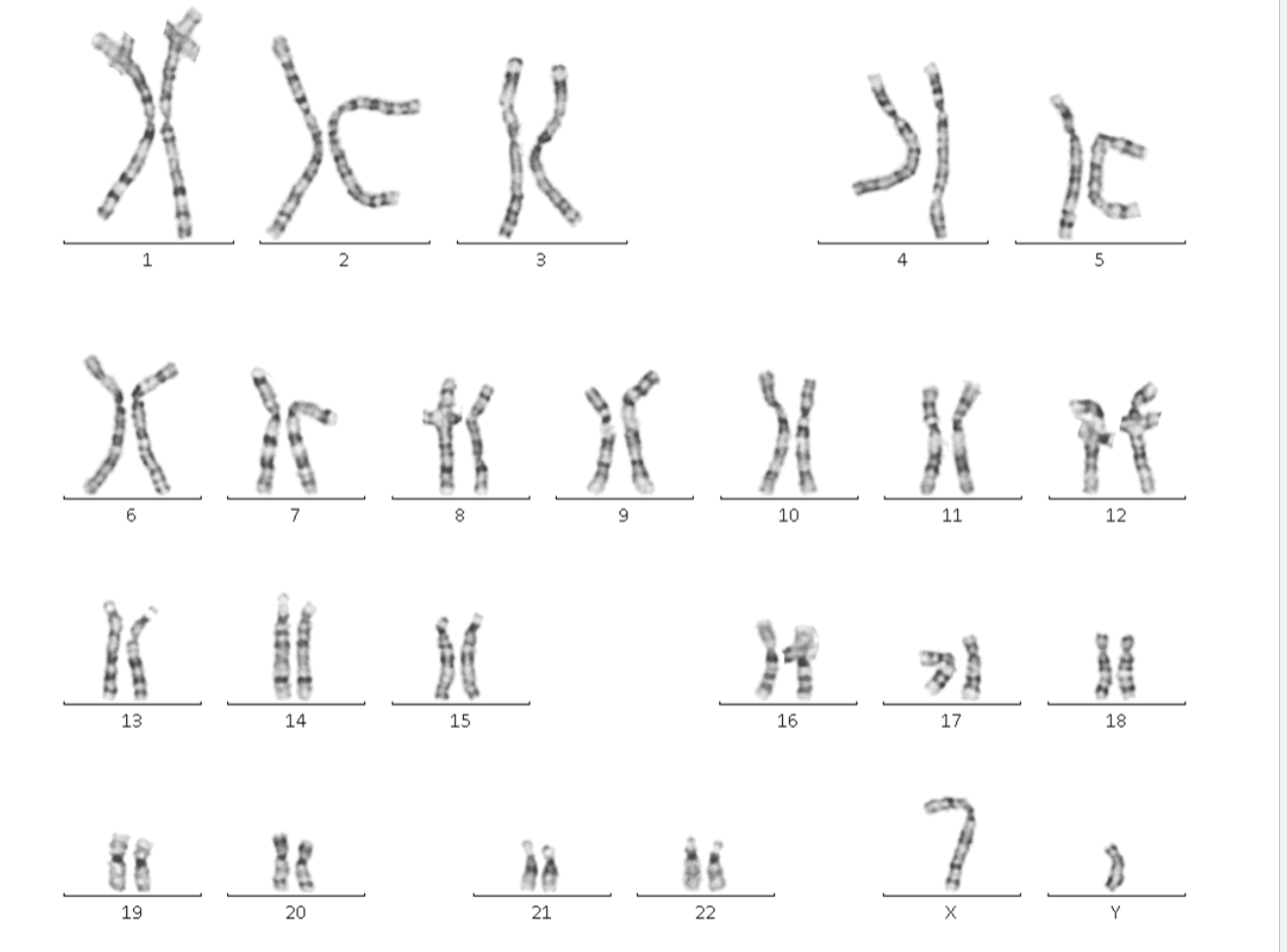
23yo male with Developmental delays + seizures
What can we tell from this karyotype?
Nothing really, it looks normal at this resolution (low resolution)
*****Which of the 3 Techniques (banding, FISH, Microarray) is used on CYTOGENOMICS??
Array-Comparative Genomic Hybridization (aCGH/ Array CGH)
aka chromosomal microarray or Microarray
VERY HIGH RESOLUTION
******ACGH are DNA based techniques. What is the SOURCE/SAMPLE taken to run ACGH?
Peripheral Blood samples in EDTA tubes
***What does it mean by ACGH are GENOME SCREENS?
a comprehensive laboratory test that evaluates a person's entire set of chromosomes at once to find missing or extra pieces of DNA
*****What do ACGH allow for the detection of?
SMALL chromosomal IMBALANCES (missing or extra genetic info) that cannot be detected by routine karyotype
CNVs
*****What are CNVs?
Copy Number Variations
microdeletions or Microduplications of segments of the genome, ranging from 50 bp to several mega bases
***What is a SPOTTED DNA microarray?
Laboratory technique where pre-synthesized DNA probes (oligonucleotides) are mechanically printed/”spotted” onto solid supports like glass slides using robotic pins
pins dip into wells containing DNA then deposits each probe at designated locations on the array surface

****HOW do ACGH’s work? what are the basic steps

****What do the COLOURS
yellow, red and green mean on a ACGH?
Yellow = control + patient = same
Red = more CONTROL DNA in a spot = Deletion in patient
Green = more PATIENT DNA in a spot = Duplication in patient


What does this result indicate about the Patient genome
CNV GAIN

What does this result indicate about the Patient genome
CNV LOSS
***How to interpret ACGH: What are the 5 International/National Guideline outcomes?
Pathogenic
Likely pathogenic
Uncertain clinical significance: VUS
Likely benign (no clinically significant abnormality detected + not reported)
Benign (no clinically significant abnormality detected + not reported)
*******How to interpret ACGH: For which of the 5 International/National Guideline outcomes are Family studies recommended?
First 3
Pathogenic
Likely pathogenic
Uncertain clinical significance: VUS
*****What are 6 Things to look for when INTERPRETING how serious the outcome of a ACGH is?
Gene content (OMIM/Refseq) + known syndromes- phenotypic correlations
If there is gene sequence in the region dup/del = more likely dangerous
Dosage sensitive gene/region
DGV (database of genomic variance) + Local databases
How often does this dup/del occur? if super often = likely benign
Size
Repetitive regions
Peer reviewed publications (amount of correlation/ # of papers)
Others
THERE WILL ALWAYS BE EXCEPTIONS
******What are 2 limitations of a ACGH/microarray?
Does not allow detection of balanced rearrangements (translocations, inversions) + low level mosaicism
Does not allow detection of regions consisting of highly repetitive sequences (Pericentromeric, heterochromatic regions, p-arms of acrocentric)
**What are the 3 possible outcomes of a microarray?
No abnormality: depends on level of resolution + area covered by test
CNV detected
Incidental findings: a potentially unrelated genetic risk
****What should be the next step if a CNV is detected?
Family studies
****What are 2 examples of possible Incidental findings of ACGH
Cancer susceptibility locus
Loci associated with adult onset

****Patient Earlier: 23yo male with Developmental delays + seizures
Could not identify what was wrong from karyotype = Ran ACGH
What is a DUPLICATION of Chromosome 8q24.3 associated with?
Neurodevelopmental disorders
Epilepsy
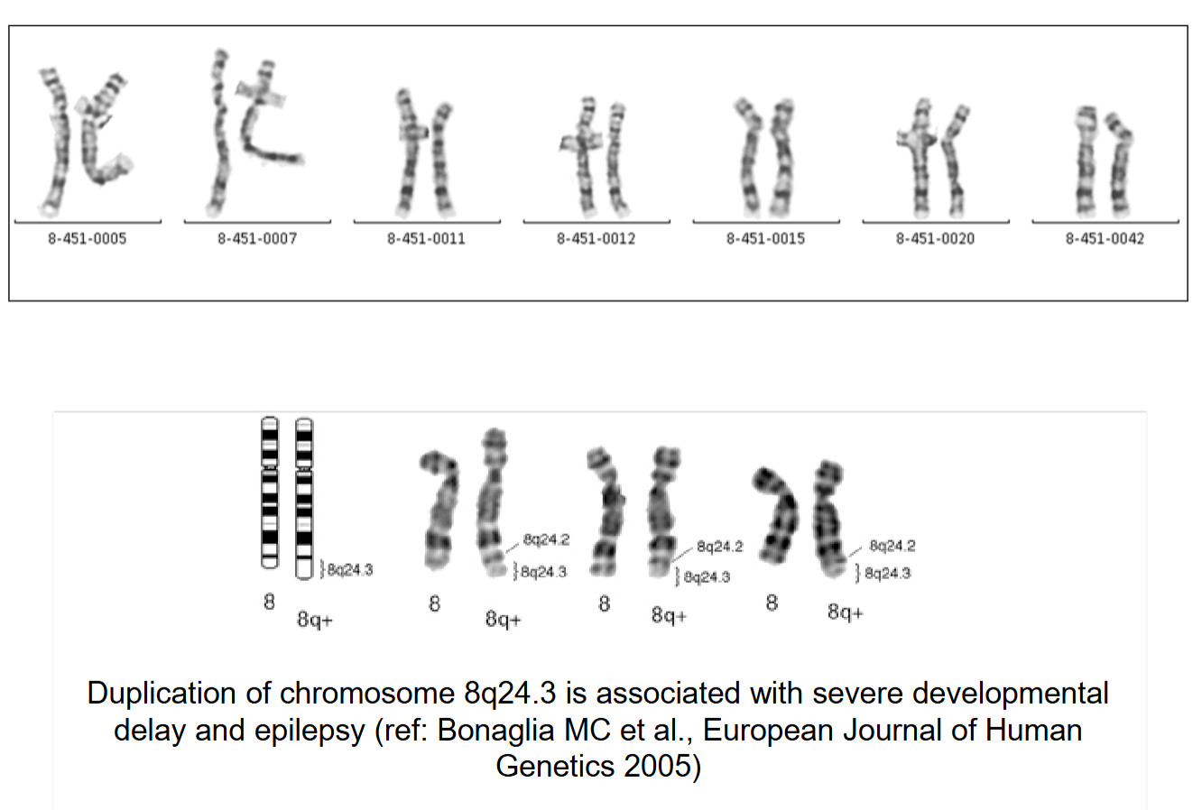

*****Now that we know the specific area we are looking for that is causing the clinical indications we can run a FISH
Patient Earlier: 23yo male with Developmental delays + seizures
Duplication of 8 is located on chr 14
and likely inherited from the parents as they have normal chr 8 and normal other chr 14
Mother karyotype also normal but fish shows der chr 14

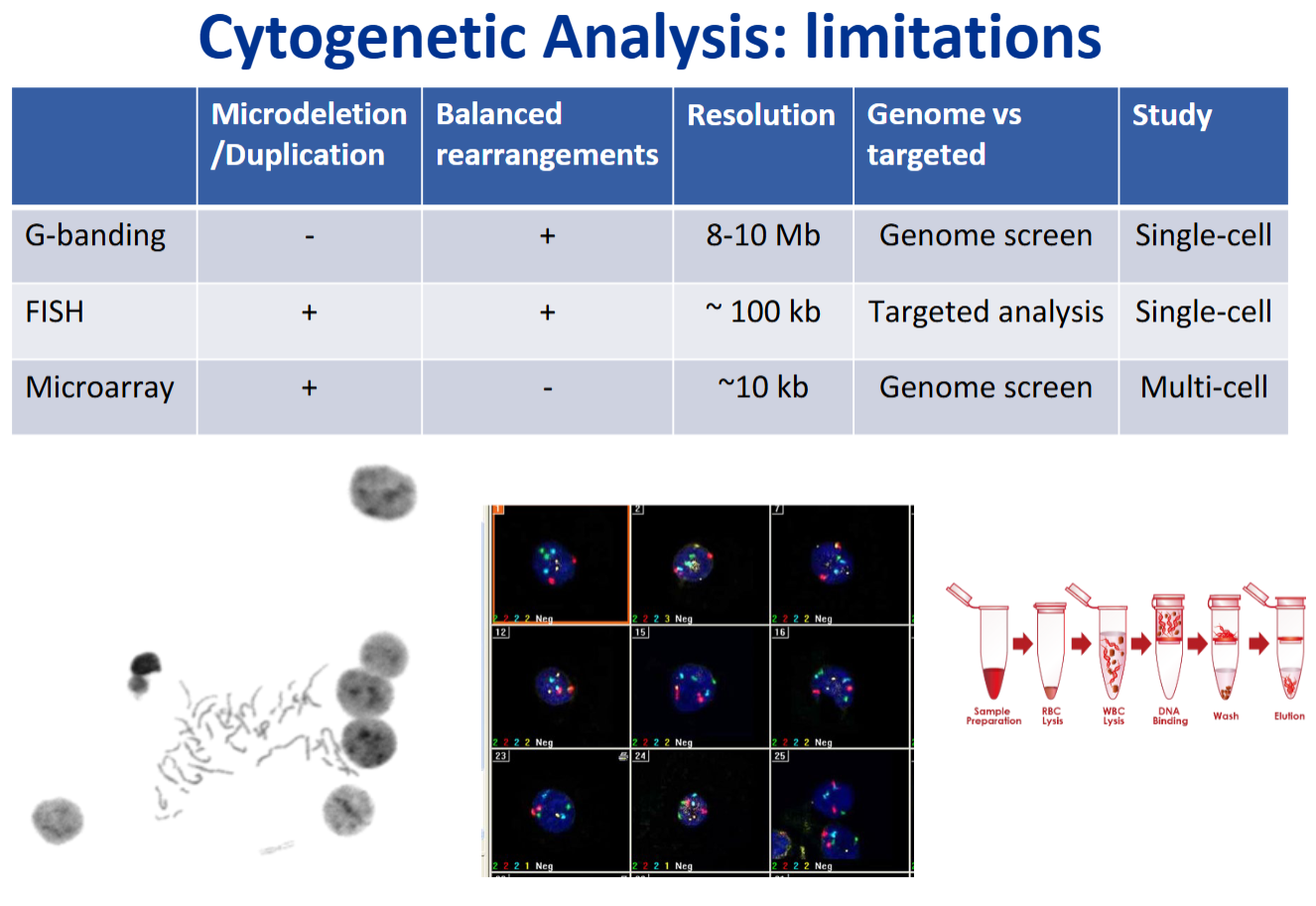
******Cytogenetic analysis RECAP: G banding, FISH, Microarray
Can it detect microdel/dup
Can it detect balanced rearrangements
Resolution
Genome screen vs. Targeted
Does it study SINGLE or MULTIPLE cells