ANS 205 Exam 1 Skeletal System
 Call Kai
 Learn
Learn Practice Test
 Spaced Repetition
 Match
Match Flashcards
 Knowt Play
1/55

There's no tags or description
Looks like no tags are added yet.
Name | Mastery | Learn | Test | Matching | Spaced | Call with Kai | Chat |
|---|
No analytics yet
Send a link to your students to track their progress
56 Terms
Skeletal system functions
1. Support
2. Protection
3. Movement
4. Storage
5. Blood cell formation
Lipid storage occurs in the
medullary cavity
Hematopoiesis
bone marrow is responsible for blood cell production (occurs in long bones)
Cancellous bone (spongy bone)
spicules arranged in a porous network, has small open spaced filled with marrow
Medullary cavity
space surrounded by a cortex of a long bone filled with red or yellow marrow
Epiphysis
ends of the bone
Diaphysis
the shaft of the bone which surrounds the medullary cavity
Articular cartilage
cushions ends of bones
Epiphysal plate
growth plate, allows for growth/lengthening of the bone
Periosteum
fibrous membrane lining the outside of the compact bone containing osteoblasts, stem cells, and osteoclasts
Endosteum
fibrous membrane lining the spongy bones containing osteoblasts, osteoclasts and stem cells
Lacunae
"lake" where the osteocytes are sitting
Canaliculius
the "canals" used to transport nutrients and waste in and out of osteocytes. can be vertical or horizontal in the osteon
Lamelli
sections between the osteocyte layers; made up of inorganic calcium phosphate. calcium leeches at the outer lamelli which degrades to give the body calcium
Five classifications of bones
1. Long
2. Short
3. Flat
4. Irregular
5. Sesamoid
Long bones
- Acts as levers, aid in support, locomotion, prehension (bringing food to mouth)
- Ex: humerus, radius, ulna, metacarpals, femur, tibia, fibula, metatarsals
Short bones
- gliding/sliding movements, absorb shock/concussion
- Ex: carpals, tarsals
Flat bones
- protect vital organs, provide large areas for attachment of muscles
- Ex: skull, pelvic bones, ribs
Irregular bones
- offer protection, support, and muscular attachment
- Ex: vertebrae
Sesamoid bones
- help in reducing friction, increase leverage, or change the direction of pull
- Ex: patella
Axial system
- skull
- vertebral column (synsarcosis)
- sternum
- ribs
Appendicular system
- thoracic limbs (forelimb)
- pelvic limbs (hindlimbs)
- shoulder
- pelvis
Order of vertebrae
- Cervical
- Thoracic
- Lumbar
- Sacral
- Coccygeal
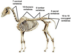
A horse has 18 thoracic vertebrae and therefore _____ ribs
18
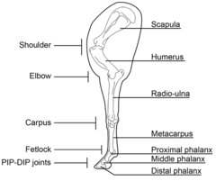
Order of bones in the front limb
- Scapula
- Humerus
- Radius/ulna
- Carpals
- Metacarpals
- Proximal phalanx
- Middle phalanx
- Distal phalanx

Joints of forelimb
- Scapular synsarcosis (scapula)
- Scapulohumeral (shoulder) (scapula+humerus)
- Elbow (Ulna+radius+humerus)
- Carpal
- Metacarpophalangeal (fetlock)
- Proximal interphalangeal (pastern)
- Distal interphalangeal (coffin)
Order of bones in the hind limb
- pelvis (os coxae)
- femur, patella
- tibia/fibula (fused)
- tarsal bones (hock)
- metatarsals (cannon/splint bones)
- phalanges (long/short pastern and coffin bone).

Joints of Hind Limb
- Coxofemoral (ilium, lechium, pubis, femur)
- Stifle/knee(femur, patella, Tibia, Fibula)
- Tarsal/hock
- metatarsophalangeal (fetlock)
- Proximal interphalangeal (Pastern)
- Distal Interonalangeal (Cofin)
osteogenic (mesenchymal) cells
stem cell whose divisions produce osteoblasts
Osteoblast
immature bone cell that secretes organic components of matrix. no well defined shape, deposits calcium onto osteoid.
Osteoid
matrix onto which calcium is deposited using alkaline phosphate. Formed from osteoblast.
Alkaline phosphatase
helps with calcium deposition onto the osteoid (organic matrix produced by osteoblasts)
Osteocyte
mature bone cell that maintains the bone matrix
Osteoclast
multinucleate cell that secretes acids and enzymes to dissolve bone matrix (contains lysosomes)
What is step one of endochondral ossification
Hyaline cartilage model forms- Mesenchymal cells differentiate into chondroblasts, creating a cartilage model of the future bone.
What is step two of endochondral ossification
Periosteal bone collar forms-
- The perichondrium becomes periosteum.
- Osteoblasts form a thin bone collar around the diaphysis.
What is step three of endochondral ossification
Primary ossification center develops in the diaphysis-
- Chondrocytes in the center hypertrophy (enlarge).
- Surrounding cartilage matrix calcifies and chondrocytes die leaving cavities.
What is step four of endochondral ossification
Blood vessels invade (periosteal bud)-
- Blood vessels, osteoblasts, and osteoclasts enter the diaphysis.
- Spongy bone begins to form in the primary ossification center.
What is step five of endochondral ossification
- Medullary cavity forms-
- Osteoclasts break down spongy bone in the diaphysis, creating the marrow cavity.
What is step six of endochondral ossification
Secondary ossification centers develop in epiphyses-
- Occur after birth.
- Similar process as the primary center but no bone collar or medullary cavity forms.
where does the cartilage still remain after endochondral ossification?
- Articular cartilage (covers joint surfaces)
- Epiphyseal (growth) plates, which allow lengthwise growth until adulthood
What is the first step that occurs during intramembranous ossification
An ossification center appears in the fibrous connective tissue membrane-
- Selected centrally located mesenchymal cells cluster and differentiate into osteoblasts, forming an ossification center.
What is the second step that occurs during intramembranous ossification
Bone matrix (osteoid) is secreted within the fibrous membrane-
- Osteoblasts begin to secrete osteoid, which is mineralized within a few days.
- Trapped osteoblasts become osteocytes.
What is the third step that occurs during intramembranous ossification
Woven bone and periosteum form-
- Accumulating osteoid is laid down between embryonic blood vessels, which form a random network. The result is a network (instead of lamellae) of trabeculae.
- Vascularized mesenchyme condenses on the external face of the woven bone and becomes the periosteum.
What is the fourth step that occurs during intramembranous ossification
Bone collar of compact bone forms and red marrow appears-
- Trabeculae just deep to the periosteum thicken, forming a woven bone collar that is later replaced with mature lamellar bone.
- Spongy bone (diploë), consisting of distinct trabeculae, persists internally and its vascular tissue becomes red marrow.
Which type of ossification forms bone directly from mesenchyme?
Intramembranous ossification
Which type of ossification uses a hyaline cartilage model?
Endochondral ossification
Which ossification process has no cartilage model?
Intramembranous ossification
Which ossification process forms flat bones of the skull?
Intramembranous ossification
Which ossification process forms long bones?
Endochondral ossification
Which type of ossification begins at multiple ossification centers?
Intramembranous ossification
Which type of ossification begins at a primary ossification center in the diaphysis?
Endochondral ossification
Which ossification process forms a medullary cavity?
Endochondral ossification
Which ossification process does NOT form a medullary cavity?
Intramembranous ossification
Where does bone growth occur in endochondral ossification?
Epiphyseal (growth) plates
Where does bone growth occur in intramembranous ossification?
Connective tissue membranes