Exam 1 (Ch.1, 2, 3, 3+5, 4, 12.3)
 Call Kai
 Learn
Learn Practice Test
 Spaced Repetition
 Match
Match Flashcards
 Knowt Play
1/39

There's no tags or description
Looks like no tags are added yet.
Name | Mastery | Learn | Test | Matching | Spaced | Call with Kai | Chat |
|---|
No analytics yet
Send a link to your students to track their progress
40 Terms
Basic Properties of Cells
Are alive
Acquire and use energy
Reproduce (cell division)
Move
Respond to stimuli
Self-regulate (e.g. when to divide)
Eukaryotes
Can be both unicellular & multicellular
Has membrane bound nucleus (which carries DNA)
Has membrane-bound organelles
Big
Cellular differentiation → specialization
Ribosome present
i.e. Animals, plants, fungi, protists
Has plasma membrane, cellulose in cell wall, cytoplasm, cell wall, flagella, lysosomes
Cell division by mitosis
Prokaryotes
Unicellular (single-celled)
No nucleus (which carries DNA)
No membrane-bound organelles
Small
Ribosome present
i.e. Bacteria & archaea
Has plasma membrane, pilus, flagella, cytoplasm, peptidoglycan in cell wall
Cell division by binary fission
No histone proteins w/ DNA
Ribosome
Present in both prokaryotes and eukaryotes
Non-membrane bound organelle
Made by proteins
RNA & protein
Rough ER → ribosome on ER
Cells evolution
Last common ancestor → 3 cell types
Prokaryotes: Bacteria, Archaea
Eucarya: Eukaryotes
Electron microscope can measure …
1 nm-100nm
Standard light microscope can measure …
1 um-100 um
Human vision can measure …
1 mm and above
Proteins
Made up of enzymes
Are polypeptides → chains of amino acids
Have backbone and R groups
Functions
Transport
Motors (help in movement)
Cell signaling (enzymes or hormones)
Gene expression (RNA pol)
Structural proteins (present on cell membranes)
How does protein shape determine function?
Unique shape/structure determined by unique amino acid sequence
R groups → amino acid properties
3 things determine shape
1) # of amino acids
2) Types of amino acids
3) Organization of amino acids
4 Levels of Protein Structure
Primary → amino acid sequence (number, types, organization of them) → determines protein shape
Secondary → backbone interactions (non covalent interactions in backbone determines secondary structure)
Tertiary → R group interactions
Quaternary → R group interactions
Covalent Bonds
Strong bonds
Shared electrons
Non-polar covalent bonds: electrons are equally shared; no partial charges
Polar covalent bonds: one atom has partial neg charge, other has partial pos charge due to unequal distribution of charge
contain 1 or more electroneg atom (O, N, S)
Formation of covalent bond is accompanied by release of energy which must be reabsorbed at some later time
large energy needed to cleave C-H, C-C, or C-O covalent bonds
stable under most conditions b/c thermal energy of a molecule = too weak to break a covalent bond

Polar molecules are …
Hydrophilic b/c can react w/ water
easily dissolves in water
Non-covalent bonds
Weak bonds in cell due to presence of water, but strong within core of protein (where water is often excluded and where they are collected together)
Ionic bonds: attraction between + and - ions (attraction between fully charged component)
dissolves in water very easily
Hydrogen bonds: attraction of partial charges on polar molecules (need partially (+) charged hydrogen atom in molecule)
weak, easily broken
form between a bonded electroneg atom (N,S) w/ a partial neg charge AND a bonded hydrogen atom that bears a partial pos charge
strength = additive
Hydrophobic interactions: association of nonpolar molecules (= hydrophobic)
clumping together minimizes exposure to polar molecules
van der Waals forces: weak attractive forces when atoms are very close/ due to close proximity
Nonpolar molecules are …
Hydrophobic b/c no charged regions that would attract them to poles of water molecule
when mixed w/ water, they clump up together which minimizes their exposure to the polar surroundings
Protein Secondary Structure
Local folding of the protein backbone which are stabilized by hydrogen bonds → backbone interacts w/ backbone
Hydrogen bonds are mainly between backbone atoms
R groups “stick out” from the backbone
2 types
1) α helix: protein backbone coils into a spiral/spring w/ R groups sticking outward
2) β pleated sheet: folded/pleated strands

Protein Tertiary Structure
Final 3D shape of one polypeptide chain (folded shape of 1 polypeptide) → R groups interact w/ R groups
Types of R group interactions that are non-covalent
van der Waals/hydrophobic interactions
H-bonds
Ionic bonds
Types of R group interactions that are covalent
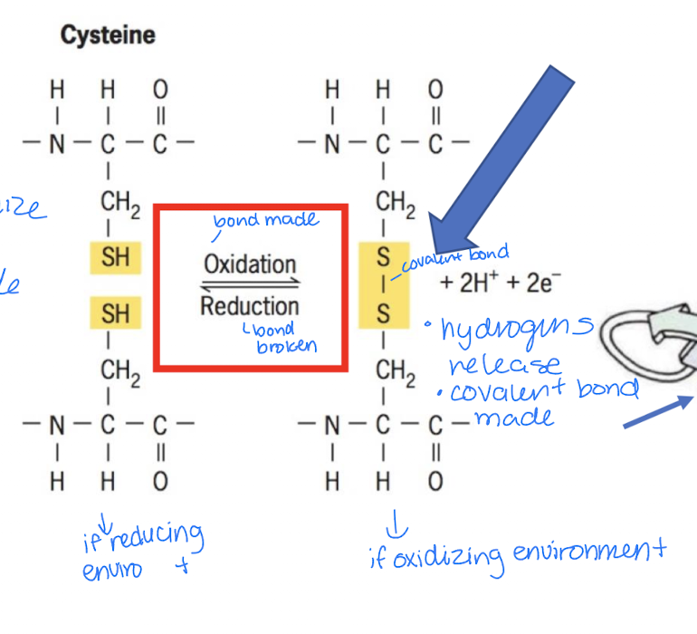
Disulfide bond between R groups of two cysteine groups
How to identify side chain?
Look for the central carbon and identify the group attached that’s not:
amino group (NH2)
carboxyl group (COOH)
hydrogen (H)
Different types of amino acids that are determined by R groups
Polar charged: clearly written (-) or (+) on side chain → name includes acid if (-) side chain
Polar uncharged: all oxygens on side chains are uncharged/no explicit written charge
Nonpolar: side chains are primarily carbons and hydrogens; have no oxygen
Side chains w/ unique properties
Glycine: has tiny H as side chain, making it very flexible
hydrophobic/hydrophilic
Cysteine: has side chain containing -SH that can form disulfide bonds which help stabilize intricate shapes of proteins (especially those present outside cells)
covalent
Proline: has a side chain that can loop to the backbone, making the protein bend/less flexible → turns = rigid
hydrophobic

How does the charge of side chains change depending on pH? (Specific to polar charged amino acids)
The side chain can gain or lose H+
at low pH (<7) → lots of H+ → groups tend to gain H+
at high pH (>7) → less H+ → groups tend to lose H+
Make sure to compare with original side chain before it gains/loses H+
Properties of side chains: Polar uncharged amino acids
Hydrophilic side chains tend to have partial (+) or (-) charge allowing them to:
participate in chemical rxns
form hydrogen bonds
associate w/ water (can form hydrogen bonds w/ water)
Often reactive

Properties of side chains: Nonpolar amino acids
Hydrophobic side chains consists almost entirely of C & H atoms (lack O & N)
Tend to form inner core of soluble proteins, buried away from aqueous medium
Play important role in membranes by associating w/ lipid bilayer
Can’t form electrostatic bonds or interact w/ water

For disulfide bonds between cysteines, bonds are …
Broken in reducing environment/after reduction
S-S bond breaks
Made in oxidizing environment/after oxidation
S-S bond forms
so, disulfide bonds play more important role in structure in proteins that function in an oxidizing environment
Soluble proteins
Soluble in water
Inside of cell compartments, it’s mostly water
cytoplasm/cytosol
inside of nucleus
mitochondrial matrix/intermembrane space
B/c of this …
Surface amino acids = hydrophilic & polar so they can associate w/ surrounding water and contribute to proteins solubility in aqueous solution
Core amino acids = hydrophobic & nonpolar, often tightly packed together
Fatty acid tails are hydrophobic → proteins touching tails = hydrophobic
Fatty acid heads are hydrophilic → proteins touching heads = hydrophilic

How can we find out the 3D (tertiary) structure of proteins?
a) X-ray crystallography ***
proteins it works on can be crystallized
Advantage: can provide higher resolution structures for larger proteins
Limits: limited by need to get any given protein to form pure crystals
b) Nuclear Magnetic Resonance (NMR) spectroscopy
Use for proteins that can’t be crystallized, provides info about dynamic changes in protein structure
Limits: more difficult to apply as size of protein increases, less resolution than x-ray crystallography
Domains
Protein tertiary structure = overall 3D folded shape of a protein → within that shape are domains (distinct folded regions)
Traits of domains
distinct modules/structure and shape → perform specific functions
fold independently from each other
diff types/combos of modules
can be shuffled to create new proteins = protein evolution
domains can change due to mutation or environment

Protein-protein interactions
Are interactions where proteins bind to and work with other proteins
Happens in multiprotein complexes → where several proteins assemble together to perform a function
Stable interaction: proteins stay together for a long time
Dynamic interaction: proteins temporarily bind, then separate
Proteins have complementary surfaces (shape, charges, hydrophobic/hydrophilic regions) so they know what to bind to
Interactomics
The complete set of interactions between proteins in a cell/organism
Hub proteins: a proteins interacting w/ several other proteins = important connection point in interaction network
Interactions can change depending on cell’s condition (presence/absence of light, nutrients or fasting, etc)
Protein folding
Final 3D structure/protein shape depends on primary structure (amino acid sequence)
Evidence: experiments w/ Ribonuclease
if you unfold a protein and then remove the chemicals that unfolded it, the protein can fold back into its original shape (Note: folded = stable structure)
Protein becomes unfolded and denatured w/ treatment of:
Urea: disrupts the weak interactions that help maintain protein’s shape by interfering w/ hydrogen bonds & hydrophobic interactions
Mercaptoethanol (reducing agent): in reducing environment, breaks/interferes w/ disulfide bonds (aka covalent bonds facilitating tertiary structure)
If both are removed, primary structure reforms and acquires tertiary structure again aka renaturation and refolding → ribonuclease is capable of self-assembly
1st and 2nd Law of Thermodynamics
1st Law
Total amount of energy is constant but can be created from one form to another
Amount of energy in a system can change vs. surroundings
System gains energy from surroundings
System loses energy to its surroundings
2nd Law
events in the universe go from high energy to low energy → spontaneous events (go from high energy to low energy; no energy needed)
energy transductions: some energy lost to surroundings
Entropy, S
Randomness/disorder
Increases w/ spontaneous events (high energy → low energy) & loss of energy → more randomness, doesn’t req energy
Spontaneous events: energetically favorable, -∆G, exergonic (releases heat)
Decreases w/ nonspontaneous events → less randomness & more orderliness, req energy input
Nonspontaneous events: energetically unfavorable, +∆G, endergonic (absorbs heat)
Reversible rxns w/ ATP
Like a rechargeable battery
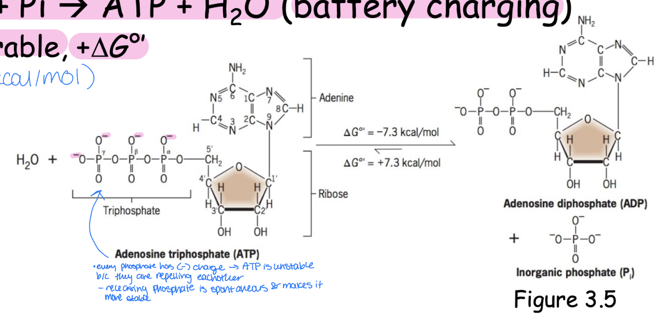
ATP hydrolysis: ATP + H2O → ADP + Pi (battery depletion)
energetically favorable, -∆G, produces/releases energy that cells use to do work, spontaneous, increases entropy
ATP synthesis: ADP + Pi → ATP + H2O (battery charging)
energetically unfavorable, +∆G, nonspontaneous, supplies energy to make ATP, decreases entropy
How do cells control chemical rxns?
In a closed system, rxns tend toward equilibrium (concentration of reactants remain unchanged but aren’t the same, rate is equal)
At equilibrium: high entropy (more stable, spontaneous) & low amount of energy to do work
ADP will be higher at equilibrium b/c is spontaneous
But after some time, ATP amount will deplete since producing ADP is favorable → no more energy production = cell death (ADP will be higher in amount than ATP)
Cells aren’t closed systems → aren’t at equilibrium, but at steady state where concentration of reactants & products are controlled by cell constantly using energy and replacing what it uses
Cells at steady state
In dynamic nonequilibrium
Use energy from surroundings to make ATP → continually obtaining energy and making ATP = plenty of free energy to do work → hydrolyze/break down ATP to do work
has low entropy (high order)
Ultimately, ATP hydrolysis can be coupled to energetically unfavorable rxns (nonspontaneous/endergonic rxn)
ATP hydrolysis supplies energy needed for nonspontaneous rxns
To make endergonic rxns go, cells couple endergonic rxns w/ exergonic rxns
Enzymes
Catalyze (speed up) chemical rxns (both exergonic and endergonic), but can’t start or end rxns
Note: in endergonic rxns, energy still must be supplied regardless of enzyme presence → can’t make unfavorable (endergonic) rxns go w/out supplied energy
Most are proteins (some RNA, ribozymes)
Exhibit properties
1) Req only in small amounts
2) Not altered irreversibly during course of rxn, so each one can participate repeatedly in individual rxns
3) Have no effect on thermodynamics of rxn
don’t supply energy → don’t determine whether its thermodynamically favorable or unfavorable
don’t determine ratio of products to reactants at equilibrium
Breaking peptide bonds
Req energy to make peptide bonds, so when broken, energy is released = exergonic
Spontaneous
Energetically favorable
Increases entropy
Produces energy
Process is slow b/c peptide bonds are stable → activation energy (EA) needed to break bond → need an enzyme to speed up rxn rate b/c it reduces activation energy (aka the minimum energy needed to overcome to start a rxn)
How? It binds to and stabilizes transition state/enzyme substrate state which is the transition between reactants & products and where bonds are being made and broken
Enzyme-substrate complex: How does an enzyme bind a substrate and turn it into products?
Substrates are reactants/what the enzyme acts on; don’t have to just be proteins
Substrate binds to enzyme’s active site due to both having complementary shapes (fit together) = enzyme-substrate complex
held together by noncovalent bonds → weak bond so easier to release substrate, but sometimes can temporarily form covalent bond
some enzymes require cofactors to function (i.e. metal ions)
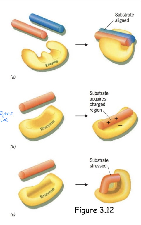
Enzyme mechanisms
a) Aligning substrates to bring them close together and position them correctly
b) Changing substrate reactivity by making it more likely to react
c) Inducing strain: enzyme binds substrate and slightly distorts/bends it so that it’s easier to break its bonds or form new bonds = more reactive
Induced fit: conformation shifts so that the complementary fir between the enzyme & reactants is improved & proper reactive groups of the enzyme move into place
Enzyme rates depend on …
Substrate concentrations (to a point)
pH
Temperature
Inhibitors
Enzyme kinetics: How does changing amount of substrate affect how fast enzyme makes products?
Velocity (v) = rate of rxn
Rate of enzymatic rxn varies w/ concentration of substrate, [S] → relationship not linear but exponential (non-enzyme catalyzed rxn is linearly increasing)
Increasing substrate concentration = increase in enzyme rxn rate until it reaches Vmax (where enzyme’s saturated w/ substrate) and plateaus since there’s no more enzyme available to couple w/ increasing amount of substrate
Km = substrate concentration when rxn’s at ½ Vmax / measure of enzyme affinity for substrate
Lower Km = less substrate req to meet ½ Vmax, so higher affinity between enzyme & substrate
How do enzyme rates also depend on pH and temperature?
There is an optimum pH and temp for enzymes to function best, so more or less than this pH/temp negatively impacts enzyme’s functions
Why?
pH: changing pH (ion concentrations) changes bonds → enzymes loses functional structure
Temp: has a denaturing effect → breaks bonds and changes enzyme shape which changes structure & function (abnormal structure = abnornal function)
Inhibitors