Lesson 1.5 Surgical Risk Assessment. ASA Classification System. Healthcare Priority Assessment.
 Call Kai
 Learn
Learn Practice Test
 Spaced Repetition
 Match
Match Flashcards
 Knowt Play
1/25

There's no tags or description
Looks like no tags are added yet.
Name | Mastery | Learn | Test | Matching | Spaced | Call with Kai | Chat |
|---|
No analytics yet
Send a link to your students to track their progress
26 Terms
Extraoral examination - specific deformities
Dentofacial deformities result from a lack of harmony between the maxilla and the mandible, leading to malocclusion (the upper and lower teeth do not meet properly). Due to this altered relationship between the maxilla and mandible, there is generally an associated aesthetic alteration.
Mandibular retrognathism→ the mandible is positioned further back than is considered normal (underdevelopment of the mandible).
Mandibular prognathism→ forward extension or protrusion of the mandible

Consequences of Facial disharmonies
Facial disharmonies can cause aesthetic and functional alterations, including malocclusion, difficulties with mastication, temporomandibular joint (TMJ) disorders, and tooth wear.
Crouzon Syndrome
Craniofacial malformation associated with the premature fusion of the cranial sutures, resulting in shortening of the skull and frontal bossing. This also affects the relationship between the teeth, causing severe dental crowding and malposition of the teeth (orthodontic problems).

Traumatic defects
Teeth can fracture in:
• The crown (the visible part of the tooth that projects above the gum).
• The root (the internal part of the tooth that is normally anchored in the jawbone).
• Injuries can also affect the tongue, cheeks (their inner surfaces), and gums.

Facial palsy
Affects the facial nerve, causing total or partial loss of voluntary muscle movement on one side of the face.
Deviation of the corner of the mouth, Inability or difficulty smiling symmetrically, Reduced facial mobility on one side, Difficulty closing the eye on the affected side.
Observation of facial asymmetries can help detect possible neurological disorders that may require medical assessment.

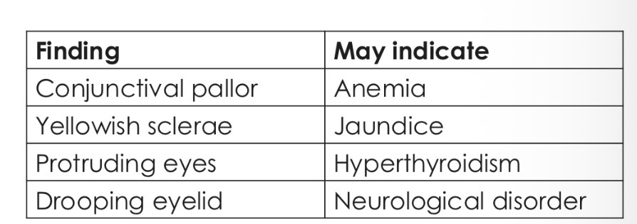
Eyes
The eyes can show signs of systemic disease
Intraoral examination structures
Lips and corners of the mouth, Cheeks, Hard and soft palate, Oropharynx, Tongue, Floor of the mouth, Gingiva / gums, Jaws, Teeth and occlusion (bite)
What do you have to consider during a Intraoral examination?
Oral hygiene
Soft tissues. The entire oral mucosa should be carefully inspected. Any ulcer of >3 weeks’ duration requires further investigation.
Periodontal condition. This can be assessed rapidly, using a periodontal probe. Pockets >5mm indicate the need for a more thorough assessment.
Chart the teeth present
Examine each tooth in turn for caries and examine the integrity of any restorations present.
Occlusion. This should involve not only getting the patient to close together and examining the relationship between the arches, but also looking at the path of closure for any obvious prematurities and displacements. Check for evidence of tooth wear
Oral ulcers
Caused by bacteria, viruses, malnutrition, immunosuppression

Herpetic stomatitis
Sores on the lips and around the mouth caused by herpes simplex virus type 1

Cheilosis
dryness, scaling, and fissures of the lips, frequently associated with nutritional deficiencies (iron, vitamin B2, folate, or vitamin B12).

Angular cheilitis
Fissures or cracks in the skin radiating from the corners of the mouth. Frequently associated with Candida albicans infection.

Leukoplakia
a white patch or plaque of the oral mucosa that cannot be removed by scraping and requires follow-up due to its potential for malignant transformation.

Dry mouth or xerostomia
decreased salivary production caused by certain medications and diseases, including autoimmune and psychological disorders.

Gum or dental problems - receding gums
Gingivitis: inflammation of the gums caused by infection or by the accumulation of bacterial plaque and calculus. Symptoms: bleeding, swelling, redness, sensitivity to cold, and bad breath.
Periodontitis: progressive infection of the gums and loss of bone around the teeth, which can eventually lead to tooth loss.

Lip and Oral Cavity Cancer
Sores on the lips or in the mouth that do not heal.
A lump or thickening of the lips, gums, or other areas of the mouth.
A white or red patch on the gums, tongue, or lining of the mouth.
Early detection of oral cancer is essential, and dentists play a key role during routine oral examinations.

What is essential to obtain at the start of a patients visit?
1. Obtain a careful medical, dental, family, social history, and make a risk assessment
2. Assess the patient’s needs and agree them with the patient
3. Obtain the patient’s valid consent to any investigations required
4. Obtain the patient’s consent to an agreed treatment plan
What is the aim of surgical risk assessment?
The aim of surgical risk assessment is to identify factors that may increase the likehood of complications during or after treatment.
Surgical risks depend on:
Patient’s general health status, Type of procedure, Duration of the procedure, Degree of surgical trauma, Level of stress generated, Urgency of treatment, Practitioner’s experience, Available resources and equipment.
Levels of risks
Low risk: minimally invasive, short procedures with a low likelihood of complications.
Examples: scaling, dental filling, pit and fissure sealants, fluoride application, intraoral radiographs, and routine local anesthesia.
Intermediate risk: procedures involving manipulation of soft or hard tissues, longer duration, or a moderate risk of complications.
Examples: root canal treatment, simple tooth extraction, surgical extraction, third molar
extraction, placement of single dental implants, and biopsy.
High risk: extensive or prolonged procedures, or procedures requiring general anesthesia and advanced monitoring.
Examples: orthognathic surgery, maxillofacial reconstruction following extensive trauma,
resection of tumors of the oral cavity or jaws, and procedures performed under general
The administration of sedative and anesthetic drugs carries potential risks
These drugs must be administered carefully, particularly those that depress the central nervous system and may affect consciousness, respiratory function, and cardiovascular function.
The risk of respiratory and cardiovascular complications generally increases as the level of consciousness decreases.
What increases the risk:
Increasing age, medical treatments, surgical treatments, prolonged dental procedures, drug use - medication or recreational
What reduces the risk:
Planned treatment, Non-invasive treatment, appropriate monitoring, safety measures, competent practiotioner
ASA (American society of Anaesthesiologists) classfication

Dental care priority assessment helps determine which patients require more urgent care based on:
Severity of the condition, Risk to overall health, Pain, Risk of progression, Impact on oral function (e.g., eating, speaking, swallowing, or opening the mouth)
Objective: to allocate healthcare resources to patients with the greatest clinical need.
Levels of priority of dental treatments
High priority
Facial cellulitis, Persistent bleeding, Dentoalveolar trauma, Severe uncontrolled pain
Intermediate priority
Active periodontitis, Extensive dental caries, Oral lesions requiring follow-up
Low priority
Routine check-ups, Dental scaling, Tooth whitening