microbio microscopy
 Call Kai
 Learn
Learn Practice Test
 Spaced Repetition
 Match
Match Flashcards
 Knowt Play
1/23

There's no tags or description
Looks like no tags are added yet.
Name | Mastery | Learn | Test | Matching | Spaced | Call with Kai | Chat |
|---|
No analytics yet
Send a link to your students to track their progress
24 Terms
light microscopes
brightfield
darkfield
phase contrasts
differential interference contrast (DIC)
fluorescent contrasts

describe brightfield microscopes
fixed, stained specimen =, 1000x-2000x oil immersion
white light , dark against light illuminated background
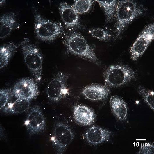
describe darkfield microscopes
opaque disk over light source, unstained + live, makes background dark

describe phase contrasts
two sets of light brought together (light source + light detracted from structures in the specimen; creates sharp images of internal structures in specimen)

describe DIC
2 beams of light from light source; almost 3D image of a specimen
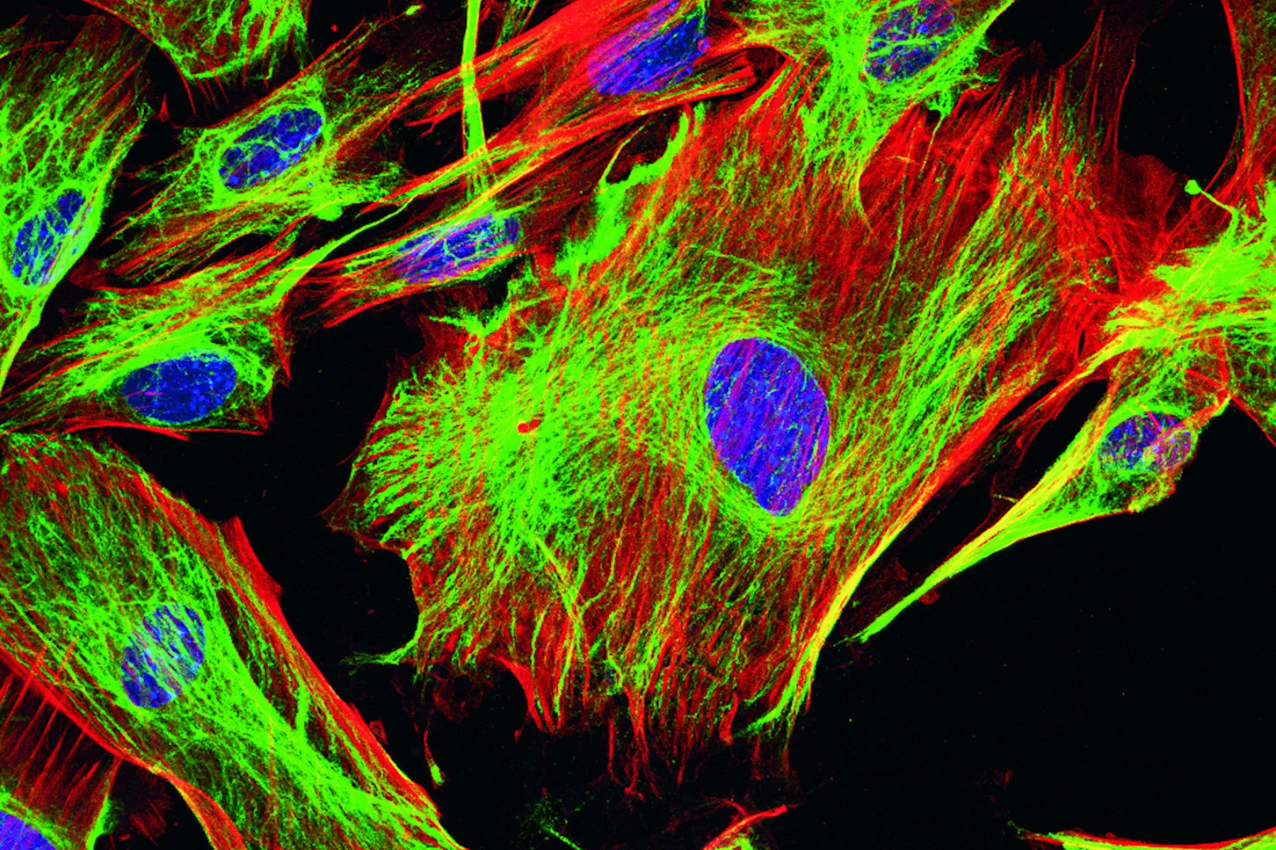
describe fluorescent contrast
fluorescent light absorbs short wavelength of light; emits light as longer wavelengths; immunofluorescence (antibodies)
types of fluorescent contrasts
confocal
two photon microscope
super resolution
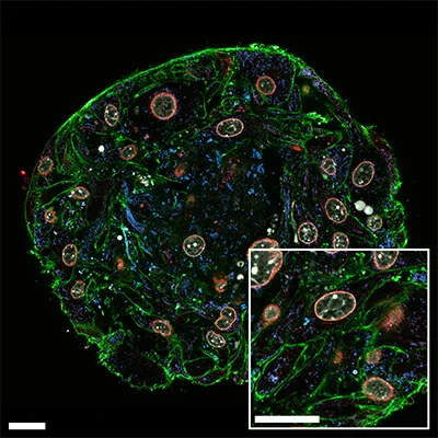
describe confocal
fluorescent, using short wavelengths of light (blue), pinhole aperture (significantly reduces the blurring of the light —> sharp image) increases resolution; 3-D images
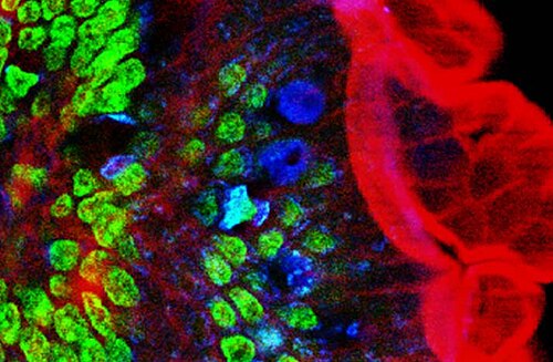
describe two photon microscope
longer wavelength (red); deeper/thicker image of specimen; can image cells up to 1 millimeter (mm) deep
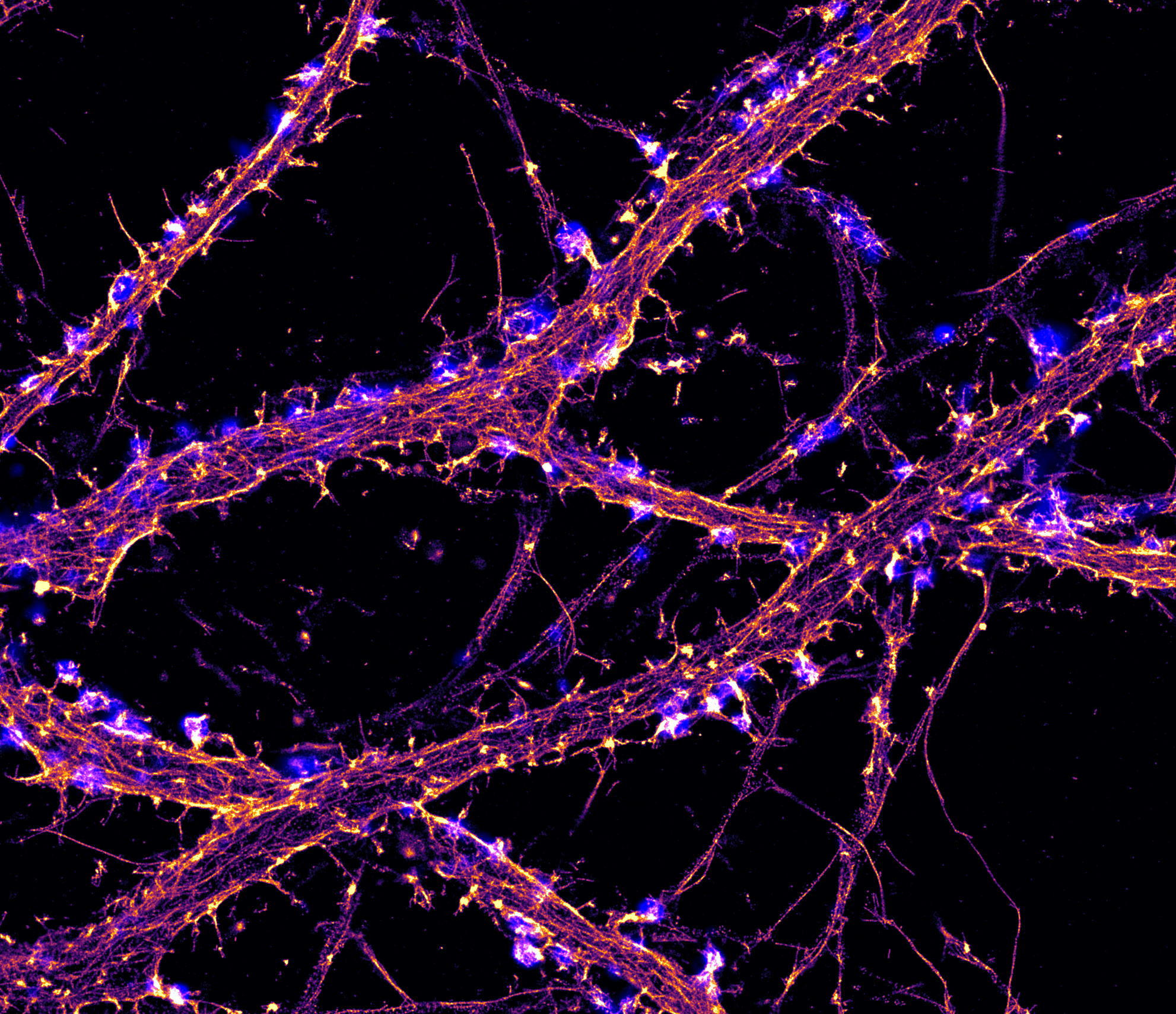
describe super resolution
nanometer scale (10^-9)

describe scanning acoustic
sound waves to create a computerized image; biofilms
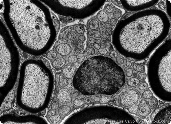
describe transmission electron (TEM)
electrons, 10,000-10,000,000 picometer scale 10^-12pm, focus on internal structure of cell (drawback: specimen is very thin, no more than 100nm thick) BLACK and WHITE
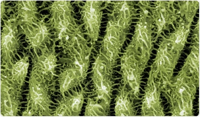
Scanning electron microscope (SEM)
1000x-500,000x ; 10nm scale surface structure and creates 3D images of specimen IN COLOR
describe scanning probe microscope
uses probe to examine specimens with electrical current, scanning tunneling, tungsten probe, observes atoms, resolution: 1/100 size of atom, detailed view of molecules

describe atomic force
metal and diamond probe to scan surface, creates 3D image

describe simple stains
single basic dye
mordant if necessary to (improve bond between specimen + stain)
types differential stains
gram-stain and acid- fast

describe gram-stains
distinguish types of bacteria cell wall, used to categorize bacteria: gram + (positive) or gram - (negative)
stains used for gram-stains
crystal violet (purple) (+) and safranin (pink/red) (-)

describe acid-fast stain
specimen stained by carbofusion (red) or methylene (blue)
types of special stains
negative stains: capsules (layer of sugar outside cell)
endospores
flagella
what is resolution
how well the microscope distinguishes 2 points as separate
what is refraction
bending (way your eye perceives) o flight as it passes from one medium to another
what is magnification
how much larger a specimen appears compared to the actual size, using 2 measurements (objective x ocular)