Neuro Exam 3
0.0(0)
Card Sorting
1/149
Earn XP
Description and Tags
Last updated 6:09 PM on 4/26/23
Name | Mastery | Learn | Test | Matching | Spaced | Call with Kai |
|---|
No analytics yet
Send a link to your students to track their progress
150 Terms
1
New cards
What is the level of motor control at the spinal cord?
Spinal cord- reflexes
2
New cards
What is the level of motor control at the brainstem?
Brainstem- vegetative function
3
New cards
What is the level of motor control at the cerebellum?
Cerebellum- coordination
4
New cards
What is the level of motor control at the basal ganglia?
Basal ganglia - regulation (inhibition/exhibition)
5
New cards
What is the level of motor control at the motor cortex?
initiation and regulation of movement
6
New cards
What is the primary function of the pre-central gyrus?
motor homunculus and contralateral control
7
New cards
What is the primary function of the premotor and supplementary motor area?
Aid the primary motor cortex in selection, planning, and generation movement
8
New cards
What is the lobe that the precentral gyrus, premotor area, and supplementary motor area located in?
The frontal lobe, primary motor cortex
9
New cards
What are the inputs to the motor cortex?
Prefrontal (intention, motivation, goals)Primary somatosensory (current muscle status/position)
Somatosensory association (complex spatial aspects, where you are in space)
Thalamocortical (excitatory loop; integration with basal ganglia)
Limbic System (motivation, goals)
\
Somatosensory association (complex spatial aspects, where you are in space)
Thalamocortical (excitatory loop; integration with basal ganglia)
Limbic System (motivation, goals)
\
10
New cards
What are all the componenets of the motor unit?
LMN and all innervated muscle fibers
11
New cards
What is the neurotransmitter involved at the neuromuscular junction?
Acetycholine (ACh)
12
New cards
What is a pyramidal tract?
direct connection between brain and muscles
13
New cards
Know the pyramidal tracts (corticobulbar & corticospinal \[both anterior & lateral\]) including the names of neurons, location of cell bodies, location of axons, location of crossover, location of synapses, and target of the pathway.
Pyramidal tract overview
Corticobulbar tracts (70%)
Upper Motor Neurons end in brainstem
Lower Motor Neurons = cranial nerves
\n
Corticospinal tracts (30%)
Upper Motor Neurons end in spinal cord
Lower Motor Neurons = spinal nerves
\
Lateral corticospinal tract overview
makes up 90% that crossover
crosses over at pyramidal decussation
contralateral spinal cord
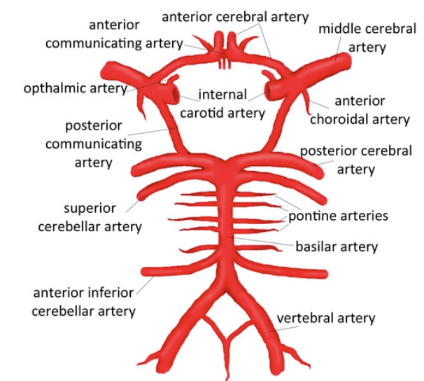
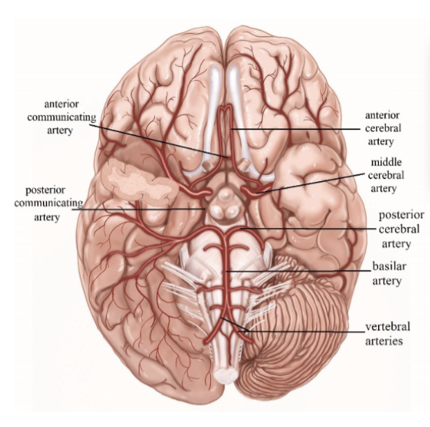
(limbs)
\
Anterior corticospinal tract overview
makes up 10% in the ventral tract
remains ipsilateral down the spinal cord
cross over prior to synapse
most innervate trunk muscle
\
Corticobulbar tracts (70%)
Upper Motor Neurons end in brainstem
Lower Motor Neurons = cranial nerves
\n
Corticospinal tracts (30%)
Upper Motor Neurons end in spinal cord
Lower Motor Neurons = spinal nerves
\
Lateral corticospinal tract overview
makes up 90% that crossover
crosses over at pyramidal decussation
contralateral spinal cord
(limbs)
\
Anterior corticospinal tract overview
makes up 10% in the ventral tract
remains ipsilateral down the spinal cord
cross over prior to synapse
most innervate trunk muscle
\
14
New cards
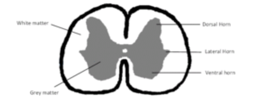
Identify the landmarks on the spinal cord - dorsal/ventral horns, dorsal/ventral roots
15
New cards
What is a myotome?
Areas/muscles innervated by efferent nerves (spinal nerves) (Motor)
16
New cards
How does a reflex arc work?
\- It is a direct sensory-motor connection
\- Stimulation of sensory nerve ending afferent signal travels through dorsal root into dorsal horn
\- Synapses on motor nerve: efferent signal travels through ventral root out to muscle
\- Reciprocal inhibition: inhibition of antagonist muscle to allow movement of primary muscle
\- Stimulation of sensory nerve ending afferent signal travels through dorsal root into dorsal horn
\- Synapses on motor nerve: efferent signal travels through ventral root out to muscle
\- Reciprocal inhibition: inhibition of antagonist muscle to allow movement of primary muscle
17
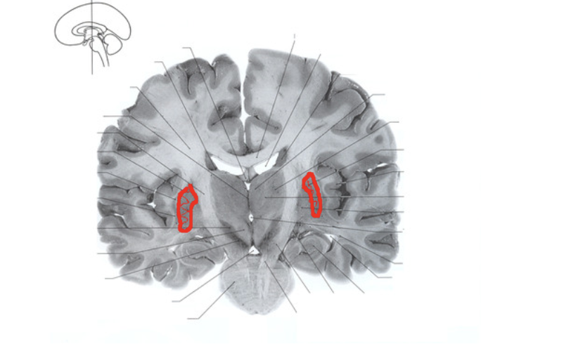
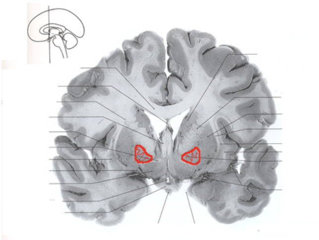
New cards
Label the caudate nucleus

18
New cards
Label the putamen
19
New cards
Label the globus pallidus
20
New cards
What is the role of the basal ganglia?
\
“Fine-tuning” motor functions, also plays a role in cognition and reward
“Fine-tuning” motor functions, also plays a role in cognition and reward
21
New cards
How does the indirect basal ganglia circuit modify movement?
\
basal ganglia: indirect circuit -> INHIBITS MOVEMENT = LESS MOVEMENT
basal ganglia: indirect circuit -> INHIBITS MOVEMENT = LESS MOVEMENT
22
New cards
How does the direct basal ganglia circuit modify movement?
\
basal ganglia: direct circuit -> FACILITATES MOVEMENT = MORE MOVEMENT
basal ganglia: direct circuit -> FACILITATES MOVEMENT = MORE MOVEMENT
23
New cards
What is meant by “comparator” in relation to the cerebellum?
\
“comparator” (compares intended movement with actual movement)
“comparator” (compares intended movement with actual movement)
24
New cards
What are symptoms of UMN lessions: contralateral?
*UMN lesions: contralateral symptoms*
\- loss of discrete muscle control
\- muscle weakness
\- loss of discrete muscle control
\- muscle weakness
25
New cards
What are symptoms of UMN lessions: bilateral?
*UMN lesions: bilateral symptoms*
\- hypertonia
\- loss of discrete motor control of head/neck
\- hypertonia
\- loss of discrete motor control of head/neck
26
New cards
What are symptoms of LMN lessions?
*LMN lesions signs*
\- flaccid paralysis (reduced/floppy muscle tone)
\- reduced reflexes
\- muscle fibrillations and atrophy
\- flaccid paralysis (reduced/floppy muscle tone)
\- reduced reflexes
\- muscle fibrillations and atrophy
27
New cards
What are symptoms of Spinal Cord lessions?
\- Complete transection
Bilateral loss of all sensory & motor below the lesion
\- Spinal Hemisection(Brown-Sequard syndrome)
Damage to 1 side of spinal cord(R or L)
Bilateral loss of all sensory & motor below the lesion
\- Spinal Hemisection(Brown-Sequard syndrome)
Damage to 1 side of spinal cord(R or L)
28
New cards
What are symptoms of Motor Programming System lessions?
Apraxia of Speech
29
New cards
What are symptoms of Cerebellum lessions?
Ipsilateral effects
Motor coordination affected
paralysis or weakness (bc cerebellum not involved in initiation)
Sensory functions intact
Hypotonia: reduced tone (appears ipsilaterally)
Ataxia: decreased coordination/order of movement
Motor coordination affected
paralysis or weakness (bc cerebellum not involved in initiation)
Sensory functions intact
Hypotonia: reduced tone (appears ipsilaterally)
Ataxia: decreased coordination/order of movement
30
New cards
What causes ALS (amyotrophic lateral sclerosis)?
Caused by degeneration of motor neurons (upper and lower)
31
New cards
What are the symptoms of ALS?
Symptoms: total loss of speech production and full movement disorder
32
New cards
What causes Parkinson’s?
Loss of dopaminergic neurons from substantia nigra
33
New cards
What are the symptoms of Parkinson’s?
Symptoms: resting tremor, hypokinetic dysarthria
34
New cards
What causes Huntington’s?
Caused by damage to caudate nucleus
35
New cards
What are the symptoms of Huntington’s?
Symptoms: Unwanted, uncontrollable movements and writhing movements
36
New cards
Where is movement initiated?
In the motor cortex
37
New cards
What pathways are involved directly in motor control?
Pyramidal Tracts–Direct Motor System
38
New cards
\
Which regions and pathways are involved in modifying motor processing?
\
Which regions and pathways are involved in modifying motor processing?
\
The extrapyramidal tracts
39
New cards
What is CNI?
olfactory
40
New cards
What is CNII?
Optic
41
New cards
What is CNIII?
Occulomotor
42
New cards
What is CNIV?
Trochlear
43
New cards
What is CNV?
Trigeminal
44
New cards
What is CNVI?
Abducens
45
New cards
What is CNVII?
Facial
46
New cards
What is CNVIII?
Auditory/Vestibular
47
New cards
What is CNIX?
Glossopharyngeal
48
New cards
What is CNX?
Vagus
49
New cards
What is CNXI?
Spinal accessory
50
New cards
What is CNXII?
Hypoglossal
51
New cards
What is cranial nerve nuclei?
cell bodies for the LMN of corticobulbar tract
52
New cards
What is the relation of cranial nerve nuclei with the corticobulbar tract?
They’re intimately connected, the corticobulbar provides the primary source of motor control to the cranial nerve nuclei
53
New cards
What is the function of CNI?
Sensory
The olfactory nerve transmits information regarding a person’s sense of smell to the brain.
\
The olfactory nerve transmits information regarding a person’s sense of smell to the brain.
\
54
New cards
What is the function of CNII?
Sensory
The optic nerve transmits information to the brain regarding a person’s vision.
\
The optic nerve transmits information to the brain regarding a person’s vision.
\
55
New cards
What is the function of CNIII?
Motor
The oculomotor nerve helps control muscle movements of the eyes.
\
The oculomotor nerve helps control muscle movements of the eyes.
\
56
New cards
What is the function of CNIV?
Motor
also has a role in eye movement.
\
also has a role in eye movement.
\
57
New cards
What is the function of CNV?
Both
The trigeminal nerve is the largest cranial nerve and has both motor and sensory functions.
The trigeminal nerve is the largest cranial nerve and has both motor and sensory functions.
58
New cards
What is the function of CNVI?
Motor
control eye movements.
\
control eye movements.
\
59
New cards
What is the function of CNVII?
The facial nerve also has both motor and sensory functions.
\
* movement of muscles that produce facial expression
* movement of the lacrimal, submaxillary, and submandibular glands
* the sensation of the external ear
* the sensation of taste
\
\
* movement of muscles that produce facial expression
* movement of the lacrimal, submaxillary, and submandibular glands
* the sensation of the external ear
* the sensation of taste
\
60
New cards
What is the function of CNVIII?
Both
helps with a person’s hearing and balance.
helps with a person’s hearing and balance.
61
New cards
What is the function of CNIX?
Both
The sensory function receives information from the throat, tonsils, middle ear, and back of the tongue. It also has a role in the sensation of taste on the back of the tongue.
The sensory function receives information from the throat, tonsils, middle ear, and back of the tongue. It also has a role in the sensation of taste on the back of the tongue.
62
New cards
What is the function of CNV?
Both
has a range of functions, providing motor, sensory, and parasympathetic functions.
has a range of functions, providing motor, sensory, and parasympathetic functions.
63
New cards
What is the function of CNXI?
Motor
provides motor function to some muscles in the neck.
provides motor function to some muscles in the neck.
64
New cards
What is the function of XII?
Motor
supplies the tongue muscles. It originates in the medulla.
supplies the tongue muscles. It originates in the medulla.
65
New cards
What does CNI innervate?
enter brain directly
66
New cards
What does CNII innervate?
ganglion cell axons from retina
67
New cards
What does CNVIII innvervate?
the rectus muscle and oblique musclesup
68
New cards
What does CNIV inneravate?
superior oblique muscle
69
New cards
What does CNV innervate?
Three sensory branches: face, teeth, dura, mucous membranes
One motor branches:muscles of mastication, tenses the soft palate
One motor branches:muscles of mastication, tenses the soft palate
70
New cards
What does CNVI innervate?
Innervates 1 of the 6 eye muscles
71
New cards
What does CNVII innervate?
Travels through internanl auditory meatus with CNVIII
72
New cards
What does CNVIII innervate?
Enter brainstem in upper medulla
73
New cards
What does CNIX innervate?
enters/exits at medulla
74
New cards
What does CNX innervate?
enters/exits at medulla
75
New cards
What does CNXI innervate?
Leaves brainstem at medulla
76
New cards
What does CNXII innervate?
leaves brainstem at lower medulla
77
New cards
What does lateral strabismus mean?
deviation of ipsilateral eye to lateral position which leads to double vision
78
New cards
What does Ptsosis mean?
Drooping eyelid
79
New cards
What does Mydriasis mean?
permanent dialation of pupil
80
New cards
Which cranial nerves are important for speech production?
CN X for voicing and movement of velum
CN XIII for movement of tongue
CN VII for movement of lips
CN XIII for movement of tongue
CN VII for movement of lips
81
New cards
Where does the blood supplying the brain come from?
• Blood supply to the brain comes directly from the heart, via the aorta
82
New cards
What is the anterior route?
• Anterior route (80%)
internal carotid arteries
from aorta, travel up the side of neck
branches supply anterior, medial & lateral structures
internal carotid arteries
from aorta, travel up the side of neck
branches supply anterior, medial & lateral structures
83
New cards
\
What is the posterior route?
What is the posterior route?
• Posterior route (20%)
vertebral arteries from aorta, travel up through vertebral column & along brain stem fuse to form basilar artery, branch to supply cerebellum, inferior & posterior surfaces/structures
vertebral arteries from aorta, travel up through vertebral column & along brain stem fuse to form basilar artery, branch to supply cerebellum, inferior & posterior surfaces/structures
84
New cards
What is the circle of willis?
structure of arteries at base of the brain
85
New cards
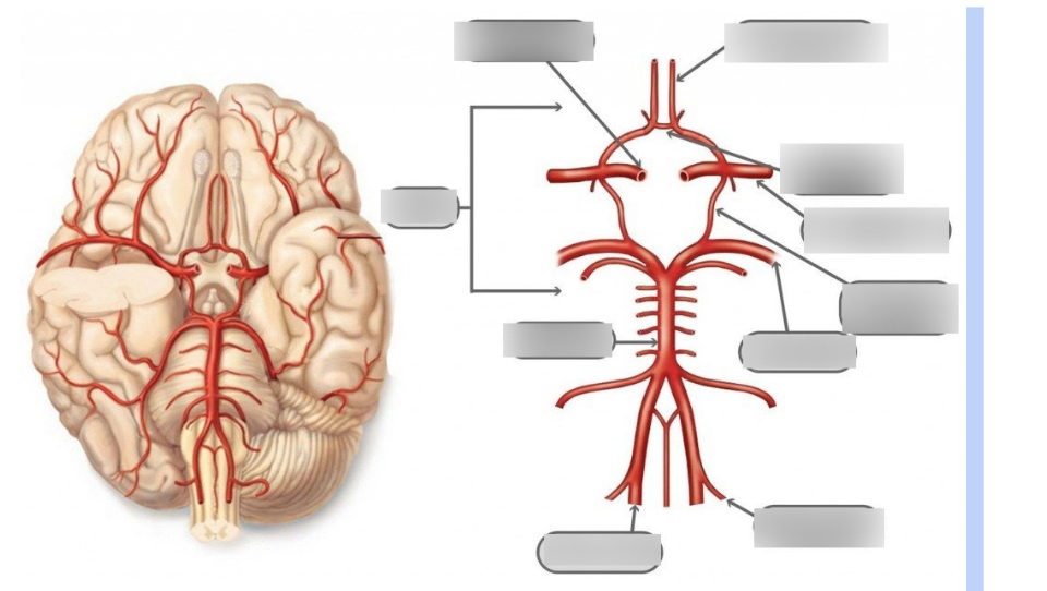
Label the circle of willis
86
New cards
Identify/label the areas of the brain supplied by the 3 cerebral arteries.
• 3 Cerebral Arteries • Anterior • Middle (supplies blood to language region of brain) • Posterior
87
New cards
What is meant by watershed region?
\
\
#### • Watershed provides double coverage for areas of overlap between two vascular territories
• Watershed vessels are small diameter (terminal branches), so they are more susceptible to small vessel disease
• Watershed vessels are small diameter (terminal branches), so they are more susceptible to small vessel disease
88
New cards
What is the difference between an artery and a vein?
\
* Arteries carry oxygen-rich blood to the brain
* Veins carry oxygen-low blood to the heart for reoxygenation
\
* Arteries carry oxygen-rich blood to the brain
* Veins carry oxygen-low blood to the heart for reoxygenation
\
89
New cards
What is a stroke?
CerebroVascular Accident (CVA)
• Interruption of blood flow to the brain that results in damage to brain tissue
• AKA stroke
• Interruption of blood flow to the brain that results in damage to brain tissue
• AKA stroke
90
New cards
What’s the difference between schemic and hemorrhagic stroke?
• Ischemic: inadequate blood flow to the brain
• Hemorrhagic: blood spills out of vessels
• Hemorrhagic: blood spills out of vessels
91
New cards
What factors determine the effects of a stroke?
Location!
92
New cards
What happens if a stroke affects the left peri-sylvian area?
Aphasia, dysarthria, apraxia of speech
93
New cards
What happens if a stroke affects the right hemisphere?
Cognitive-communication deficits
94
New cards
What happens if a stroke affects the occipital lobe?
Visual field cuts
95
New cards
What happens if a stroke affects the cerebellum?
Ataxia, balance/coordination deficits
96
New cards
What is the difference between a stroke and a TIA?
• CerebroVascular Accident (CVA)
* Interruption of blood flow to the brain that results in damage to brain tissue
* AKA stroke
• Transient Ischemic Attack (TIA)
* “mini stroke”
* Temporary (transient) arterial blockage
* Interruption of blood flow to the brain that results in damage to brain tissue
* AKA stroke
• Transient Ischemic Attack (TIA)
* “mini stroke”
* Temporary (transient) arterial blockage
97
New cards
What are controllable risk factors of a stroke?
• Hypertension
• Heart disease
• Diabetes
• High cholesterol
• Smoking, drug use, excessive alcohol use
• Heart disease
• Diabetes
• High cholesterol
• Smoking, drug use, excessive alcohol use
98
New cards
What are warning signs of a stroke?
All occur suddenly
• Weakness/numbness in face, arm, leg unilaterally
• Dizziness, loss of balance/coordination
• Loss of vision (one or both eyes)
• Sudden, severe, unexplained headache
• Difficulty speaking and/or understanding speech
\
• Weakness/numbness in face, arm, leg unilaterally
• Dizziness, loss of balance/coordination
• Loss of vision (one or both eyes)
• Sudden, severe, unexplained headache
• Difficulty speaking and/or understanding speech
\
99
New cards
Why is it important to get immediate help if you suspect a stroke?
In one second 32,000 neurons can be lost so it’s important to get help right away and preserve as many neurons as possible
100
New cards
What is hemispheric specialization of the left hemisphere?
Speech and language perception and production (phonology, morphology, syntax)