Module 4
 Call Kai
 Learn
Learn Practice Test
 Spaced Repetition
 Match
Match Flashcards
 Knowt Play
1/36

There's no tags or description
Looks like no tags are added yet.
Name | Mastery | Learn | Test | Matching | Spaced | Call with Kai | Chat |
|---|
No analytics yet
Send a link to your students to track their progress
37 Terms
Joint
Any location where adjacent bones or bone and cartilage
Also called an Articulation
Have Functional and Structural Classifications.
Cannot freely rotate in a complete circle in the body.
Due to tangle of blood vessels, nerves, and muscles.
Functional Classification
Describes the amount of movement available between the bones.
Ex. Synarthrosis, Amphiarthrosis, and Diarthrosis
Structural Classification
Examines the anatomical structure of the joints.
Considers how adjacent bones are connected.
Directly Connected → Fibrous Connective Tissue or Cartilage.
Contact → With a fluid-filled joint cavity.
Ex. Fibrous, Cartilaginous, and Synovial
Fibrous Joints
Bones united by fibrous connective tissue.
Adjacent bones that are directly connected to each other by fibrous connective tissue.
No joint cavity.
Gap between bones may be narrow or wide
Ex. Suture, Syndesmosis, and Gomphosis.
Cartilaginous Joints
Adjacent bones joined by Hyaline cartilage or Fibrocartilage.
Lack of joint cavity.
Ex. Synchondrosis and Symphysis.
Synovial Joints
Articulating surfaces of the bones are not directly connected.
Bones interact within a joint cavity, filled with a lubricating fluid.
Allow for free movements between bones.
Most common joint.
Ex. Plane, Condylar, Hinge, Saddle, Pivot, and Ball-Socket.
Synarthrosis
Immobile joint
Provide for a strong union between the articulating bones.
Important where bones provide protection for internal organs.
Immobile or nearly
Syn - together or with
Ex. Sutures and Manubriosternal Joint
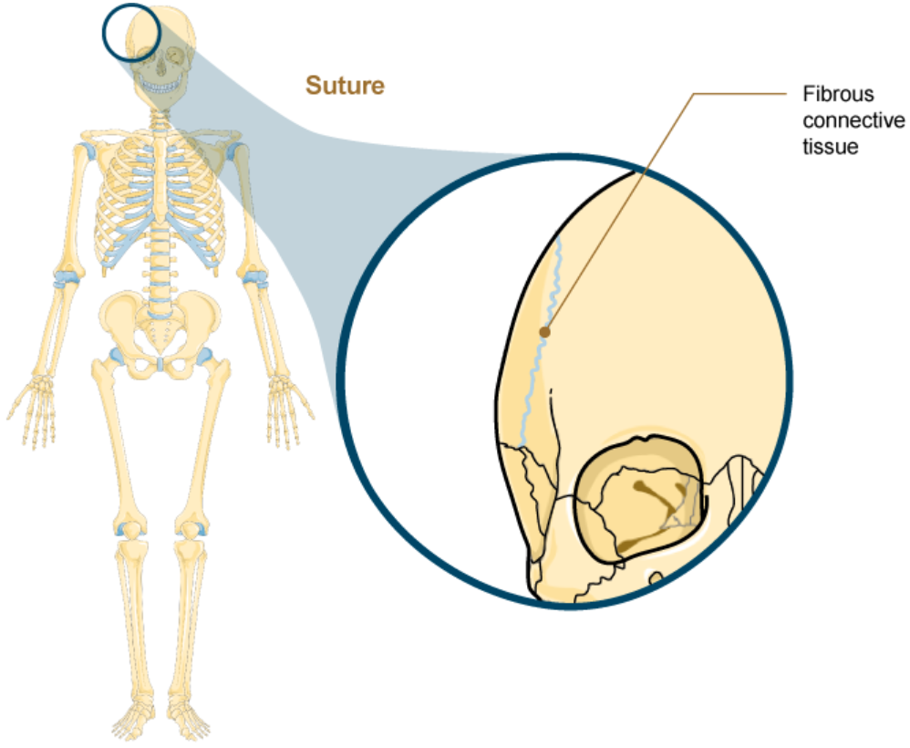
Suture
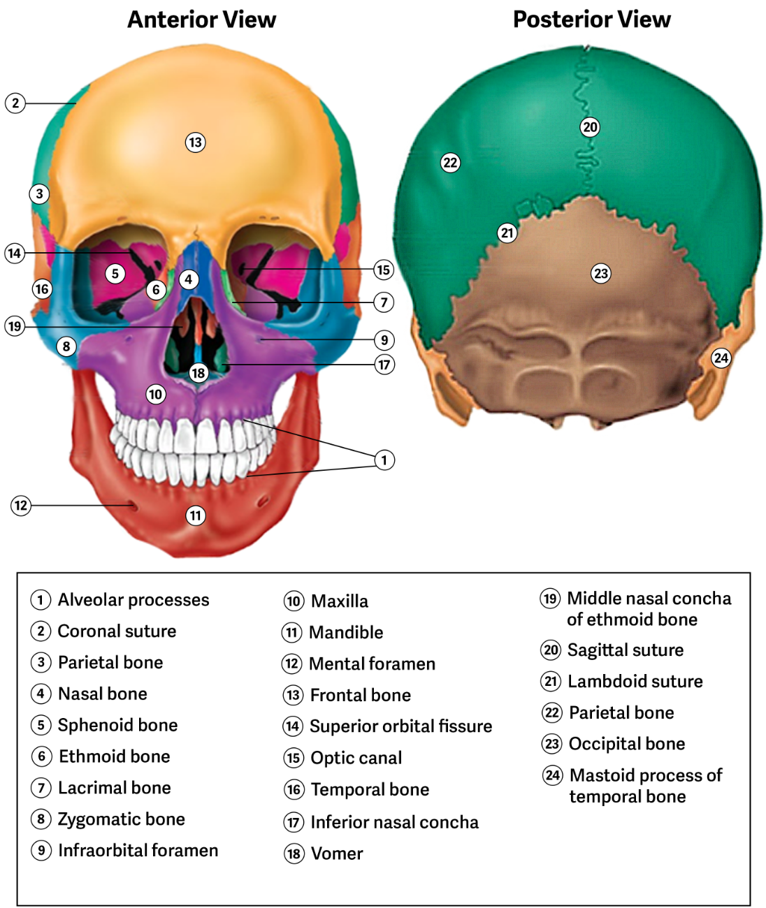
Fibrous joints between the bones of the skull.
A narrow strong fibrous connective tissue joint.
Surround and protect the brain.
All bones of the skull except the mandible joined together.
Strongly unites the adjacent skull bones.
Forms the face.
Synarthrosis Joint.
Undergo Synostosis.
Short Fibers.
Manubriosternal joint
Cartilaginous joints that unites the manubrium and body of the sternum
Protection of the heart.
Amphiarthrosis
Limited mobility, small amounts of movements.
Amphi - Both.
Ex. Intervertebral Disc and Pubic symphysis.
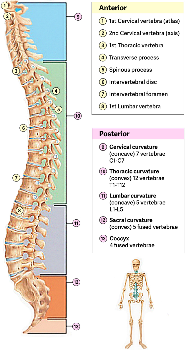
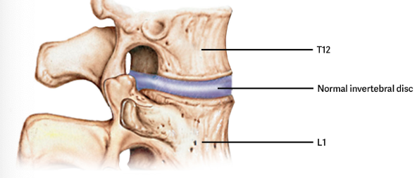
Intervertebral Disc
Cartilaginous joint in between each vertebrae
Thick pad of fibrocartilage.
Fills the gap between vertebrae.
Each disc units the vertebrae.
Allows limited amount of movement.
Small amounts of each each = large movement of the spinal column.
Intervertebral Symphysis
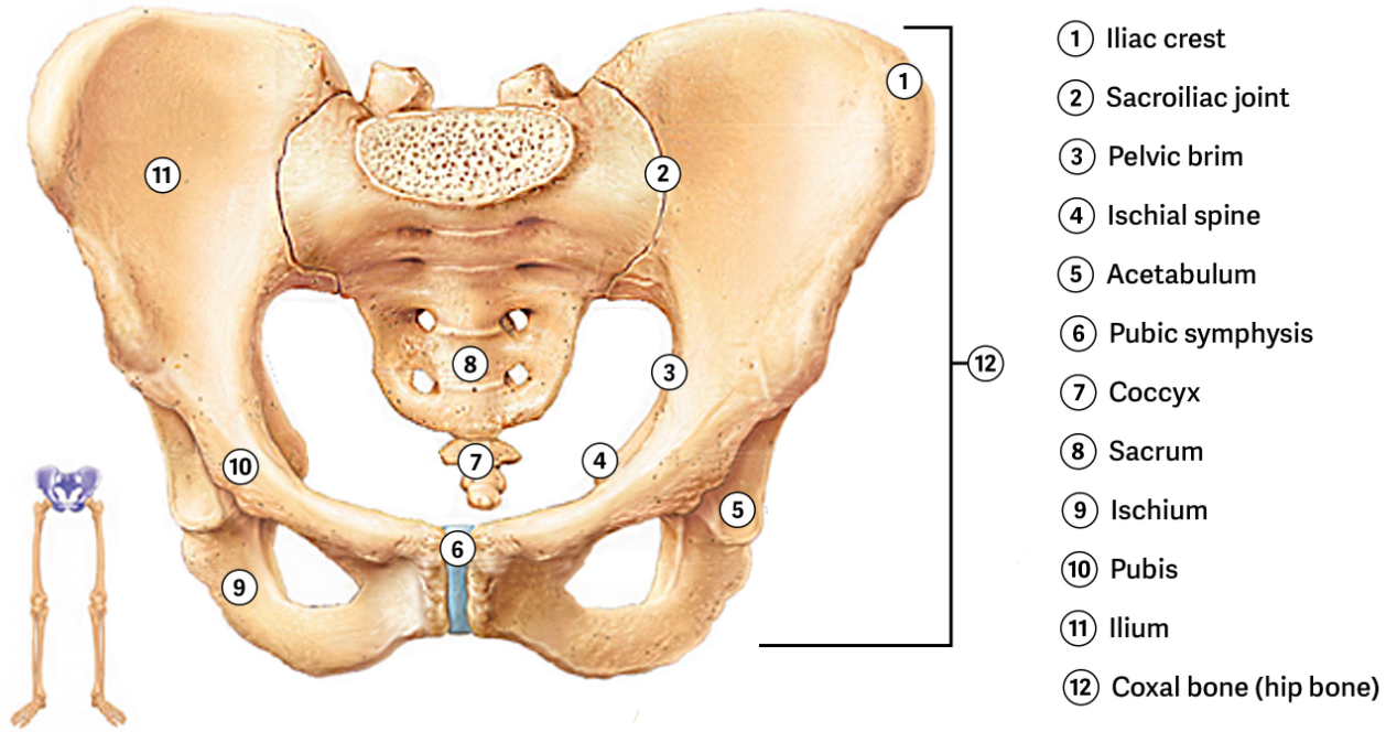
Pubic Symphysis
A cartilaginous joint between pubic regions of the right and left hip bones.
Strongly anchored to each other by fibrocartilage.
Little mobility (usually), but great strength.
Maintains stability of the body through the pelvis.
Diarthrosis
Freely mobile
Di - Apart.
All synovial Joints.
Most found in the appendicular skeleton.
Gives limbs wide range of motion.
Movement along an Axis.
Can also be described as types of motion available at a joint.
Ex. Linear, Angular, Rotation motion.
Axis
Movements in reference to the three anatomical planes
Transverse, Frontal, and Sagittal.
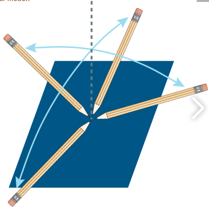
Linear Motion
Holding pencil upright, without security the point
Called gliding. (Point can move anywhere)
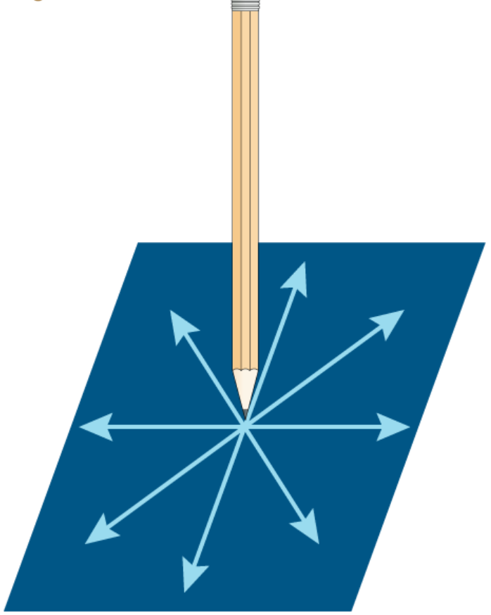
Angular Motion
Pencil point holds still.
Shaft can move
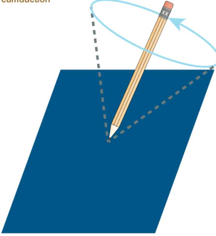
Ex. Circumduction
Circumduction Motion
More complex angular motion
Pencil point remains still, but eraser can move through a complete circle.
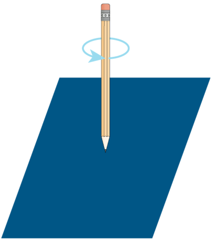
Rotation Motion
Keep pencil tip still, but rotate the shaft vertically around longitudinal axis.
Can also be classified as Uniaxial, Biaxial, and Multiaxial joints.
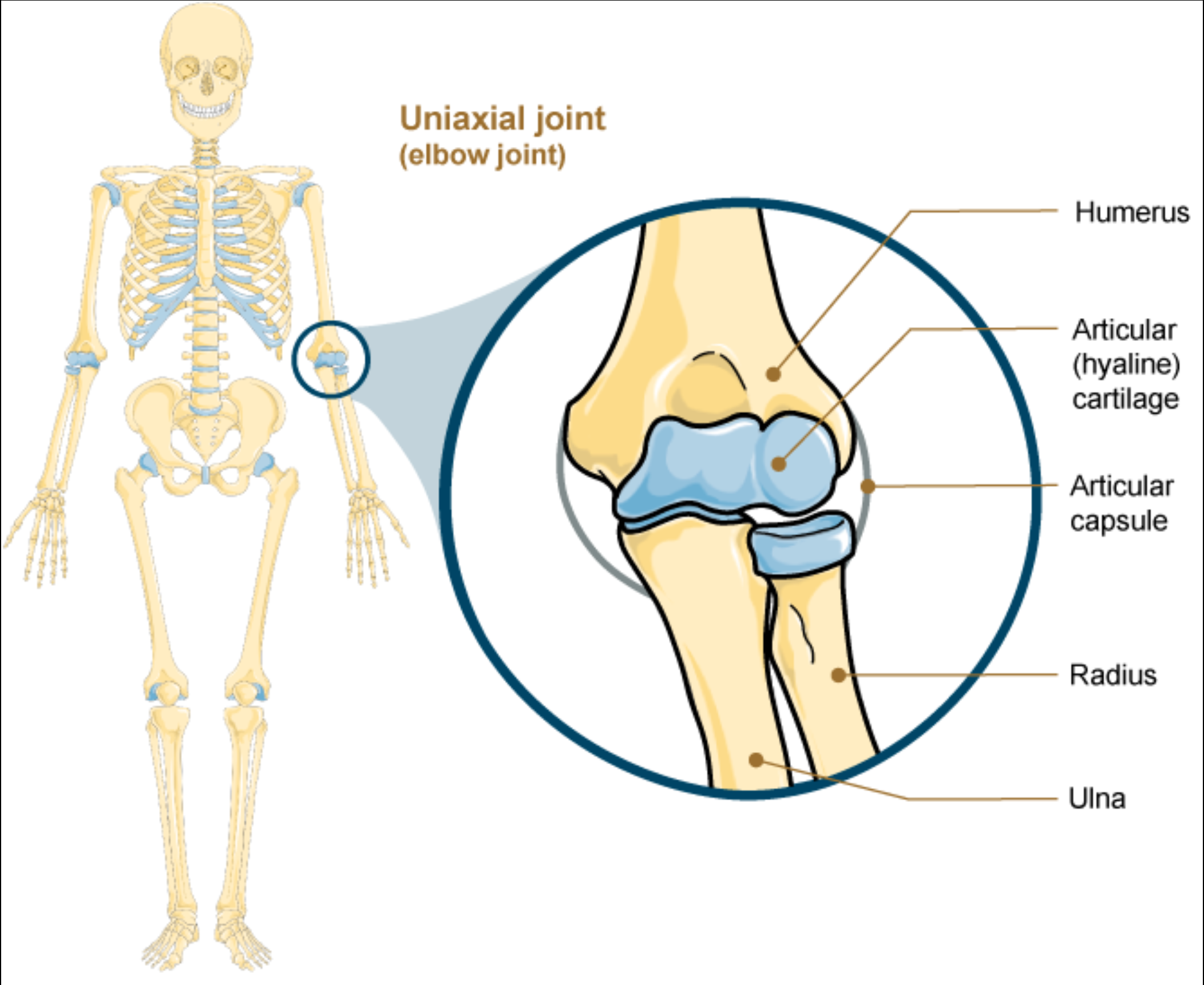
Uniaxial
Motion in one plane.
Only allow Flexion or Extension.
Ex. Elbow Joint and Knee Joint
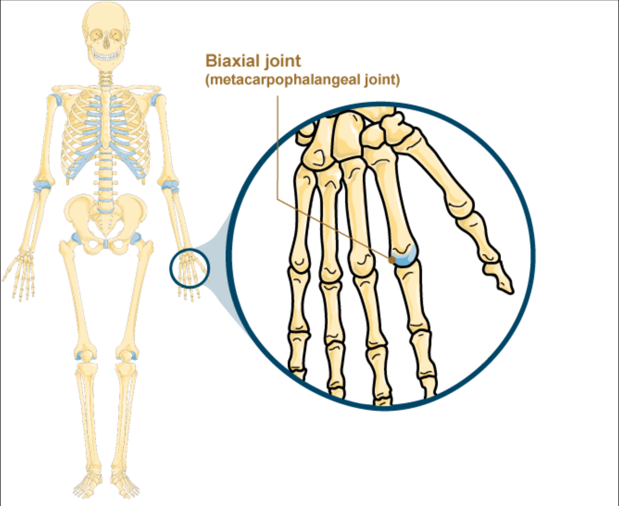
Biaxial
Motion in two planes
Axis 1 - Flexion/Extension.
Axis 2 - Spreading fingers away and bringing them together.
Ex. Metacarpophalangeal Joint.
Metacarpophalangeal Joint
Knuckles
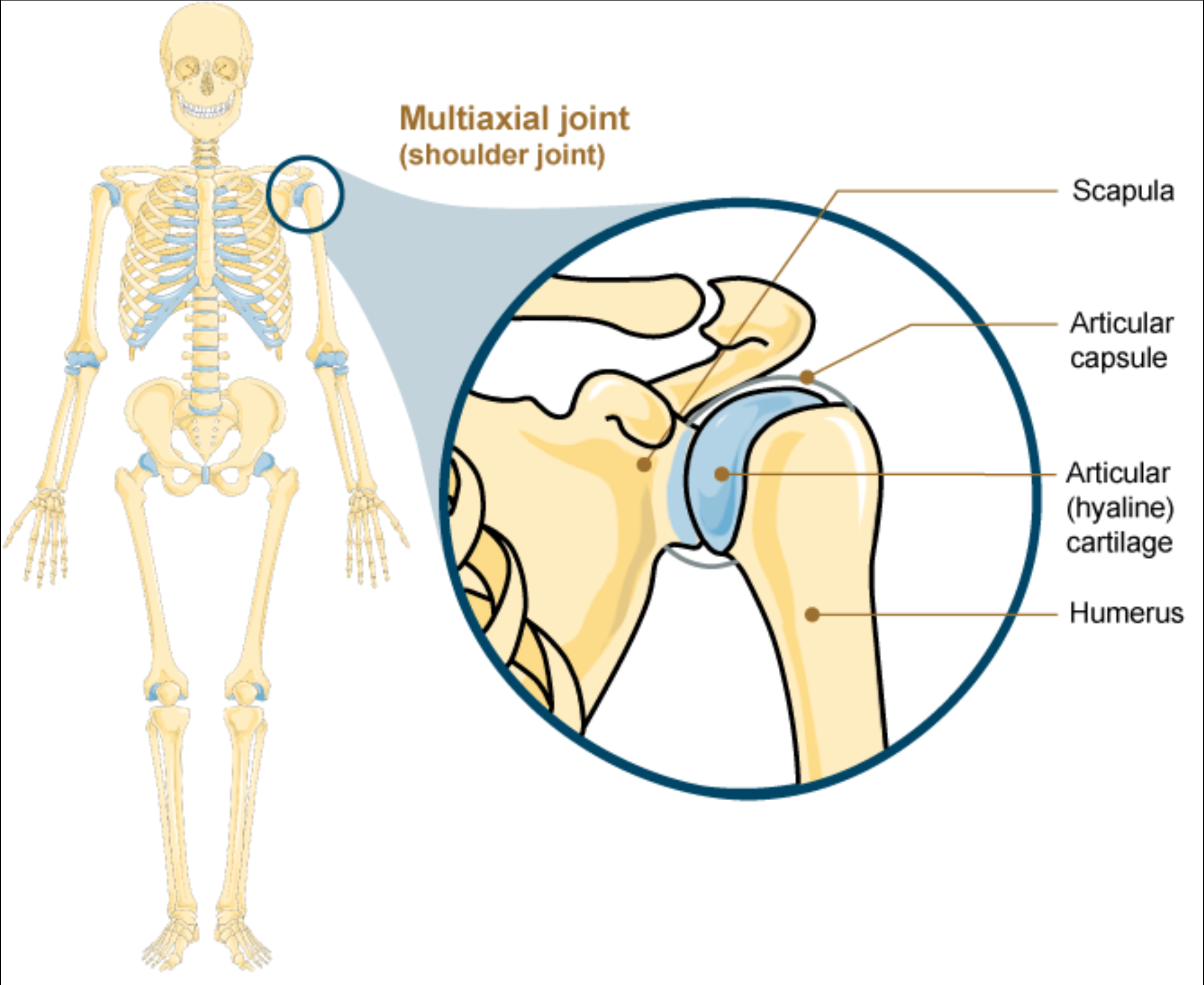
Multiaxial (triaxial)
Motion in three planes
Anterior-posterior direction
Medial-lateral direction.
Rotating around its long axis.
Ex. Shoulder and Hip
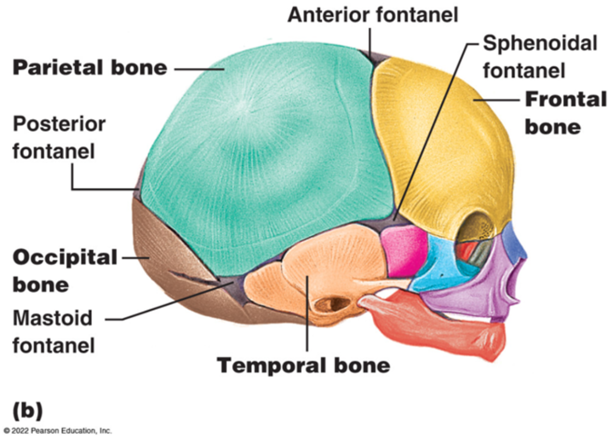
Fontanelles
Wide area between bones of connective tissue.
Provide flexibility through birth canal.
In Newborns and Infants.
Allow for rapid growth of the brain.
Decrease During the first year and closed by age 2.
Called sutures once gap is a narrow layer.
Synostosis
Joined by bone
At some sutures the connective tissue is converted to bone
Fusion of bone.
During adulthood and gradually disappear.
Ex. Sagittal, Coronal, and Lambdoid Sutures.
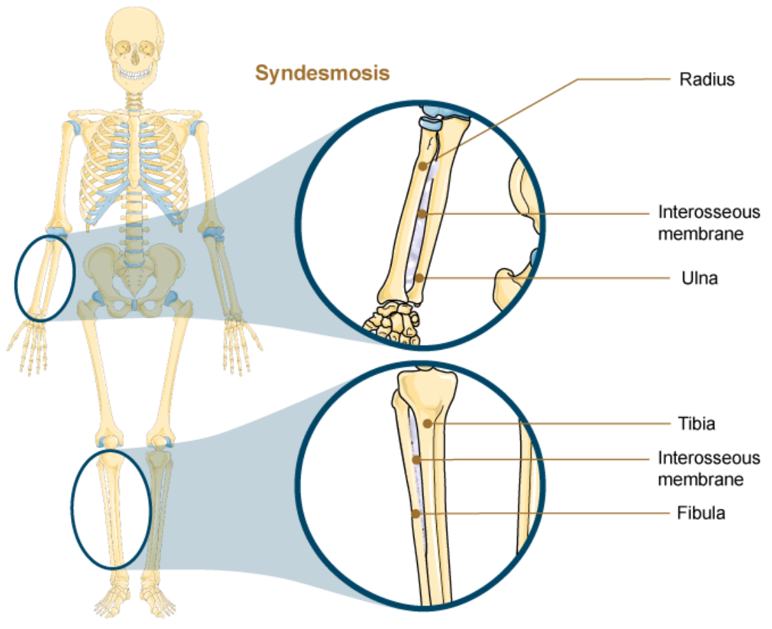
Syndesmosis
Two parallel bones joined by fibrous connective tissue.
Gap can be narrow, wide, or filled by Interosseous Membrane.
Bone joined by ligaments.
Amphiarthrosis Joint
Does not prevent all movement.
Damage to joint results from bone fracture.
Can tear Interosseous Membrane
If not properly immobilized muscles can pull bones out of proper alignment.
Long fibers.
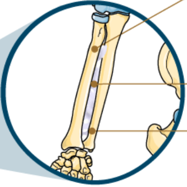
Interosseous Membrane
A wide gap filled in by a broad sheet of connective tissue.
Unites bones and prevents separation.
Allows for muscle attachment.
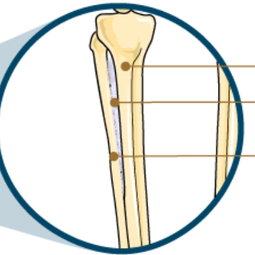
Ex. Between shaft of Radius and Ulna and Between Tibia and Fibula.
Interosseous Membrane of the Radius and Ulna
Flexibility of the membrane allows rotation of the radius (Supination/Pronation).
Interosseous Membrane of the Tibia and Fibula
Provides strength and stability for walking, standing, running.
Allows less movement.
Allows more muscle attachment.
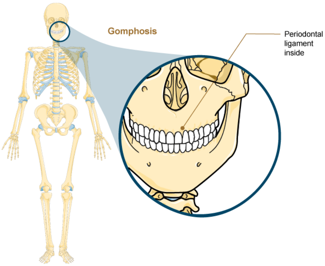
Gomphosis
Specialized fibrous joint that anchors the root of a tooth into it’s bony socket.
Within the maxillary or mandible bones.
Bony sockets contain Periodontal Ligaments.
A Synarthrosis Joint.
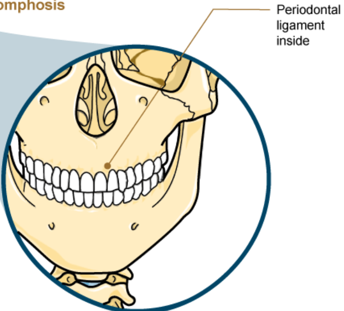
Periodontal Ligaments
Span between the bony walls of the socket and the root of the tooth.
Contains numerous shortt bands of dense connective tissue.
Synchondrosis
A cartilaginous joint where bones are joined directly by hyaline cartilage.
Synarthrosis Joint.
Ex. Temporary and Permanent.
Temporary Synchondrosis of Epiphyseal Plate
Region of growing bone = Hyaline Cartilage unites the Diaphysis to the Epiphysis.
Bone lengthening
Cartilage is replaced by bone.
Adds to the length of the bone.
During Puberty.
Bone lengthening slows as cartilage growth slows and stops.
Epiphyseal plate converted to bone.
Synostosis
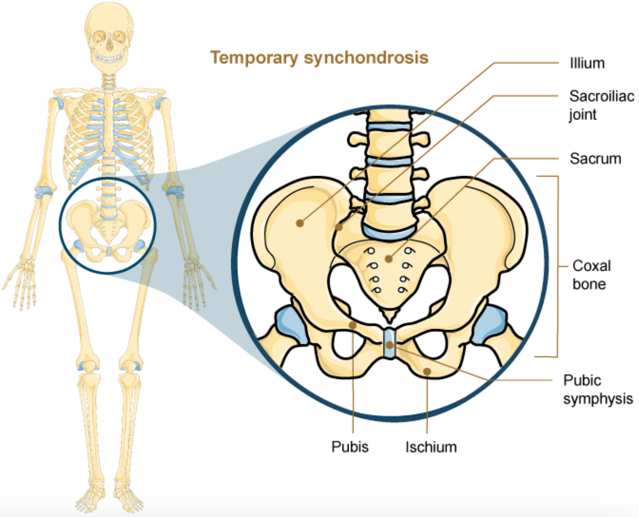
Temporary Synchondrosis of Formation of Coxal bones
Fusion of the bones of the ilium, ischium, and pubis into one adult bone.
Synostosis
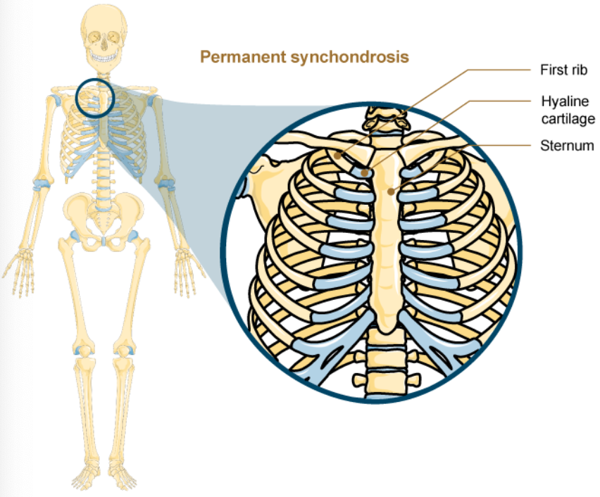
Permanent Synchondrosis of Rib Cage
First sternocostal joint. ( Between first rib and sternum )
First rib is anchored to the manubrium by costal cartilage.
Does not ossify. ( Stays as cartilage )
Retains hyaline cartilage with age.
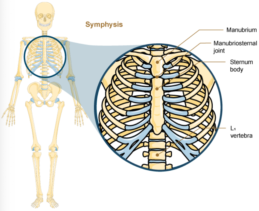
Symphysis
A cartilaginous joint where bones are joined by fibrocartilage.
Allows for a strong attachment between bones.
Bundles of thick collagen fibers.
Better to resist pulling and bending than hyaline cartilage.
Amphiarthrosis Joint
Ex. Pubic Symphysis, Manubriosternal Joint, and Intervertebral Symphysis.
Intervertebral Symphysis
A wide symphysis located between the bodies of adjacent vertebrae of the vertebral column.
Filled with Intervertebral Discs.
Important for carrying heavy objects, high impact activities. (Running/Jumping)
Manubriosternal Joint
A cartilaginous joint that joins the manubrium and body of the sternum by fibrocartilage.
Synarthrosis: Depending on degree of mobility.