HN220 Lab Midterm Possible Questions
1/58
There's no tags or description
Looks like no tags are added yet.
Name | Mastery | Learn | Test | Matching | Spaced |
|---|
No study sessions yet.
59 Terms
L1: List three signals that can be collected by Lt Labstation
EEG (Electroencephalogram)
ECG (Electrocardiogram)
EMG (Electromyogram)
L1: EEG (Electroencephalogram)
Measures electrical activity of the brain.
L1: ECG (Electrocardiogram)
Measures electrical activity of the heart.
L1: EMG (Electromyogram)
Measures electrical activity produced by skeletal muscles.
L1: What are the buttons on the data collection screen?
Delete
Select Point
Start Recording
Marker
Auto Scale
Compression
Comment
L1: Delete
Deletes the data collected during a session.
L1: Select Point
Allows the user to select specific points on the data graph for analysis.
L1: Start Recording
Begins the collection of data.
L1: Marker
Place a marker on the data graph for reference during analysis.
L1: Auto Scale
Adjusts the graph’s scale to fit the collected data for better visualization.
L1: Compression
Compacts data to fit more on the screen or into a smaller time frame.
L1: Comment
Lets the user add annotations or notes to specific parts of the data.
L1: When would you use a signal conditioner versus a transducer?
Transducer: Converts biological or physical signals (e.g., pressure, temperature) into an electrical signal.
Signal Conditioner: Processes the electrical signal (e.g., amplification, filtering) to make it more suitable for analysis or recording.
Transducer is biological whereas a signal conditioner adds extra processing to signal.
L1: Why is it important that the hand be in a supinated (palm-up) position when recording using the pulse transducer?
In a supinated position:
There is less gravitational force acting on the transducer.
Reduced pressure on the transducer ensures an accurate reading of pulsatile blood flow in arteries.
L1: What happens to the signal when the volunteer twitches their fingers?
Noise is introduced into the recording.
The signal becomes less reliable as the noise may mask the actual physiological data.
Finger twitching creates additional electrical and mechanical signals that interfere with the primary signal (e.g., pulse).
L1: What signal is each channel collecting?
This depends on the specific setup:
Channel 1: ECG (heart activity).
Channel 2: EMG (muscle activity).
Channel 3: EEG (brain activity) or pulse data (e.g., using a pulse transducer).
Ensure that each channel is calibrated and labeled properly during the experiment.
L2: You measure the membrane potential of a cell and find that it is -60mV. What does this mean?
It means that the inside of the cell is 60 millivolts more negative than the outside.
L2: Ion X has an equilibrium potential of -94mV. Use one or two sentences to explain what this really means.
An equilibrium potential of -94 mV for ion X means that at this membrane potential, the electrical and chemical (concentration) forces acting on ion X are balanced because they are at equilibrium. Hence this is why there is no net movement of the ion across the membrane.
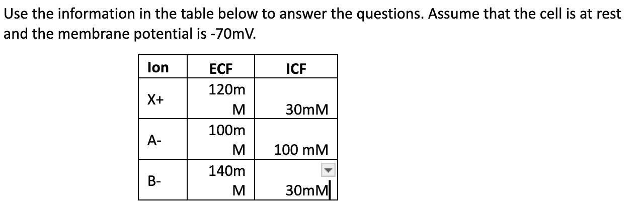
L2: Which ion(s) have a net chemical force acting to move the ion(s) into the cell?
Since X⁺ is more concentrated outside the cell (ECF=120>ICF=30), the chemical force will drive X⁺ into the cell.
Since the concentrations are equal inside and outside the cell (ECF=100=ICF=100), there is no net chemical force acting on A⁻. It is at equilibrium.
Since B⁻ is more concentrated outside the cell (ECF=140>ICF=30), the chemical force will drive B⁻ into the cell.
Therefore, the ions with a net chemical force acting to move them into the cell are X⁺ and B⁻.
L2: Which ion(s) have a net electrical force acting to move the ion(s) into the cell?
The membrane potential is -70 mV, meaning the inside of the cell is more negative relative to the outside. So, only ions charged positively can move into the cell.
X⁺ is positively charged so the negative membrane potential will attract X⁺ into the cell. A⁻ and B⁻ are both negatively charged so the negative membrane potential will repel A⁻ and B⁻, so the electrical force acts to move A⁻ out of the cell.
The ion with a net electrical force acting to move it into the cell would only be X⁺.
L2: Would the equilibrium potential for X+ be a positive or negative value?
The equilibrium potential for an ion depends on the direction of its concentration gradient. X⁺’s concentration is higher outside (120) than inside (30), so X⁺ tends to move into the cell down its concentration gradient (higher to lower always). To prevent this movement, the inside of the cell must be positive relative to the outside so the electrical driving force matches the chemical driving force, to repel further influx of X⁺. The equilibrium potential for X⁺ would be a positive value.
L2: If the concentration of sodium in the extracellular fluid increased, then the equilibrium potential would (become more positive / become less positive / remain the same)?
Concentration gradient always goes from higher concentration to lower concentration so if the ECF is increased, the ions from there would move inside the cell (ICF), increasing ICF concentration and because Na+ is positive, the equilibrium potential will become more positive inside the cell as more positive ions move into the cell.
L2: Assuming that for Na+ (higher concentration outside cell) , ENa+ = +60 mV and VM is -70 mV, what is the direction of the electrochemical gradient?
Equilibrium potential (ENa+ = +60 mV) This is the membrane potential at which there is no net movement of Na+ because the electrical and chemical forces are balanced; it is at equilibrium. Voltage movement (VM= -70 mV) which is the actual voltage across the membrane, indicating the inside of the cell is more negative than the outside.
Therefore, the chemical gradient drives Na+ into the cell. The membrane potential (-70 mV) is more negative than equilibrium potential (+60 mV) meaning the inside of the cell is very negative. Since sodium is positively charged, the electrical gradient also attracts Na+ into the cell. Both the chemical and electrical forces act in the same direction (into the cell). Thus, the electrochemical gradient drives Na+ into the cell.
L2: Anion Q has an equilibrium potential of -60 mV. Q is located in greater concentration (inside / outside) the cell. When a cell is at rest (-70 mV), the direction of the electrochemical force acting on Q is to move it (into / out of) the cell? If channels for Q suddenly opened, the membrane potential would become (more negative / less negative / or not change)?
Q is located in greater concentration outside the cell.
The electrochemical force acts to move Q out of the cell.
The membrane potential would become less negative.
L2: If a negatively charged ion is more concentrated outside the cell, the forces required to balance the chemical gradient would be directed ________. Thus, the equilibrium potential for this ion would be ________ charged.
outward : positively
outward : negatively
outward : neutrally
inward : negatively
inward : positively
outward : negatively
L2: An anion is found in greater concentration inside the cell than outside. Which of the following statements best describes forces acting on the anion at the resting membrane potential (-70 mV)?
Both the chemical and electrical forces are directed out of the cell.
The chemical force is directed out of the cell and the electrical force is directed into the cell.
Both the chemical and electrical forces are directed into the cell.
There is insufficient information to answer this question.
The chemical force is directed into the cell and the electrical force is directed out of the cell.
Both the chemical and electrical forces are directed out of the cell.
L3: What was the relationship between stimulus amplitude and response amplitude in the Evoked EMG activity?
As the stimulus amplitude increases, so does the response amplitude.
Response amplitude: The strength of the response measured in relation to the stimulus amplitude.
Stimulus amplitude: The strength or intensity of a stimulus applied to elicit a response.
L3: What is the physiological basis of the “stimulus amplitude increasing as the response amplitude increases” relationship? (What is actually happening in the body to cause the response, and why is the response changing as the stimulus strength changes)?
In the body: the neurons are reaching threshold and this elicits a response.
Why response increases as strength increases: as strength increase, more neurons in each nerve reach threshold, this elicits a greater response.
L3: In the Evoked EMG activity, what would the physiological explanation be for a stimulus that elicited no response?
Don’t reach the -55 action potential (all or nothing response).
L3: In the Evoked EMG activity, what would the physiological explanation be for multiple stimuli of varying strength eliciting roughly the same strength of response to each stimulus?
If ALL of the muscle fibers were stimulated (and all of the neurons as well) the response strength would no longer increase because it has reached its max
All motor neurons have been activated…no more to be activated
Within the muscle fiber, you have many neurons. Once you have elicited a response in every single one, the response will plateau because every muscle fiber has been stimulated, and there are no more to increase the response. The only way you could get a bigger response at this point would be to build up more muscle.

L4: What must happen for your participant to feel two distinct points of contact?
In order for a person to feel two points, two separate neurons must be stimulated by their respective receptive fields. When this happens, two points are felt.
L4: What are the physiological properties of the receptive fields in the area with the highest two-point threshold (smallest distance) vs. the lowest two-point threshold (largest distance)?
The two points of contact must be far enough apart to be perceived as separate. This minimum distance is known as the two-point discrimination threshold, which varies across different body areas (e.g., fingertips have a much lower threshold than the back or thighs).
Smaller receptive fields = smaller distance between two points threshold
Larger receptive fields = larger distance between the two points threshold
Receptive fields
Areas of sensory neurons that respond to stimuli within a specific region.
L4: You tap on the subject’s patella ligament with a tendon tap hammer. List the physiological responses that occur to elicit a slight extension of the leg (make sure to name specific structures).
When you tap the patellar ligament, it stretches the quadriceps muscle, activating muscle spindles (stretch receptors).

The monosynaptic patellar reflex.
(1) a tap to the patellar tendon stretches the quadriceps muscle
(2) resulting in activation of the muscle spindle
(3) the primary afferent neuron of the muscle spindle, detecting stretch, sends a signal to the spinal cord
(4) the primary afferent neuron synapses/stimulates directly with a motor neuron to extensor muscle
(5) motor neuron causes the quadriceps muscle to contract
In parallel, an inhibitory message is sent via an interneuron to the hamstrings (6) primary afferent neuron stimulates inhibitory interneuron
(7) interneuron inhibits motor neuron to flexor muscle
(8) resulting in hamstrings relaxation
The reflex at the hamstrings is polysynaptic.
Monosynaptic reflex
A reflex that involves a single synapse between a sensory neuron and a motor neuron.
Polysynaptic reflex
A reflex that involves one or more interneurons in addition to the sensory and motor neurons.
Muscle spindles
Stretch receptors located within the belly of muscles that detect changes in muscle length.
Latency period
The time it takes for a nerve signal to travel from the point of stimulation to the muscle contraction.
Patellar reflex
A monosynaptic reflex that causes slight extension of the leg when the patellar tendon is tapped.
Afferent neuron
A neuron that carries sensory signals to the central nervous system.
Interneuron
A neuron that transmits impulses between other neurons, often within the central nervous system.
What action do quadriceps do?
Extend the leg
What action do hamstrings do?
Flex the knee
Motor neuron
A neuron that transmits impulses from the central nervous system to muscles.
Reflex
An involuntary and nearly instantaneous movement in response to a stimulus.
L4: During the Knee-jerk reflex exercise, what does latency indicate about what is happening in the body?
The latency period indicates the time it takes for the nerve signal to travel from the point of stimulation (the tap on the patellar tendon) to the muscle contraction, indicating how quickly the nervous system processes and responds to stimuli, neural conduction speed, synaptic efficiency and muscle activation.
Normal function of knee-jerk reflex
A short latency indicates a healthy reflex arc, where the signal travels quickly through the sensory neuron to the spinal cord and back to the motor neuron, resulting in a prompt muscle contraction.
Since the knee-jerk reflex is monosynaptic, it has minimal delay, making it one of the fastest reflexes in the body.
Increased latency?
A longer latency could suggest damage to the sensory or motor neurons involved in the reflex pathway, potential nerve compression, or issues with the spinal cord itself. This reflex plays a crucial role in maintaining posture and balance.
Absent reflex?
If there is no knee-jerk response at all, it could indicate a more severe neurological problem affecting the reflex arc.
L4: While performing the Jendrassik manoeuvre does the participant’s latency period change? Why did or why didn’t it change?
The latency period of the knee-jerk reflex does not significantly change during the Jendrassik maneuver, but the strength of the reflex response increases.
The latency period does not change because of the fixed neural pathway:
The knee-jerk reflex is a monosynaptic reflex arc, meaning the signal travels directly from sensory neurons to motor neurons in the spinal cord without extra synapses. Since the conduction velocity of neurons remains constant, the time required for signal transmission and muscle activation does not change. The Jendrassik maneuver primarily affects reflex strength rather than signal transmission speed.The spinal cord still processes the reflex at the same rate, so the latency remains the same.
Why Does Reflex Strength Increase?
The Jendrassik maneuver suppresses inhibitory signals from the brain to the spinal cord. Normally, the brain sends inhibitory signals to regulate reflexes. By engaging in the maneuver, the brain's focus shifts, reducing its ability to dampen the reflex. As a result, more motor neurons fire, leading to a stronger quadriceps contraction and a more pronounced leg extension.
L4: During the knee-jerk reflex exercise, what does the response amplitude indicate about what is happening in the body?
The response amplitude indicates the strength of the muscle contraction. This indicates the overall health of the neural pathway involved in the reflex; a larger amplitude means that there is a stronger muscle contraction, that indicate exaggerated reflex (hyperreflexia) that could mean that there may be damage to the central nervous system, while a smaller amplitude could suggest a diminished reflex (hyporeflexia) which could mean there is nerve damage along the reflex arc.
Exaggerated reflex
A larger amplitude means a stronger muscle contraction, indicating exaggerated reflex (hyperreflexia) that could mean damage to the central nervous system.
Diminished reflex
A smaller amplitude could suggest a diminished reflex (hyporeflexia), which could mean there is nerve damage along the reflex arc.
L4: While performing the Jendrassik maneuver does the participant’s response amplitude change? Why did or why didn’t it change?
Yes, during this maneuver, the participant's response amplitude will increase while the latency stays the same. The Jendrassik maneuver will heighten (exaggerate) the patellar (knee-jerk) reflex by countering some of the normal descending inhibitory brainstem inputs to reflex arc interneurons.
L5: What is the relationship between the Stimulus Strength and the Twitch Peak Force according to your data in the twitch response and recruitment exercise?
The data from the twitch response and recruitment exercise typically shows a direct relationship between stimulus strength and twitch peak force. As stimulus strength ↑, the twitch peak force also ↑, but only up to a certain point. Initially, small stimulus strengths result in low-force responses, but as stimulus intensity rises, more motor units are recruited, leading to a higher peak force. Once all available motor units are recruited, further increases in stimulus strength do not significantly increase force output. At this point, the force plateaus or fluctuates slightly, indicating that maximal motor unit recruitment has been reached, and additional stimulation cannot generate a stronger contraction.
L5: What is the physiological basis of this relationship in the twitch response and recruitment exercise? (What is happening in the body to cause the response, and why is the response changing as the stimulus strength changes)?
The physiological reason behind this relationship lies in motor unit recruitment. When a stimulus is applied:
Low-intensity stimuli activate only small, low-threshold motor units composed of slow-twitch (Type I) fibers → fewer motor units are recruited→ leading to lower force output.
As stimulus strength increases, larger motor units with fast-twitch (Type II) fibers→ more motor units are activated→ leading to a greater twitch force.
Once maximal recruitment is achieved, all motor units in the muscle are firing so all available motor units are recruited, causing a plateau where additional stimulus strength does not further increase force because there are no additional motor units left to activate.
This follows Henneman’s Size Principle, where small motor units (slow-twitch fibers) are recruited first, followed by larger motor units (fast-twitch fibers) as needed.
L5: During data collection, at the lower stimulus strengths, did you have any occurrences where you applied a stimulus, but no response was observed? What is the physiological reason for this?
Yes, this occurs because the applied stimulus does not reach the minimum threshold potential → the stimulus is too weak; the neuron’s membrane potential does not depolarize enough to reach this threshold → so motor neurons are not depolarized → they cannot trigger an action potential → without an action potential, no muscle contraction occurs → no twitch response.
During data collection, at the higher stimulus strengths, did you have any occurrences where multiple stimuli of varying strength elicited roughly the same response? What is the physiological reason for this?
Yes, at higher stimulus strengths, multiple stimuli resulted in similar peak forces.
Once all motor units in a muscle are activated/recruited → the muscle has reached its maximum contractile capacity→ even if the stimulus strength/intensity increases → the muscle’s maximum force output has already been reached → results in a force plateau, where additional electrical input does not translate to a stronger contraction.
L5: According to the Summation Exercise, why does the peak force of the 2nd force response increase as the time between stimulation pulses decreases? Specifically, what happens within the muscle fiber to make the second and third responses bigger than the first response?
When two or more stimuli are applied in close succession, the peak force of the second response increases because of temporal (wave) summation.
A single stimulus leads to a muscle twitch, during which calcium ions (Ca²⁺) are released from the sarcoplasmic reticulum and initiate contraction.
If a second stimulus occurs before complete relaxation of the first twitch, residual Ca²⁺ remains in the sarcoplasm → greater cross-bridge formation → results in wave summation: successive stimuli build upon each other (Ca²⁺ levels accumulate) which prevents full relaxation and actually allows the muscle fibers to generate greater force with each successive contraction (2nd and 3rd contraction).