A&P The Brain Nervous System
5.0(1)
5.0(1)
Card Sorting
1/23
Earn XP
Description and Tags
A&P Exam
Study Analytics
Name | Mastery | Learn | Test | Matching | Spaced |
|---|
No study sessions yet.
24 Terms
1
New cards
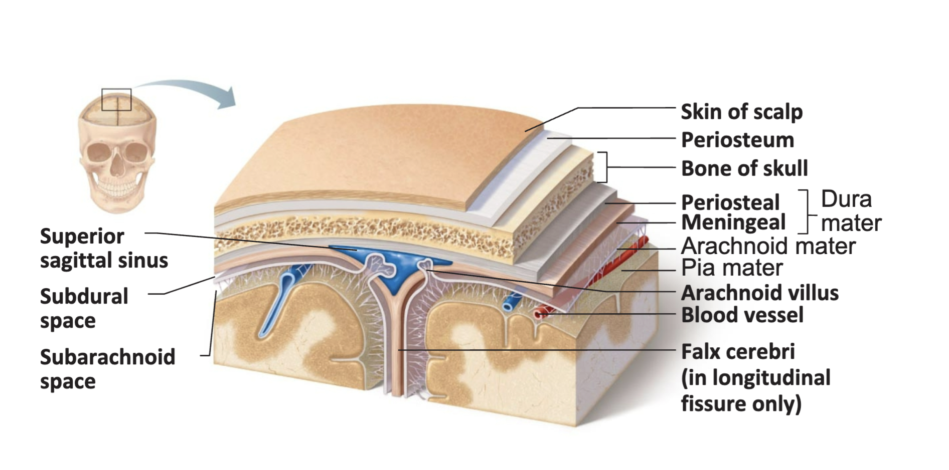
How brain is protected? List the structures protect your brain (from outside to inside)
• Bone (skull) \n
• Membranes (meninges)
-dura mater
-arachnoid mater
-pia mater \n
• Watery cushion (cerebrospinal fluid) \n • Blood-brain barrier
• Membranes (meninges)
-dura mater
-arachnoid mater
-pia mater \n
• Watery cushion (cerebrospinal fluid) \n • Blood-brain barrier
2
New cards
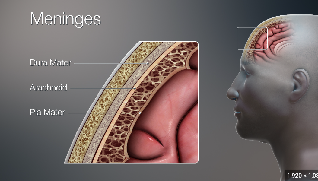
Know about three meninges. What happens in hematoma, in which space CSF is present?
\
* **Dura Mater** \n • Strongest outermost meninx
* **Arachnoid Mater** \n • Separated from the dura mater by the subdural space \n • __*Subarachnoid space contains CSF*__ and blood vessels
* **Pia Mater** \n • Layer of delicate connective tissue that clings tightly to the brain
\-A subdural __hematoma__ is a blood clot that forms between layers in the protective coverings of the brain (meninges), when veins tear as a result of sudden movement of the brain against the skull.
3
New cards
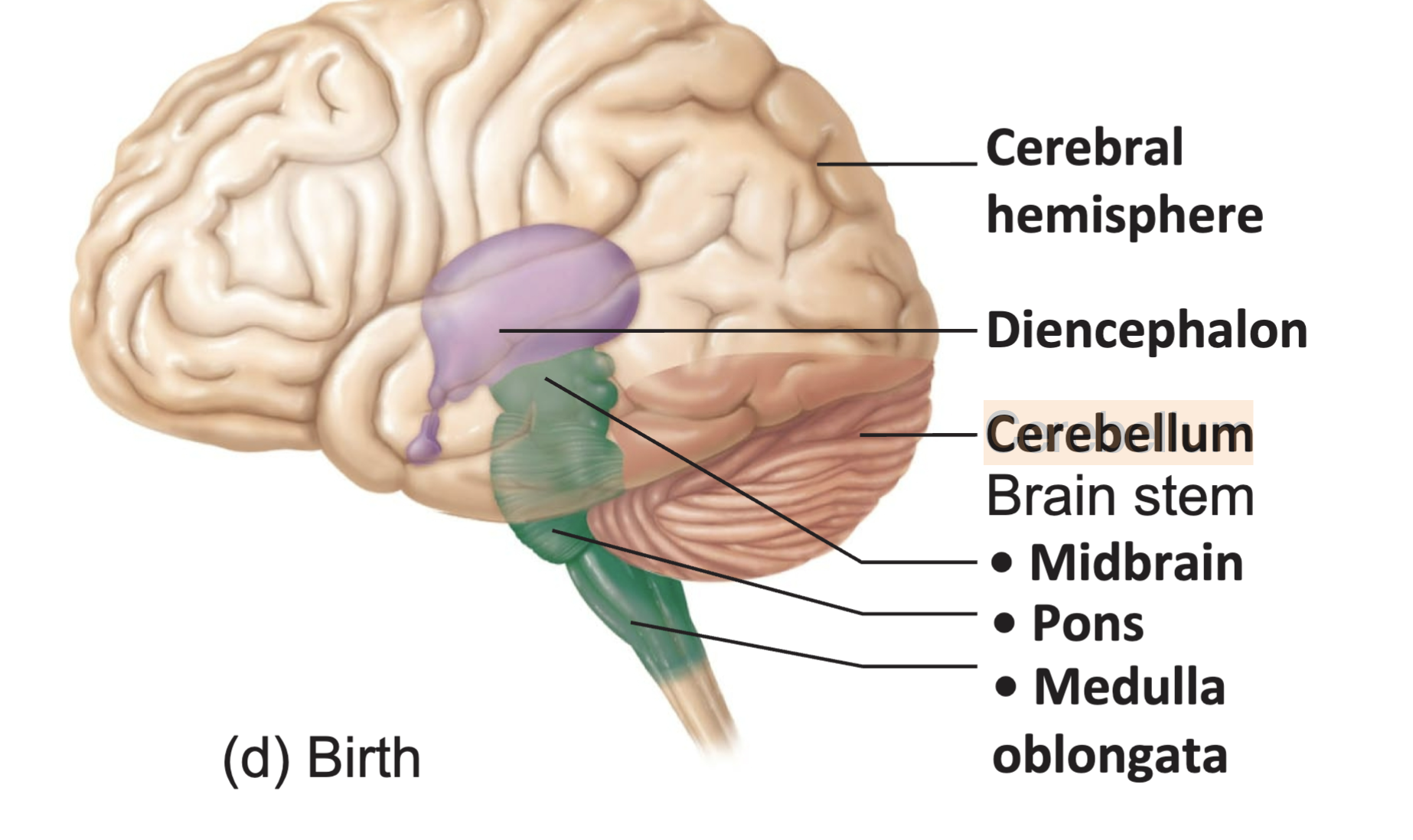
Four main parts of the brain
1. Brain stem
2. Cerebrum
3. Diencephalon
4. Cerebellum
4
New cards
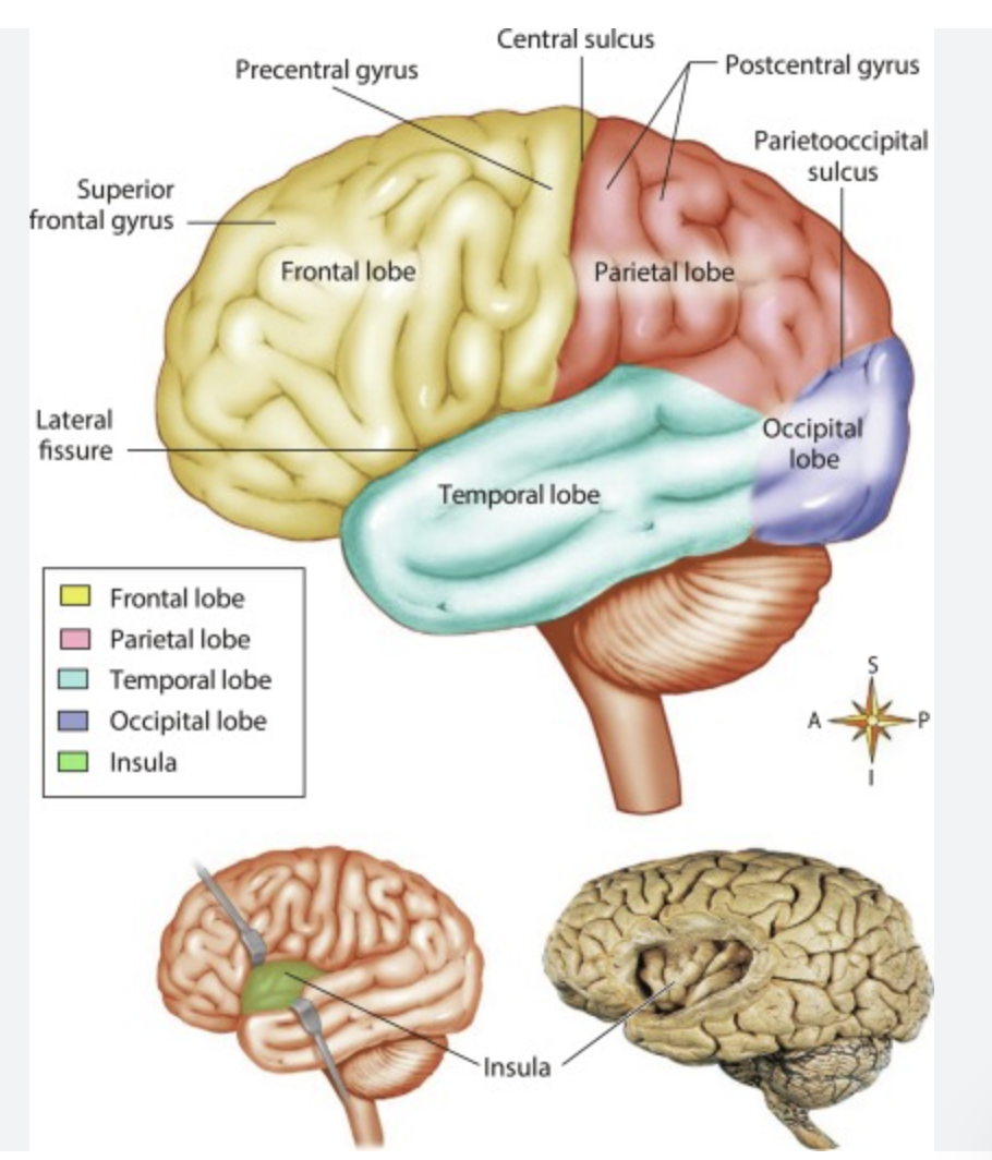
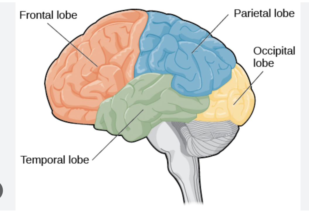
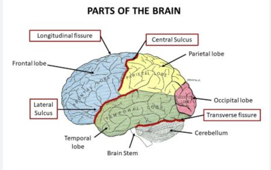
parts of cerebrum
frontal lobe, parietal love, occipital lobe, temporal lobe,
5
New cards
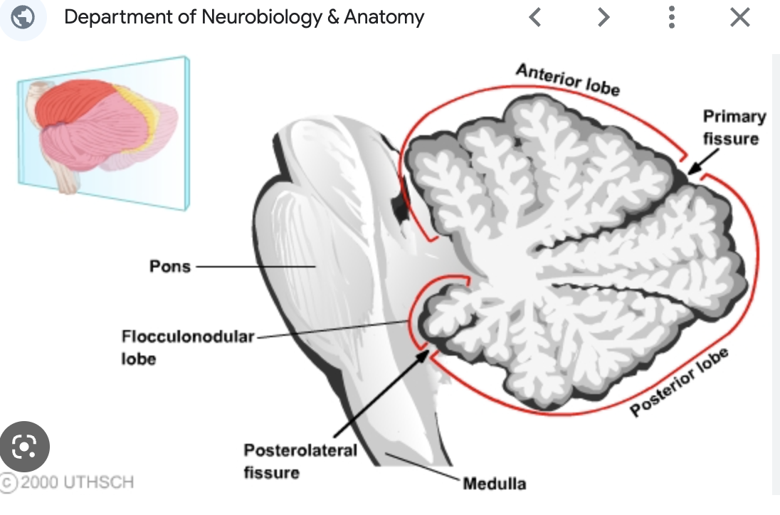
parts of cerebellum
There are three anatomical lobes that can be distinguished in the cerebellum; the anterior lobe, the posterior lobe and the flocculonodular lobe. These lobes are divided by two fissures - the primary fissure and posterolateral fissure.
6
New cards
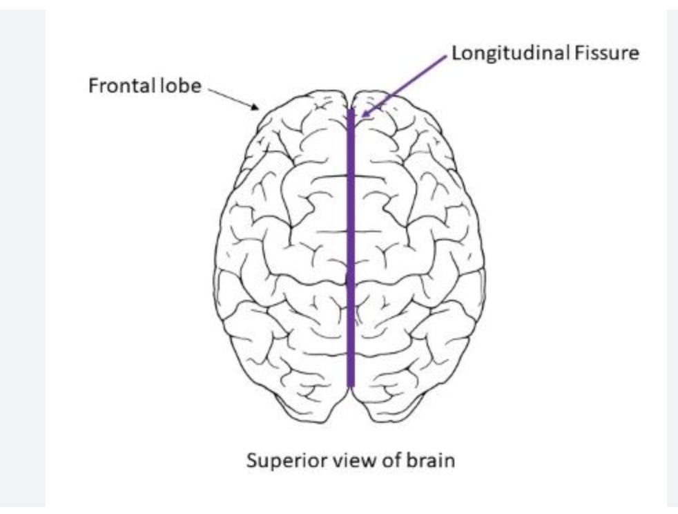
How the hemispheres are connected, composition of cerebral cortex, sulci, fissures
* A fissure or groove that separates the two hemispheres is called the great longitudinal fissure. The two sides of the brain are **joined at the bottom by the corpus callosum.**
* **Sucli**: a groove or furrow, especially one on the surface of the brain.
* **Fissure**: A fissure is a deeper grove and is often used interchangeably with sulcus. The cerebrum is divided into a left and right hemisphere by a longitudinal fissure that goes by many different names: longitudinal fissure, cerebral fissure, median longitudinal fissure, interhemispheric fissure.
* **Cerebral cortex:** The **cerebral cortex** is the thin outermost layer of the cerebral hemisphere. It is the main control center and information processing unit.
7
New cards
Four lobes in each hemisphere
frontal lobe, parietal lobe, temporal lobe, occipital lobe
8
New cards
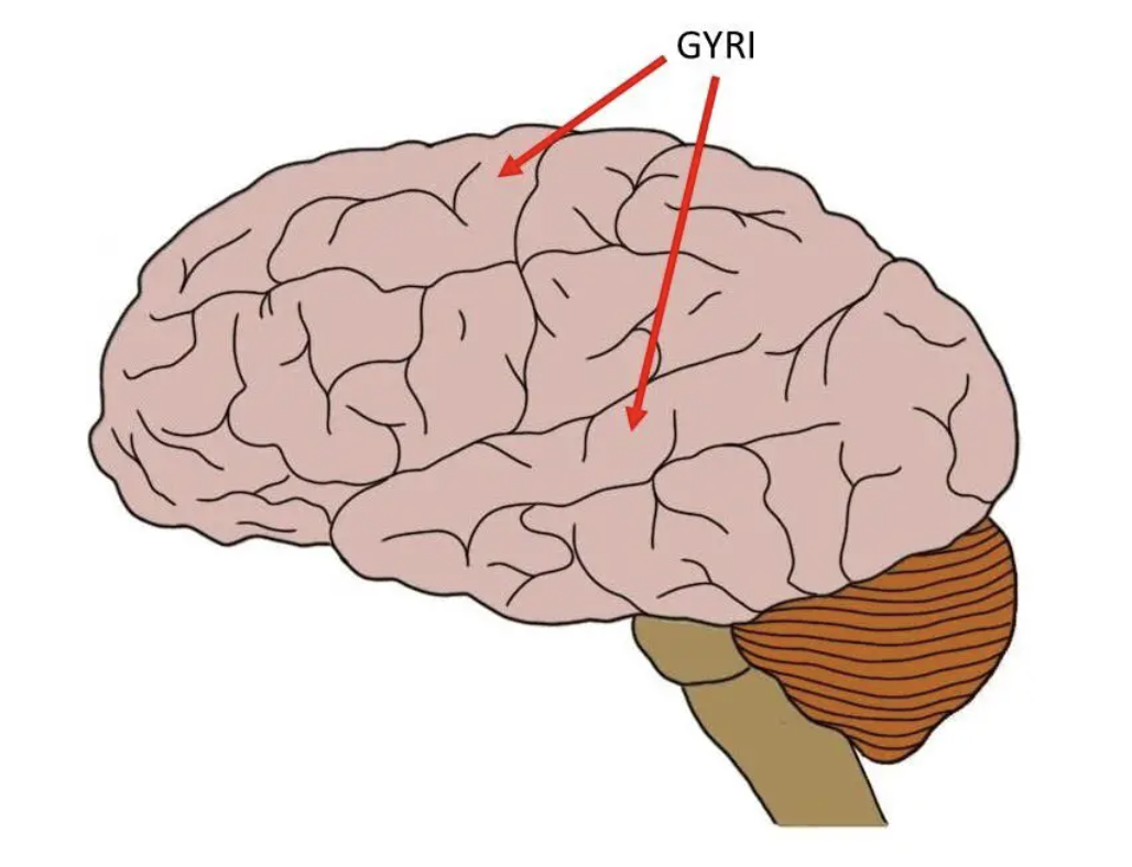
Gyri
a ridge on the surface of the brain
\-they increase the surface area of the brain
\-they increase the surface area of the brain
9
New cards
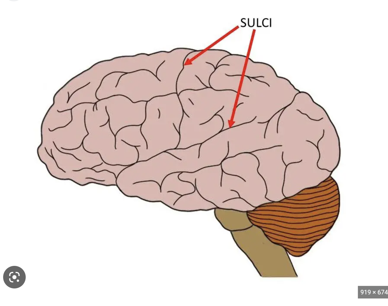
Sulci
a groove or furrow, especially one on the surface of the brain.
10
New cards
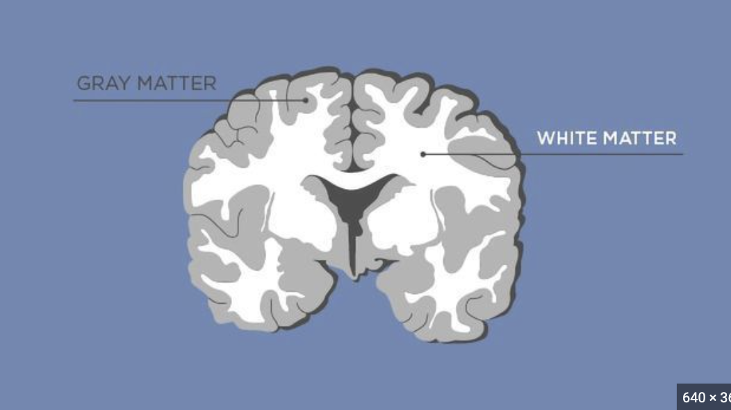
Grey matter & White matter
The tissue called "gray matter" in the brain and spinal cord is also known as substantia grisea, and is made up of cell bodies. "White matter", or substantia alba, is composed of nerve fibers.
11
New cards
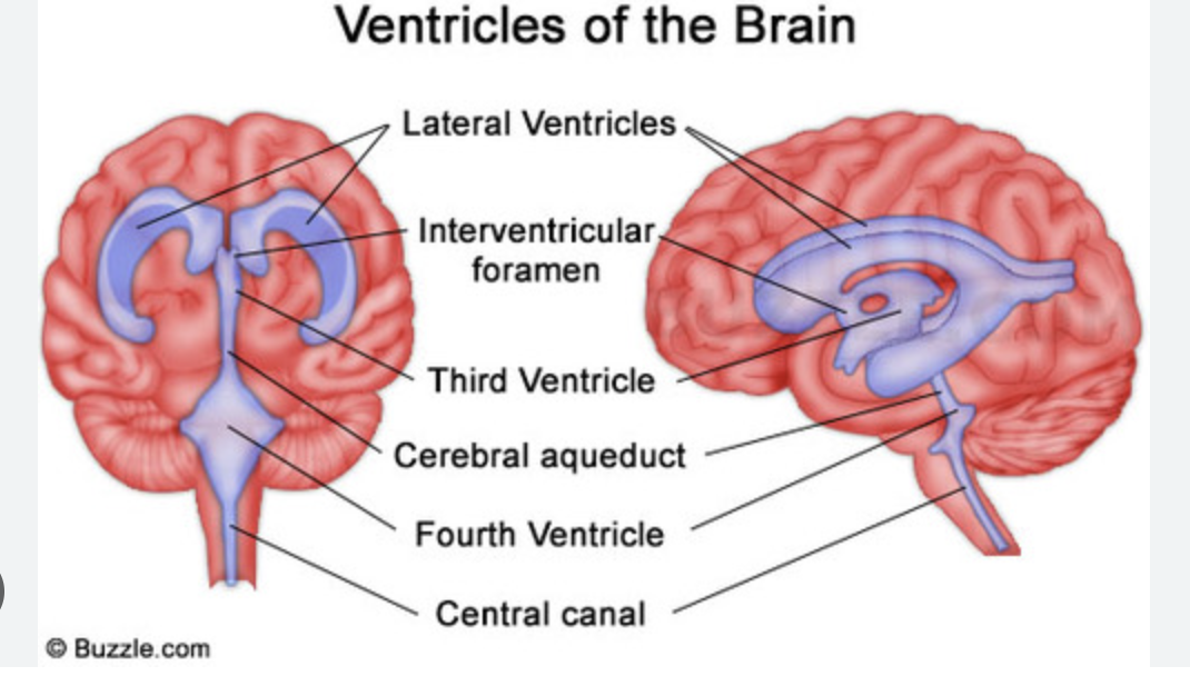
Ventricles and how they are connected.
The 2 interventricular foramens (or foramina of Monro) connect the lateral ventricles with the third ventricle.
12
New cards
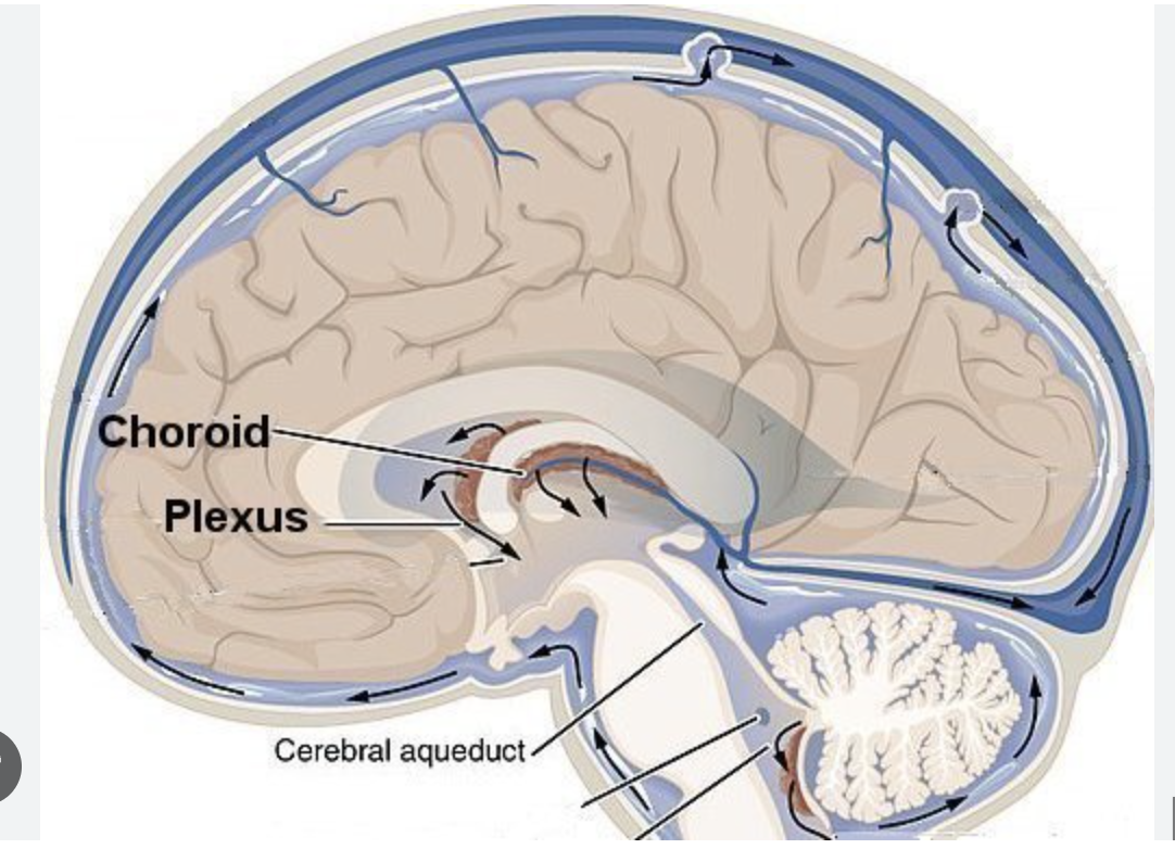
what is choroid plexus?
The choroid plexus is a complex network of capillaries lined by specialized cells and has various functions. One of the primary functions is to produce cerebrospinal fluid (CSF) via the ependymal cells that line the ventricles of the brain.
13
New cards
Function of CSF
**Cerebrospinal fluid (CSF)** is made by tissue that lines the ventricles (hollow spaces) in the brain. It flows in and around the brain and spinal cord to help cushion them from injury and provide nutrients.
14
New cards
What is hydrocephalus?
### __***A build-up of fluid in the cavities deep within the brain.***__
* The extra fluid puts pressure on the brain and can cause brain damage. It's most common in infants and older adults.
* Hydrocephalus is characterized by head enlargement in infants. Adults and older children experience headache, impaired vision, cognitive difficulties, loss of coordination, and incontinence.
* Treatment is often a tube (shunt) inserted surgically into a ventricle to drain excess fluid.
15
New cards
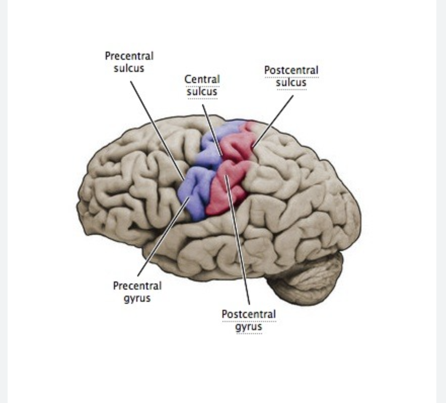
Central sulcus- precentral and post central gyri and their functions
* **The central sulcus:** separates the parietal lobe and the frontal lobe.
* **Precentral gyri:** controls voluntary motor movements
* **Postcentral gyri:** controls involuntary functions
* **Precentral gyri:** controls voluntary motor movements
* **Postcentral gyri:** controls involuntary functions
16
New cards
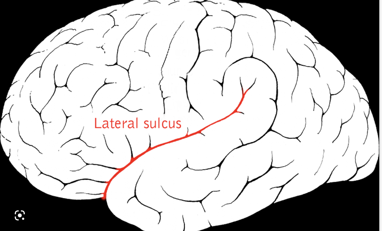
Lateral sulcus
The lateral sulcus (Sylvian fissure) is a very deep fold seen on the lateral surface of the hemisphere running in an anterior to posterior direction and serves to separate the temporal lobe from the frontal and parietal lobes.
17
New cards
Transverse fissure
divides the cerebrum from the cerebellum
18
New cards
longitudinal fissure
divides the cerebrum into two hemispheres.
19
New cards
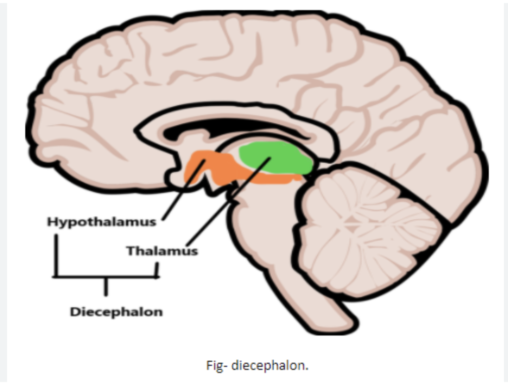
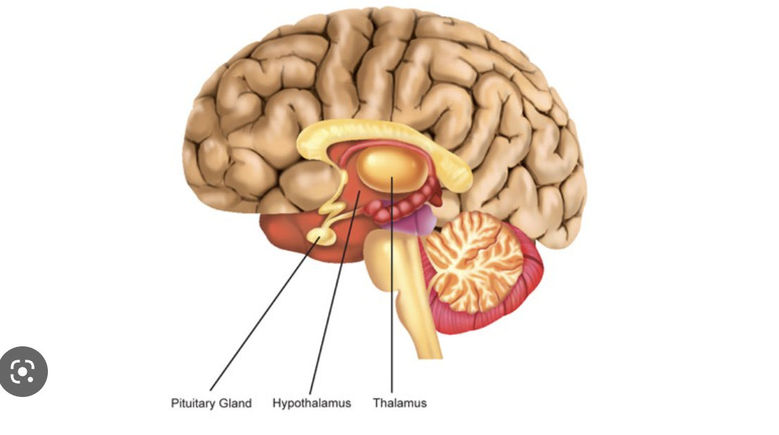
Parts of a diencephalon
The diencephalon connects the midbrain to the forebrain. It is located deep within the brain and comprises the **epithalamus, thalamus, subthalamus and hypothalamus.**
20
New cards
Know the functions of the thalamus, hypothalamus, pituitary gland
**Thalamus**: carries messages from the sensory organs like the eyes, ears, nose, and fingers to the cortex
**Hypothalamus**: controls your pulse, thirst, appetite, sleep patterns, and other processes in your body that happen automatically.
**Pituitary Gland:** regulates growth, metabolism, and reproduction through the hormones that it produces.
**Hypothalamus**: controls your pulse, thirst, appetite, sleep patterns, and other processes in your body that happen automatically.
**Pituitary Gland:** regulates growth, metabolism, and reproduction through the hormones that it produces.
21
New cards
Function and location of the cerebellum
The cerebellum is important for making postural adjustments in order to **maintain balance.**
22
New cards
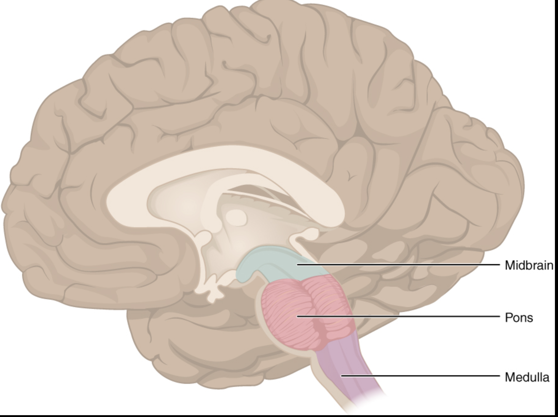
Location and parts of the brainstem and important functions of the brainstem
* **Midbrain**: The top part of the brainstem is crucial for regulating eye movements.
* **Pons**: The middle portion of the brainstem coordinates facial movements, hearing and balance.
* **Medulla oblongata**: The bottom part of the brainstem helps regulate your breathing, heart rhythms, blood pressure and swallowing.
* **Pons**: The middle portion of the brainstem coordinates facial movements, hearing and balance.
* **Medulla oblongata**: The bottom part of the brainstem helps regulate your breathing, heart rhythms, blood pressure and swallowing.
23
New cards
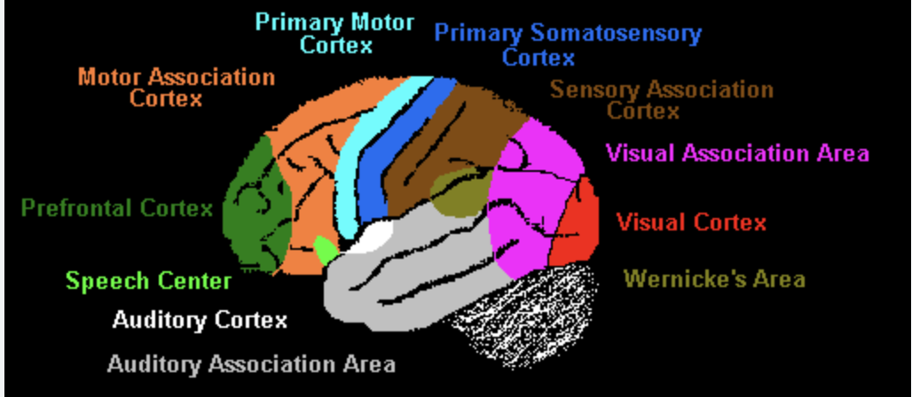
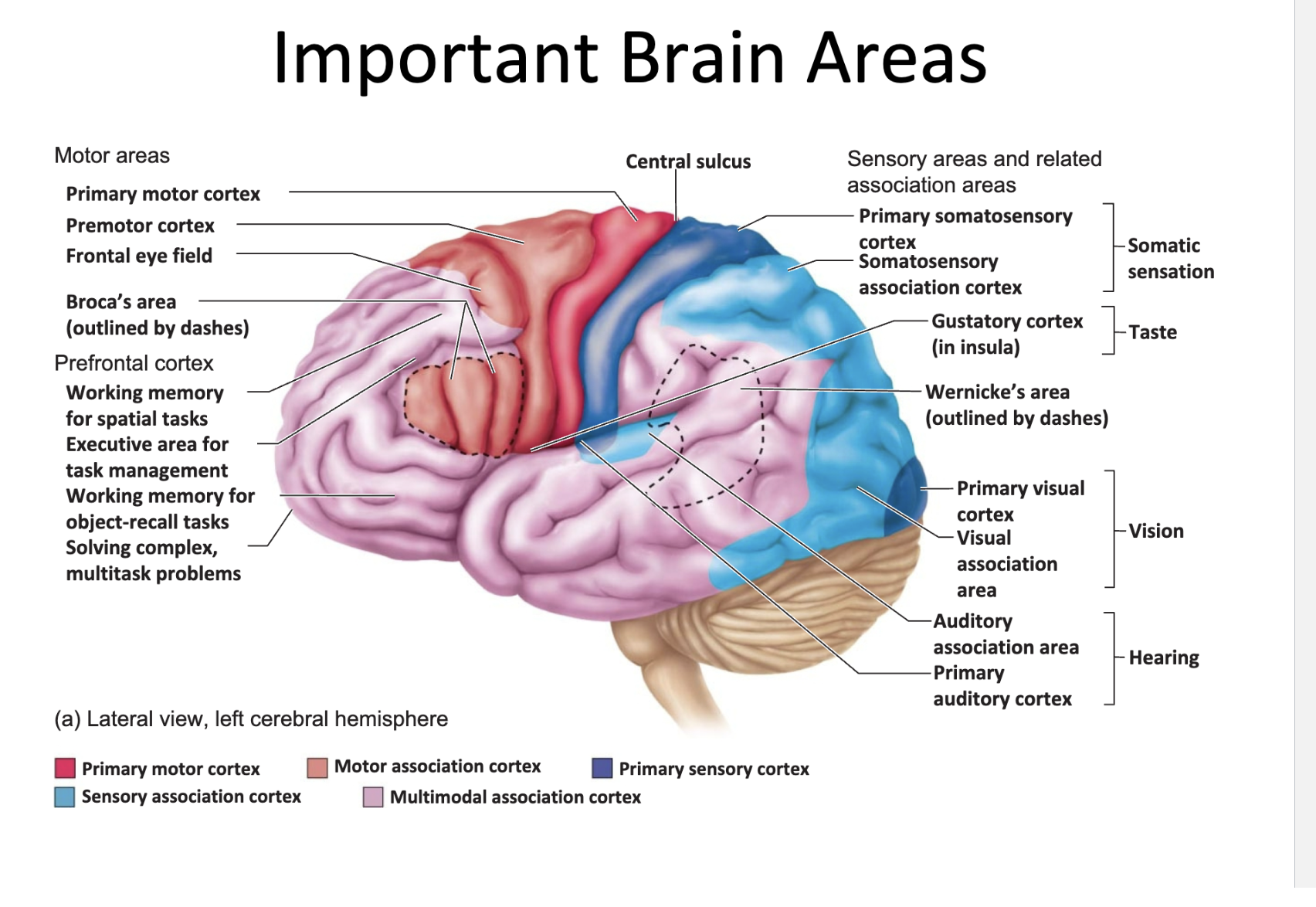
Important functional areas in the cerebral cortex (Know the location and function): primary visual \n cortex, primary auditory cortex, primary motor cortex, primary somatosensory cortex, Broca’s are, \n Wernicke’s area)
STUDY PICTURE
24
New cards
\
What is an association area? Why do we need them?
What is an association area? Why do we need them?
* The association areas integrate information from different receptors or sensory areas and relate the information to past experiences. Then the brain makes a decision and sends nerve impulses to the motor areas to elicit responses.
\
* Association areas produce a meaningful perceptual experience of the world, enable us to interact effectively, and support abstract thinking and language
\
* Association areas produce a meaningful perceptual experience of the world, enable us to interact effectively, and support abstract thinking and language