U2: Cells
1/141
There's no tags or description
Looks like no tags are added yet.
Name | Mastery | Learn | Test | Matching | Spaced |
|---|
No study sessions yet.
142 Terms
What is magnification?
How much bigger an image gets when under a microscope
What is resolution?
A measure of the microscope's ability to distinguish between two points which are close together on an object
What are the types of microscope?
Optical microscope
Scanning electron microscope (SEM)
Transmission electron microscope (TEM)
How does an optical microscope work?
Light comes from a bulb and penetrates through the specimen
Specimen is magnified using a glass lens
How does a scanning electron microscope work?
A beam of electrons are scanned across the specimen
This beam bounces off the surface of the specimen and the electrons are detected, forming an image
How does a transmission electron microscope work?
Electromagnets used to focus a beam of electrons
This beam of electrons is transmitted through the specimen
Denser parts of the specimen absorb more electrons
What are the advantages/disadvantages of an optical microscope?
Advantages:
Cheap
Can study living cells
Disadvantages:
Produces a 2D image
Low resolution
What are the advantages/disadvantages of a scanning electron microscope?
Advantages:
Can be used on thick or 3D specimens
Allow the external, 3D structure of specimens to be observed
Disadvantages:
Lower resolution image than TEMs
Cannot observe live specimen as all specimen have to be in a vacuum
They do not produce a colour image
What are the advantages/disadvantages of a transmission electron microscope?
Advantages:
High resolution
Disadvantages:
Can only be used with very thin specimen
Cannot observe live specimen as all specimen have to be in a vacuum
Lengthy treatment required to prepare specimens so artefacts can be introduced
They do not produce a colour image
What is the magnification equation?
Magnification= Image size/object size
What is cell fractionation/ultrafugation used for?
Separating cell organelles from each other
What are the conditions needed for cell fractionation/ultrafugation?
Cold: to reduce enzyme activity that may break down the cell
Isotonic: to maintain water potential inside and to prevent water from moving into the organelles via osmosis
Buffered: maintain pH so enzymes are not denatured
What is the process of cell fractionation/ultrafugation?
The sample is homogenized in a homogenizer, breaking open the plasma membrane and releases organelles into a solution called the homogenate
Homogenate is filtered through a gauze to remove any whole cells that were not homogenized
This leaves the filtrate containing a mixture of organelles
The filtrate is centrifuged
The largest, heaviest organelles (such as the nuclei) to settle at the bottom of the tube, where they form a thick sediment known as a pellet
The rest of the organelles stay suspended in the solution above the pellet (supernatant)
The supernatant is drained off and placed into another tube, which is centrifuged at a This causes the heavier organelles (such as the mitochondria) to settle at the bottom of the tube, forming a new pellet and leaving a new supernatant
This process is repeated at increasing speeds until all the different types of organelle present are separated out (or just until the desired organelle is separated out)
How is an eyepiece graticule calibrated?
Line up the stage micrometer and eyepiece graticule while looking through the eyepiece
Count how many divisions on the eyepiece graticule fit into one decision on the micrometer scale
Each division on the micrometer is 10 micrometres, so this can be used to calculate what one cisision of the eyepiece graticule is at that current magnification
What is an artefact?
A substance that is not actually part of the specimen when looking at a prepared sample (e.g, dust, air bubbles, fingerprints)
How do artefacts occur?
Samples squashed or stained during preparation
Can be prevented by more careful preparation of samples
Why were artefacts problematic for early research and how was this problem combatted?
They were unable to easily distinguish between artefacts and organelles
To distinguish, scientists had to repeatedly prepare specimen in different ways using different techniques
What is the function of the nucleus?
Stores genetic material in the form of chromosones
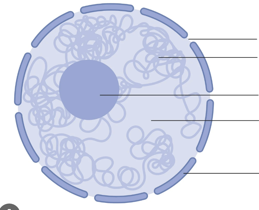
Label the diagram
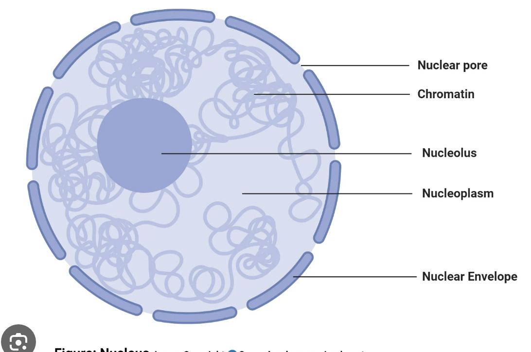
What is the function of the nuclear pores?
Allows mRNA into the cytoplasm to be translated
Allows ribosomes to move to form the cytoplasm
What is the function of the nucleolus?
Involved in making ribosomes
Contains densely packed DNA associated with histones
What is chromatin?
A complex of DNA wound around histones
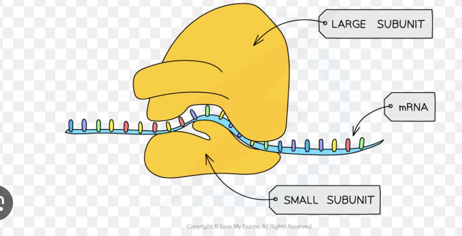
What is the function of ribosomes?
Carry out protein synthesis by translating mRNA to proteins
Can either be free loading in the cytoplasm, or bound to the rough endoplasmic reticulum
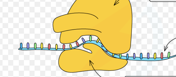
Label this diagram
What is the function of mitochondria?
Carrying out aerobic respiration to synthesise ATP
The number of mitochondria vary due to the energy demands of the cell
2-5 micrometres in size
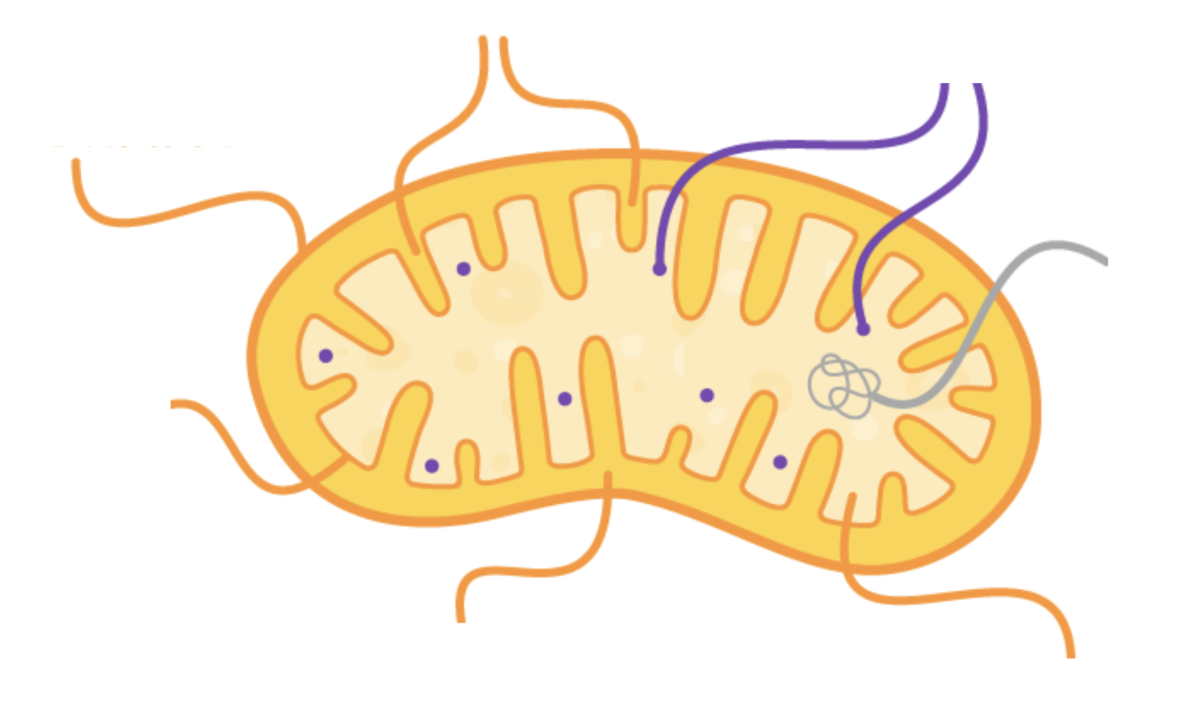
Label this diagram

What is the function of the inner membrane?
Contains proteins for ATP synthesis
Highly folded into cristae
What is the function of cristae?
Increases surface area for ATP production
What is the function of the matrix?
Contains enzymes, ribosomes, lipids and mitochondrial DNA
What is the function of mitochondrial DNA?
There is a high demand for proteins for respiration so there is no need to return to the nucleus to produce ribosomes
What are the differences between mitochondrial and nuclear DNA?
Mitochondrial DNA is circular, nuclear DNA is linear
Mitochondrial DNA has fewer genes (37), nuclear DNA has 19,000
What is the function of the cytoplasm?
Site of many chemical reactions and is aqueous so substances can dissolve into it, and these substances can interact
What is the function of the cell membrane?
Controls what enters/leaves cells
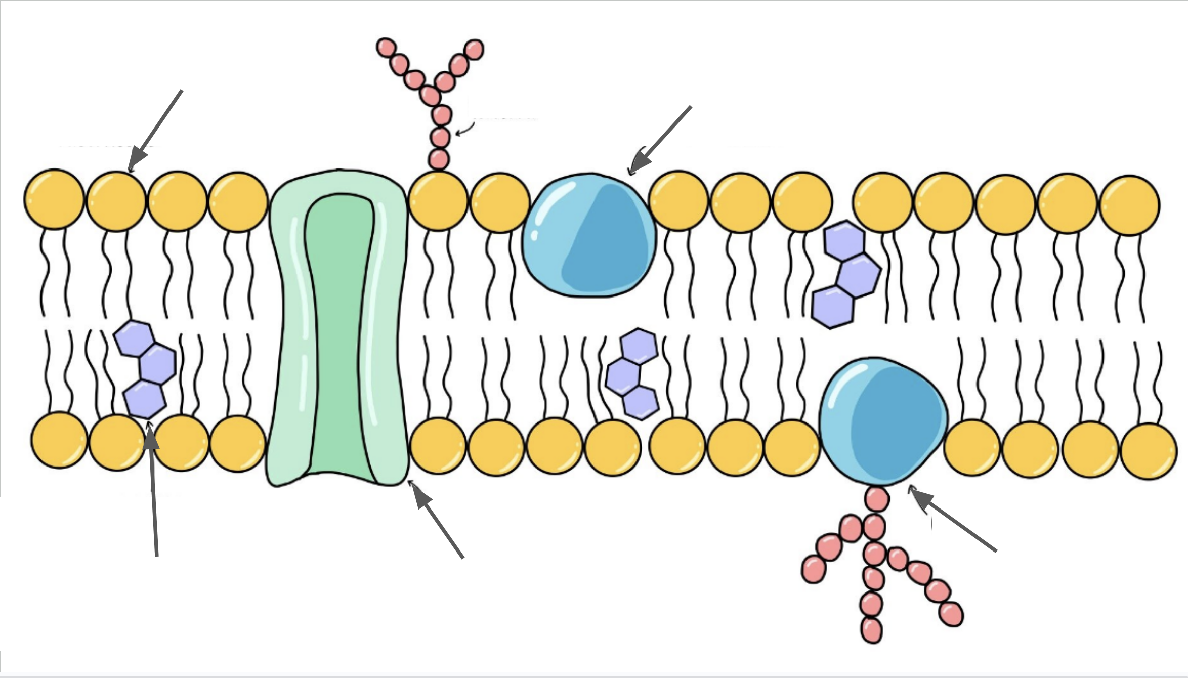
Label this diagram
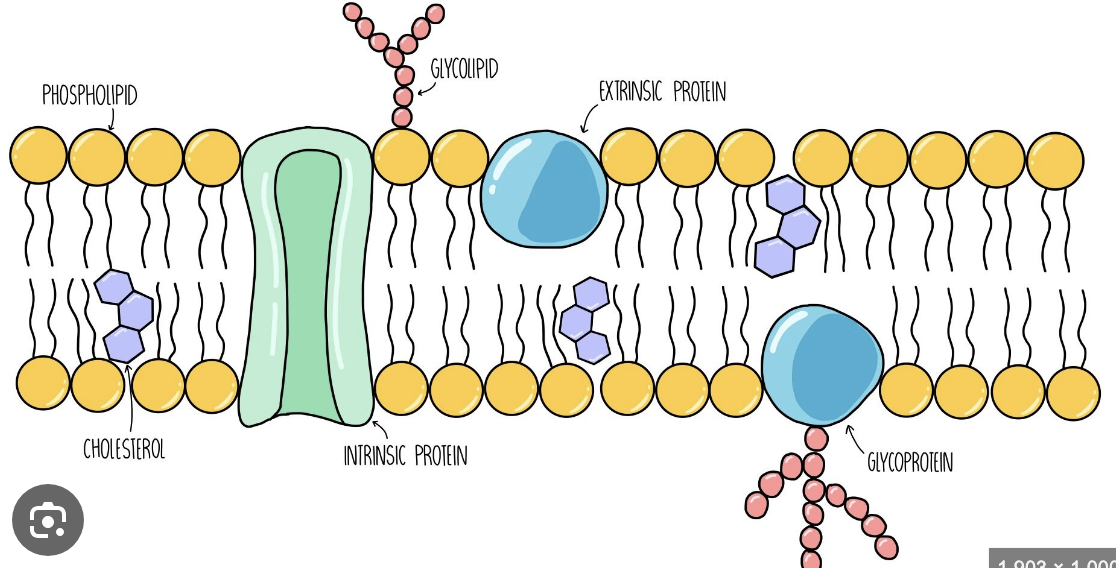
What is a extrinsic protein and what is its function?
A protein that is only present on side side of the bilayer
Provides support using signalling
What is an intrinsic protein and what is its function?
A protein that is present on both sides of the bilayer
Used for transport
Why does every organelle have its own bilayer?
Organelles need to be kept seperate
Each organelle can perform their own functions in their own optimum conditions
What are glycoproteins ad glycolipids used for?
Cell adhesion
Cell recognition
Cell signalling
What is a lysosome and what is its function?
Vesicles filled will digestive enzymes (protease, lipase and lysozymes)
Catalyse the destruction of the cell walls of certain bacteria
They must be in vesicles otherwise they would break down lipids/proteins in other cells
Functions:
Phagocytosis
Digesting worn out organelles and recycling certain parts
Apoptosis
Digesting material outside the cell
What are the features of the endoplasmic reticulum?
Often continuous with the nucleus
Network of fluid filled cavities (cisternae)
RER contain ribosomes
What is the function of the smooth endoplasmic reticulum (SER)?
No ribosomes attached to its membrane
Cisternae contain enzymes to synthesise lipids and carbohydrates
Lipids and carbohydrates are stored in the SER until needed
What is the function of the rough endoplasmic reticulum?
Contains ribosomes attached to the membrane that is not in the cisternae to translate mRNA to protein
Large SA so lots of ribosomes can be housed and proteins can be formed quickly
Once protein has been translated, a vesicle is then pinched off and enters the golgi apparatus
What is the function of the golgi apparatus?
Modify vesicle contents
Repackage modified contents
How does a golgi apparatus work?
Receives vesicles from the RER and SER
Vesicles fuse with the golgi membrane
Vesicles become part of the golgi membrane
They then release their contents into the cisternae
What modifications to the golgi apparatus make to vesicles?
Folding and joining proteins with other molecules
Adding molecules to lipids
Forming quaternary structures
What is the function of a chloroplast?
Carries out photosynthesis
7 micrometres
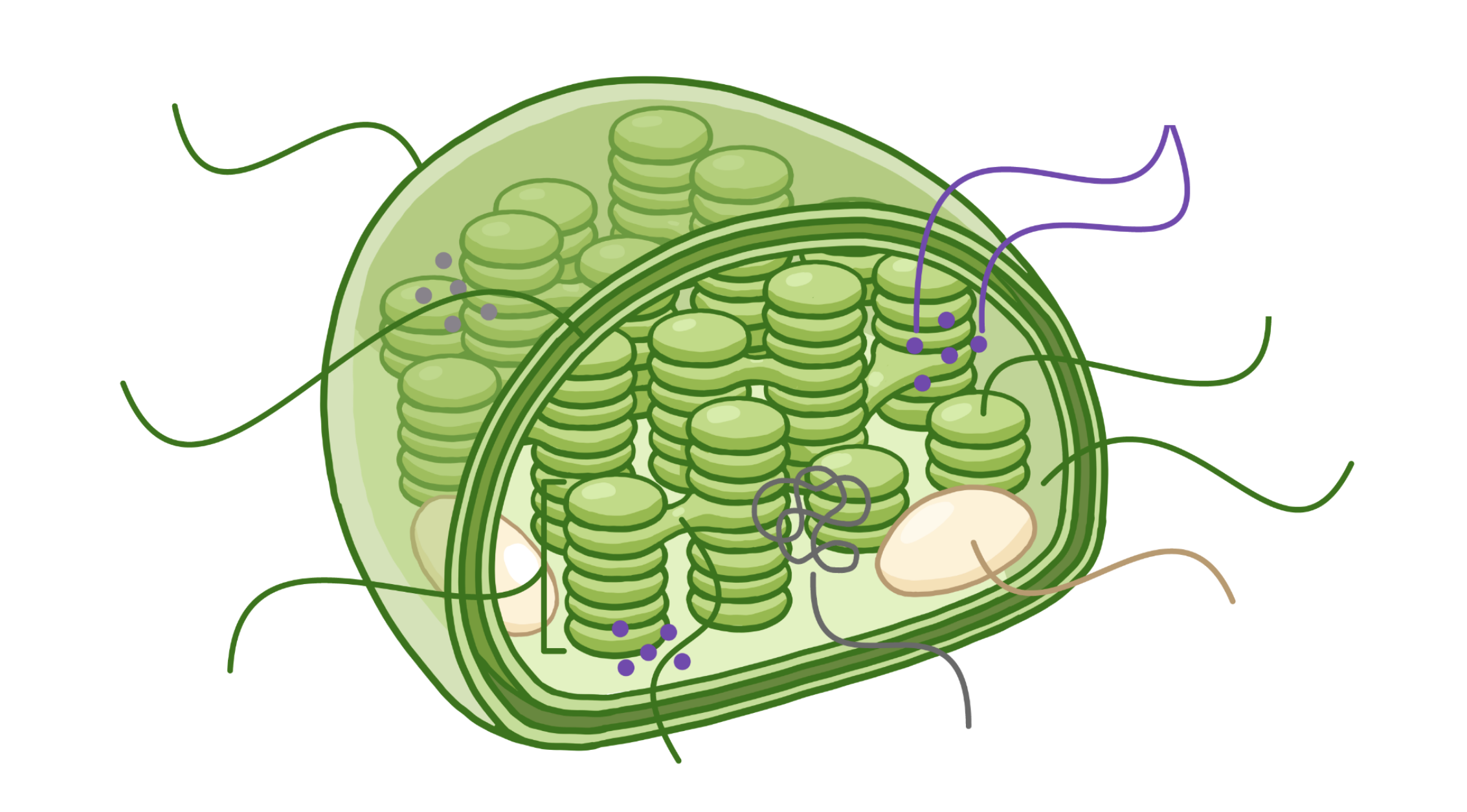
Label the diagram
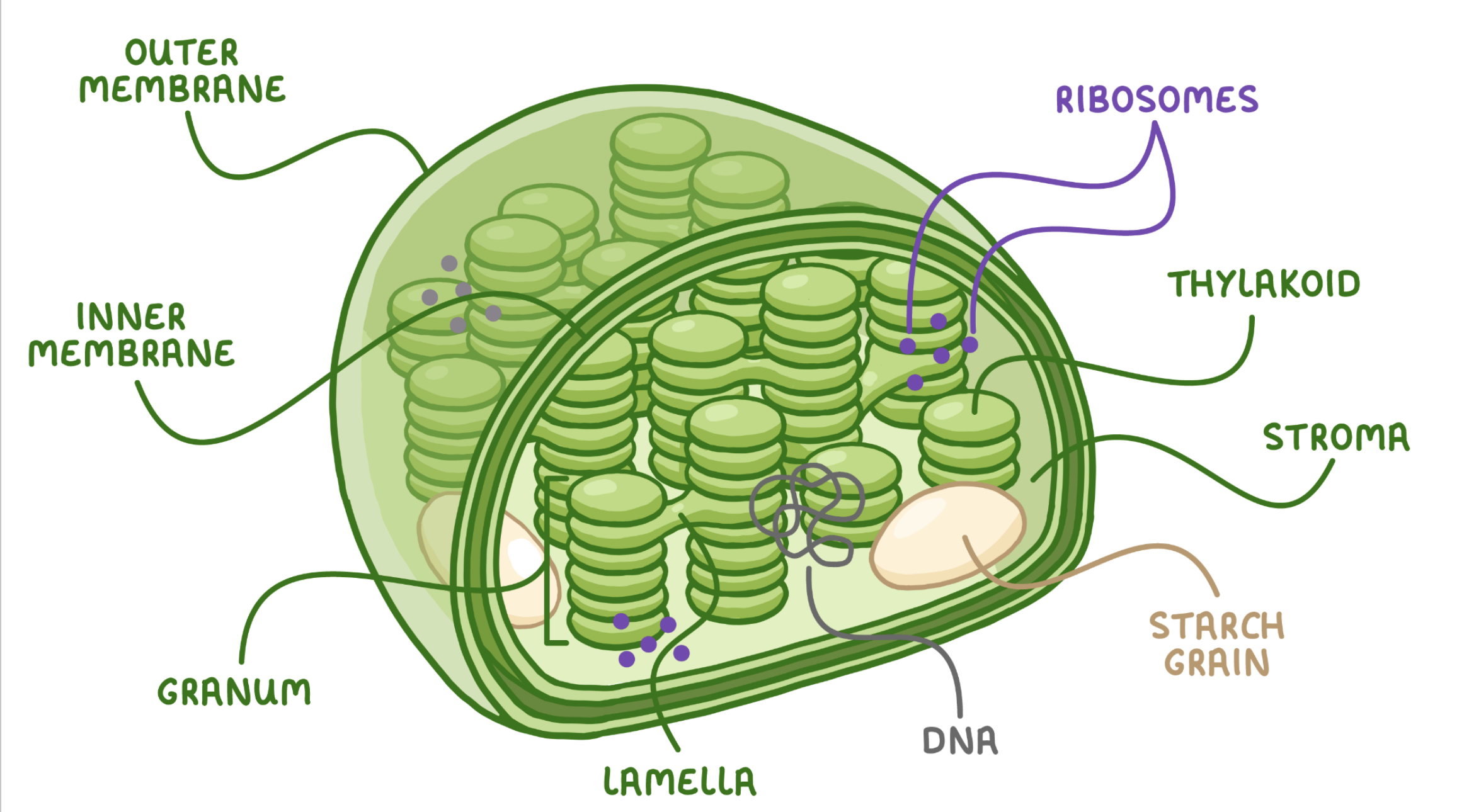
What is the function of the chloroplast double membrane?
Creates a chloroplast envelope what enters and leaves the cell
What is the function of the chloroplast inner membrane?
It is highly folded, creating stacks of flattened disks (thylakoids)
Where the first stage of photosynthesis takes place
What is a granum?
A stack of thylakoids
What is the function of the stroma?
Fluid filled chamber where the second stage of photosynthesis happens
Contains enzymes, starch grains, DNA and ribosomes
Starch grains contain glucose for photosynthesis
DNA and ribosomes allow protein synthesis to occur
What is the function of the vacuole?
Fluid filled sack that contains dissolved solutes
Surrounded by a single membrane (tonoplast)
Helps the cell maintain its structure and is able to resist pressure and external forces
Sometimes acts as a temp
What is the function of the cell wall?
Made of cellulose and is semi-permeable
Helps the cell maintain its structure and is able to resist pressure and external forces
Helps cell remain turgid even when water potential changes
What is the function of chitin?
Fungal cell walls
It is a polysaccharide of sugars that form fibres
Contains glycan and some glycoproteins
What do foreign antigen detecting molecules allow the immune system to identify?
Pathogens
Cells from other organisms of the same species
Abnormal body cells
Toxins
What is an antigen?
Molecules present on the surface of cells which trigger an immune response
What is the difference between a self and non-self antigen?
Self antigen: produced by the organism’s body cells- does not stimulate an immune response
Non-self antigen: not produced by the organisms own cells-stimulates an immune respnse
What is antigen variability?
The ability of antigens to frequently change due to genetic mutations
How does antigen variability effect disease and disease prevention?
Lymphocytes produce a specific immune response
Surface receptors on lymphocytes and memory cells are complementary to only 1 antigen
When the antigen on the pathogen changes, lymphocytes and memory cells can no longer bind to the antigen
Therefore there is no secondary immune response so the host will become infected
What are the different types of phagocytes and what are their roles?
Neutrophils: first cells response to detection of foreign cells
Macrophage: large phagocytes that engulf pathogens and present antigens on cell surface to lymphocytes
Both create a non-specific immune response
What are the differences between neutrophils and macrophages?
Neutrophils act faster
Neutrophils are smaller
Neutrophils have a shorter lifespan
What is the process of phagocytosis?
Chemicals released by pathogens attract phagocytes by chemotaxis
Phagocyte recognises antigens as foreign
Phagosome fuses with a lysosome to make a phagolysosome
Lysozyme hydrolyse proteins, breaking down the pathogen
Phagocyte (only macrophages) then present the antigens pathogens on its surface, producing antigen-presenting cells
What is the difference between T and B lymphocytes?
T-lymphocytes are primarily used in cell-mediated immunity, B lymphocytes are used in humoral immunity
T-lymphocytes kill pathogens (cytotoxic) and help other cells (helper), B lymphocytes produce antibodies to neutralize pathogens
What is the response of T-Lymphocytes to a foreign antibody (the cellular response)?
Pathogen enters and phagocytosis occurs so macrophages present antibodies on the cell surface
Specific helper t-cells have complimentary receptors to the antibody (clonal selection)
T-lymphocytes are activated when they bind to their specific antigen that is being presented, stimulating it to divide by mitosis (clonal expansion)
The newly produced cells differentiate into killer, memory, regulatory and helper t-cells
Killer: produces perforin which makes holes in cells, allowing water to enter via osmosis and causing the cell to burst
Memory: remembers specific shape of antigen providing long-term immunity
Regulatory: Stops healthy cells from destructing
Helper: stimulates b-cells to divide and encouraging phagocytosis
What is the response of B lymphocytes to a foreign antigen (humoral response)?
Pathogens antigens are specific to a specific B cell
B cell engulfs the pathogen and presents the antigen on it cell surface membrane
Helper T cells bind to the presented antigen activates the B cell to divide by mitosis (clonal expansion)
The B cells differentiate into memory and plasma cells
Memory cells: remembers the complimentary antibody and provides long term immunity
Plasma cells: Secretes large amounts of monoclonal antibodies specific to the anitgen
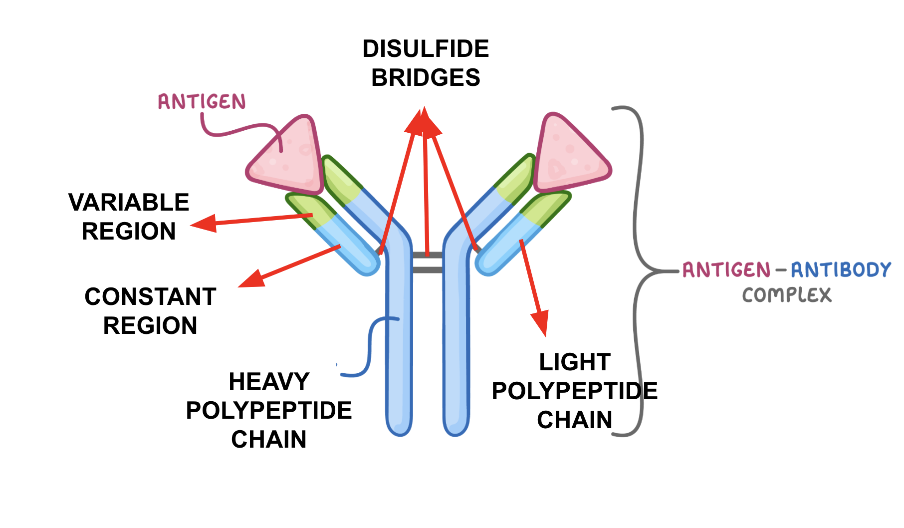
What is an antibody?
A protein released by plasma cells that can attach to pathogenic regions
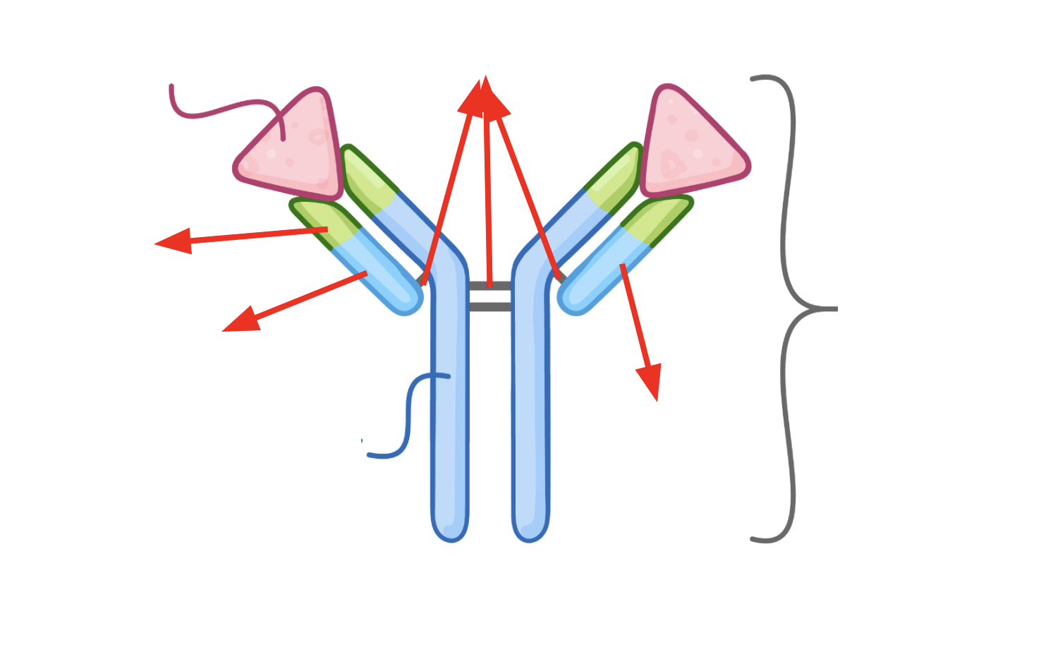
What is the structure of an antibody?
How does the antibody-antigen complex destroy the antigen?
Agglutination and phagocytosis
What is agglutination?
The clumping together of cells caused by antibodies, assisting phagocytosis
What is the secondary immune response?
Memory B cells recognise a specific antigen from re-entered pathogens
They differentiate rapidly into plasma and more memory B cells
Plasma produces specific and complimentary monoclonal antibodies in a high concentration
Memory T-cells may recognise an antigen on an antigen-presenting cell
They differentiate into helper and killer t-cells
High concentration of antibodies prevents symptoms from developing
How do vaccines provide immunity?
A small amount of dead, weakened, or inactive version of a specific pathogen or a foreign antigen is injected into the body.
The foreign antigens on the pathogen stimulate and immune response.
When the correct B cell is selected (clonal selection) due to it having a complementary receptor to the antigen the B cell divides by mitosis (clonal expansion) and differentiate into plasma cells and memory cells.
Plasma cells produce complementary antibodies that bind to the pathogens antigen
Memory B cells are able to remember the shape of the antigen and remember to correct complementary shaped antibody that can be used to destroy the pathogen if it were to enter again.
If the pathogen were to enter again memory cells can quickly differentiate into plasma cells which quickly releases large volumes of the correct antibody
What is herd immunity?
When a sufficiently large portion of the population has been vaccinated so there are not enough susceptible individuals for infection to be able to spread
What are the differences between active and passive immunity?
Active immunity has exposure to a pathogen, passive immunity has no exposure to a pathogen
Active immunity is slow, passive immunity is fast
Active immunity produces memory cells, passive immunity does not
Active immunity is long lasting, passive immunity is short term
What are the ethical issues associated with vaccinations?
Animal testing
Human testing
Side effects
Epidemics (which groups received priority over vaccinations)
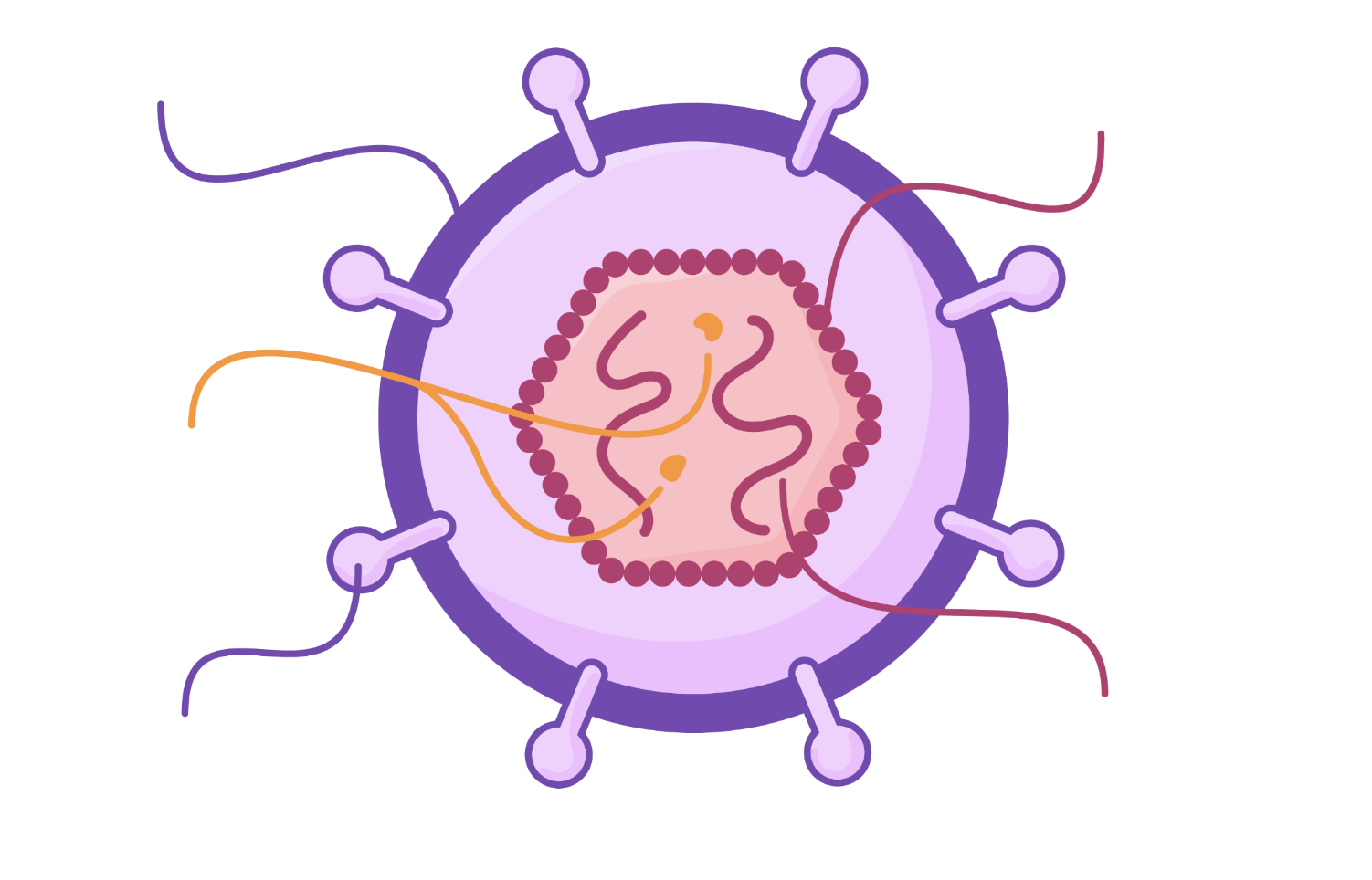
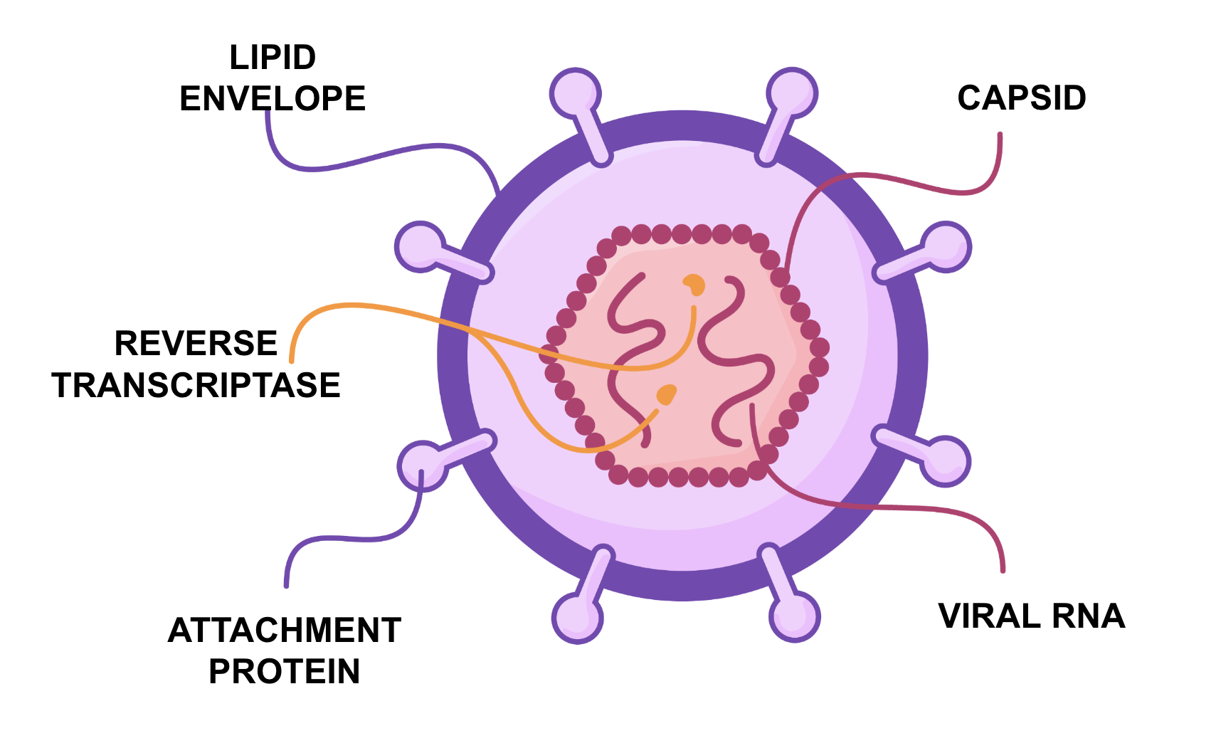
What is the structure of HIV?
How is HIV replicated?
Viral RNA enters cell
Viral reverse transcriptase produce a DNA copy of the RNA
DNA copy is inserted into the host cell genome
HIV proteins are produced from viral RNA
Proteins are synthesised to build new HIV moleculesHIV burts out of helper T cell once a sufficient amount have been produced, killing it
How does HIV cause the symptoms of AIDS?
HIV gradually reduces the number of helper t cells in the immune system
B cells are no longer activated so antibodies are no longer produced
This defeats the body’s ability to fight of infections, leading to AIDS
Why are antibiotics ineffective against viruses?
Viruses are nonliving and are found inside host cells
What is a monoclonal antibody?
Antibodies with the same tertiary structure produced from cloned plasma cells
What are some uses for monoclonal antibodies?
Diagnostics (ELISA/pregnancy testing)
Targeting medication to specific antigens (cancer)
How does the ELISA test work?
There are capture antibodies attached to the wells
Add sample to the well and if any proteins are present they attach to the capture antibodies
The well is then washed to get rid of any unbound proteins
Complementary detection antibodies are then added to the well which bind to the protein which is bound to the capture antibodies
The well is washed again to remove any un attached detection antibodies
The detection antibodies have enzymes attached
A substrate is added to the well and when it is broken down by the enzyme it causes a colour change.
The change in colour corresponds to the amount of protein in the original sample e.g. deeper the colour the more protein present
How does a pregnancy test work?
A sample of urine is applied to the test strip.
If hCG is present, it binds to the mobile monoclonal antibodies that are conjugated with a color indicator.
The hCG-antibody complex moves along the strip by capillary action.
At a specific point on the strip, immobilized antibodies capture the hCG-antibody complex.
A nutrient conjugated with the same colour indicator always bids to the control strip, ensuring that the pregnancy test is functioning
The color indicator produces a visible line, indicating a positive result.
What are the properties of the phospholipid bilayer?
Partially permeable membrane so certain molecules can pass through however others (such as charged molecules) cannot
Fluid but stable- allows movement of phospholipids so substances can pass through and the cell can change shape
What is the fluid-mosaic model?
The cell membrane contains not only phospholipids but proteins, glycoproteins, glycolipids and lipids
What is an intrinsic protien?
Proteins that are embedded into the cell membrane
What is a transmembrane protein?
Proteins that span the entire bilayer, held in place by hydrophobic interactions
What is an extrinsic protein?
Proteins only found on one side of cell
Not embedded into the bilayer (held in place by ionic bonding- phosphate heads and + R groups opn amino acids)
What is the function of a channel protein?
Enables substances to pass to/from the protein that otherwise wouldn’t be able to
What is a carrier protein?
Enables substances to cross the plasma membrane by changing shape once a specific molecule has attached itself
What are the functions of the proteins in the phospholipid bilayer?
Receptors for hormones and drugs to bind to
Recognition sites to detect foreign cells
Cell adhesion
What is a glycoprotein/glycolipid?
A protein/lipid with a chain of carbohydrate molecules attatched
What are the functions of glycoproteins/glycolipids?
Recognition sites to detect foreign cells
Cell adhesion
Stability and structure for cell membrane
Cell receptors so cells can communicate (glycoproteins only)
What are the functions of cholesterol?
Regulating fluidity of membrane
Prevents stiffness by making sure phospholipids are not too close together
Prevents membrane becoming too stiff by interacting with fatty acids
What is simple diffusion?
The net movement of a substance from a region of higher concentration to a region of lower concentration.
Passive process
Can only be carried out by small, non-polar molecules or molecules with a slight charge
What are the factors affecting diffusion?
Steepness of concentration gradient: greater difference=greater number of molecules passing in the two directions= faster rate of diffusion
Temperature: higher temperature= more kinetic energy=higher rate of diffusion
Surface area: greater surface area=greater number of molecules that can cross= faster diffusion
Properties of molecules/ions: large molecules diffuse slowly as they require more energy to move, uncharged and nonpolar molecules diffuse directly across bilayer, non-polar molecules diffuse faster than polar molecules as they are soluble in the bilayer
What are the limitations of simple diffusion?
Lack of controla as it is a passive process entirely dependent on concentration gradients
Not all molecules can undergo simple diffusion
What is facilitated diffusion?
Diffusion via a channel/carrier protein for larger and charged molecules
Passive movement
Moves down the concentration gradient
Why do some molecules require facilitated diffusion?
Too large
Too polar
Too charged
So they cant pass through the phospholipid bilayer and require transport proteins
What is the role of channel proteins in facilitated diffusion?
Water-filled pores that allow charged substances to diffuse through the cell membrane
They can be opened/closed as they are gated to control the exchange of ions