FINAL Biochem Boards Superset
 Call Kai
 Learn
Learn Practice Test
 Spaced Repetition
 Match
Match Flashcards
 Knowt Play
1/269

There's no tags or description
Looks like no tags are added yet.
Name | Mastery | Learn | Test | Matching | Spaced | Call with Kai | Chat |
|---|
No analytics yet
Send a link to your students to track their progress
270 Terms
Amino Acid Homeostasis
There has to be a balance of our intake (sources) and output (products) of A.As.
Sources
Dietary protein: intake via intestine provides essential and non essential A. As
Tissue Catabolism: periods of fasting or metabolic stress trigger breakdown of tissue; largest protein repository is muscle
Synthesis: metabolic pathways for non-essential AAs
Products
Anabolism: incorporation into complex proteins
Conversion: into Non-essential or metabolic intermediates (Keto or gluccogenesis)
Excretion: in urine through conversion of nitrogenous waste to urea
5 Major Aminoacidopathies
When there are defects in the enzymes of the metabolism of Amino Acids, they result in a group of disorders call Aminoacidopathies: (the most common and well understood of the group)
Phenylketonuria (PKU)
Tyrosinemia
Maples Syrup Urine Disease (MSUD)
Homocystinuria
Non-Ketonic Hyperglycinemia (NKH) —> not needed for boards
Phenylketonuria (PKU)
Found in individuals with Phenylpyruivic in their urine; had mental retardation
Inheritance:
Autosomal recessive
Incidence: 1/10-25,000 (Higher in turkey, Ireland, Japan, Africa)
Genetic Basis: Always a mutation in Phenylalanine Hydroxylase (PAH) Deficiency
Most PKU are compound heterozygotes with >500 mutant alleles (no one allele has overwhelming frequency)
Metoblic Pathway of PKU
Conversion of Phenylalanine to Tyrosine via PAH enzyme
we get all out Phenylalanine from Diet: we do no syntheses ,it is an ESSENTIAL AMINO ACID
PAH has a cofactor: Tetrahydrobiopterin, necessary for peak PAH activity
Insufficient tetrahydrobiopterin (THBP) can also cause accumulation of Phenylalanine as well as other intermediates in the THBP synthesis (and also the recycling) pathway
A DEfeicny of TYROSINE
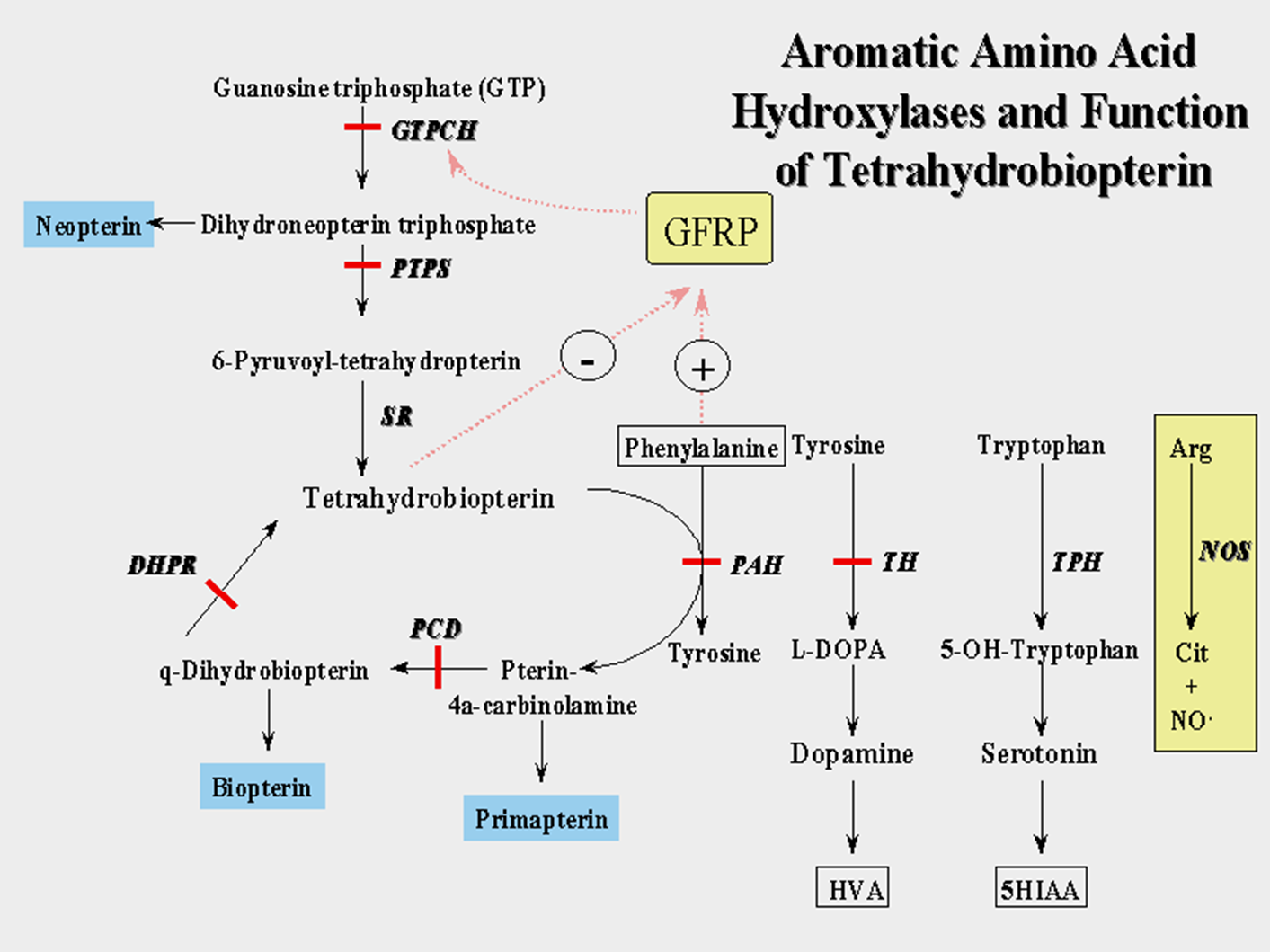
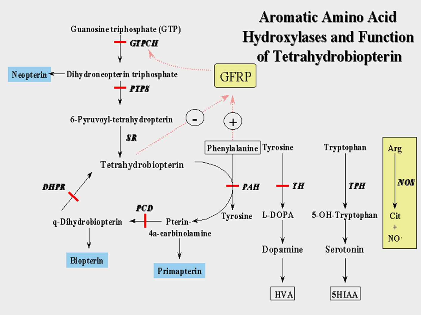
Tetrahydrobiopterin (THBP)
A cofactor that is important in maximizing the activity of some Amino Acid enzymes
Phenylalanine —> Tyrosine (affecting the PAH enzyme)
Tyrosine —> L-DOPA {—> Dopamine} (affecting the Tyrosine Hydroxylase enzyme)
Tryptophan —> 4-OH-Tyrpothan {—> Serotine} (Affecting the Tryptophan Hydroxylase enzyme)
Arginine —> Nitric Oxide (Affecting Nitric Oxide Synthase)
A defiecny of THBP = A defeicny in all these pathways (Overlap with PKU for high Phenylalaine, but also low Dopamine, Seratoin and Nitoric Oxid)
Untreated Classic PKU
At Birth: asymptomatic; moms enzyme activity avoids accumulation of phenylalanine in the fetus
Neonatal period: take-in of phenylamine via diet; accumulation begins; demonstrate symptoms
No fatal neonatal encephalopathy (unlike other metabolic disorders)
Fair skin and hair pigmentation: PHE inhibition of tyrosine hydroxylase = Melanin deficiency
Urine w musty order: Phenylpyruvic acid in urine
Infancy onward: intellectual disability and delays manifest
irreversible ID: Severe in 50%, moderate-mild in 50%
Autism, microcephaly, behavior issue
Large % with epilepsy, eczema
Diagnosing PKU
Newborn Screening: Tandem Mass Spectrometry on heel-prick newborn blood sample, give PHE levels
Normal PHE: <120 umol/L (<6 mg/dL)
Classic KU >1,200 umol/L (>20 mg/dL)
Hyperpheylalaniemia 120-1200 umol/L (2-20 mgdL)
False negative (<1%): sample collection to early, mishandling or lab error
Fals positive (frequent): liver disease/immaturity (PAH active mainly in liver), high protein load, sample collection or lab error
Confirmatory Biochemical testing:
Plasma amino acid analysis: we should see Elevated PHE AND elevated PHE/TYR ratio (due to the PHE→TYR BLOCK)
(RULE OUT LIVER DIEASE OVERLAP: WE WOULD SEE PHE AND OTHER AAs)
THBP testing
Genotyping
PAH gene sequencing: genotype / phenotype correlation incomplete but can predict some treatment response
Treating PKU
Anyone with PHE >360-600 umol/L ( >6-10 mg/dL)
Dietary PHE restriction: Limit dietary protein intake to keep PHE <360umol/L (,6mg/dL) - Classic PKU <1500-250 mg/day PHE intake (not all Hyperpheylalaniemia pts need dietary PHE restriction)
Dietary PHE-free protein formulas: PHE-free protein formulas to avoid amino acid deficiency (TYR)
Outcome with treatment if started by 3 weeks old)
Normal IQ
Increased risk for subtle Neurocognitive deficits and signs
Effect of treatment withdrawal (why it is LIFELONG TX)
increased ADD/ADHA, depression, anxiety, agoraphobia
Progressive demyelination on MRI
Decreased IQ scores (5-30 points)
Maternal PKU Embryopathy
Elevated maternal PHE highly teratogenic (Females with PKU need even MORE dietary control)
PHE level in developing fetus is 2x maternal level: (75-90% teratogenicity vs 50% with thalidomide)
Goal: Maternal PHE levels <360 umol/l (<6 mg/dL)
Higher PHE levels correlate with risk for
Microcephaly, Mental + Growth retardation, Congenital heart defects
Offer prenatal ultrasound and echocardiography to look for these issues
Newer PKU Treatments
Sapropterin Powder
Phenylalanine Ammonium Lyase SQ injection
Large Neutral Amino Acids Tablets/GMP
PKU Carrier Detection
If mutations are detected in parents
Molecular testing of PAH gene in affected individual can facilatate carrier testing: if no mutation identifiable linkage analysis (dup/del anayslsi needed)
Enzyme analysis not practical as PAH is essentially only exressed in liver (need a liver biopsy which is NOT typically done)
Prenatal implantation diagnosis: Available when molecular testing informative
Tyrosinemia Type I
Also called “Hepatorenal” Tyrosinemia due to progressive liver and kidney failure if untreated : type II and III dont present the same way
Inheritance: Autosomal Recessive
Incidence: 1/120,000 (higher in Quebeec)
Genetic defect in: Fumarylacetoacetate Hydrolase (FAH) Deficney
Tyrosinemia Type I Metabolic Pathway
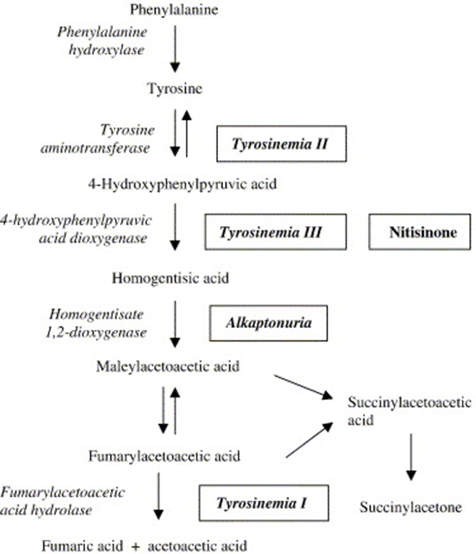
Phenylalanine → Tyrosine via Phenylalanine Hydroxylase (PAH, PKU remember?)
Tyrosine → Fumaric Acid + Acetoacetic Acid via 5 step reaction
Step 1 Enzyme Block: Tyrosinemia II (Eye irritation, skin changes
Step 2 Enzyme Block: Tyrosinemia III (cognitive disability in some but not all)
Step 3 Enzyme Bock: Alkaptonuria (Black pee disorder first IBEM identified)
Step 4: Fumarylacetoacetic Acid Hydrolase (FAH) Block : Tyrosinemia I
High levels of Fumarylacetoaecetic acid accumalte -→ conversion to Succinylacetosectic acid which would normally not accumulate: both Cause TOXICITY AT HIGH LEVELS
Untreated Tyrosinemia I
At birth: Asymptomatic
Infancy: Tyrosine, Fumarylacetoaecetic acid (FAA), Succinylacetone (SAT) accumulate
Cabbage-like odor in urine
Liver dysfunction progression (from FAA)
Renal dysfunction progressing to failure
Rickets, poor growth
Neurologic “crisies”; episodic abdominal +neuropathic pain, weakness, repository failure (from SAT inhibition of porphyrin pathways)
Adolescence: Death typically by age 10 due to
liver failure,
respiratory fialure in neurologic crisis,
hepatocellular cancer
Diagnosing Tyrosinemia I
Newborn Screening: on heel-prick blood sample
Elevated Tyrosine and SAT on tandem Mass spect.
some false negative: may take time for sufficient elevations to elevations
Often false positive: TYR elevation also with liver disease, transient benign or the other forms of tyrosinemia
Confirmatory Biochemical Testing: Plasma amino acid profile
High TYR in plasma A.As: non-specific to Type I
High SAT in blood or urine organic acids: Specific to Type I
Rarely skin fibroblast FAH enzyme activity: PBG in blood (Quebec)
Genotyping
FAH gene sequencing with del/dup analysis (>95%): cancer be targeted in some populations (French Canadians)
Genotype: Phenotype correlation no consistent
Treating Tyrosinemia I
Treatment
Dietary restriction of PHE and TYR
Dietary supplementation: PHE and TYR-free protein formulas
Medications : Orfadine - inhibits enzyme prior to FAH (p-HPPD) reducing FAA and SAT
Liver Transplantation: reserved for liver failure or hepatocellular carcinoma despite treatment
Outcome with treatment:
with NBS: 90% survival into adulthood with normal growth and function
Screening for Tyrosinemia I
Carrier Screening
Moelcualr tesing for FAH gene if mutation idefineted
Linkage analysis if mutation unidentifiable
Prenatal/Preimplantation diaognsis
availbe if moclaur testing infromative
if molecualar testing uninformative: SAT acumination can be Measured in the Amniotic fluid when molecular basis in family unknown but not 100% accurate
Branch Chain Keto-Acid Dehydrogenase Deficiency (Maple Syrup Urine Disease)
Urine smells like Maple Syrup / Sweet
Inheritance Recessive
Incidence 1:185,000 (increased in Mennonite pop.)
Genetic Basis
Mutations in genes coding Branch Chain Keto-Acid Dehydrogenase complex subunits: multiple subunits, multiple genes that can cause MSUD)
4 subunits in BCKD complex: E1a, E1b, E2, E3
3 MSUD genes:
BCKDHA (E1a)
BCKDHB (E1b)
DBT (E2)
***DLD (E3) mutations cause different disease***
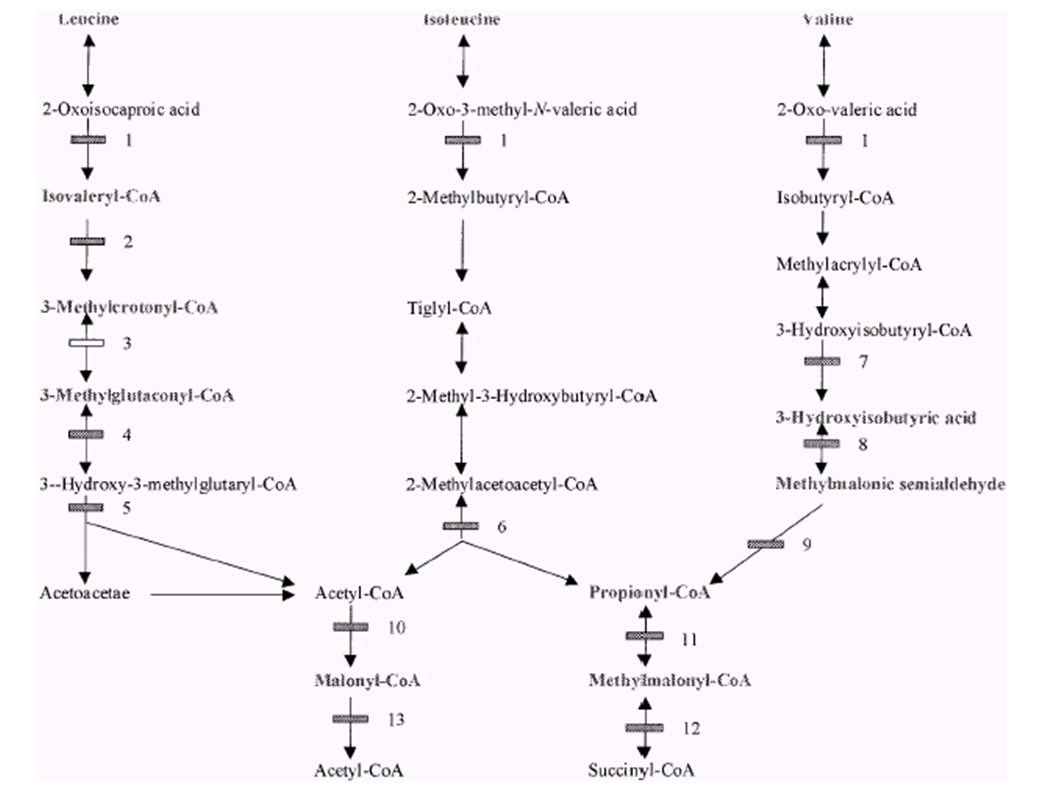
Branch Chain Amino Acid (BCAA) Metabolism
The Branch Chain Amino Acids: Leucine, Isoleucine, Valine
These three are converted via multiple steps to energy intermediates Acctly-CoA and Succinyl-CoA
First Step is via BCAA: When blocked, accumulation of the three branch chain amino acids + their associated keto-acid forms (which are what BCAA actually works with)
Untreated Classic MSUD
At Birth Asymptomatic
Within 12-24 hrs after birth: Maple syrup urine and ear wax (from Isoleucine)
Dietary intake OR Catabolism in the newborn
Within 5 Days
progressive irritability and decreased feeding
progressive encephalopathy/cerebral edema (from leucine: neurotoxic)
Within 7-10 days after birth
massive cerebral edema, coma respiratory failure
death within weeks
Some milder “intermediate” or “intermittent” forms
may only experience “decompensation” during catabolic stress
MSUD unlike PKU and Tyrosaenmia I: No threat of neo-natal death + symptom onset longer vs. rapid onset and death in MSUD
Diagnosing MSUD
Newborn Screening: Heel prick blood tandem Mass spec. analysis
Elevated Leucine and BCAA ratios
rarely false negative: early sample collection (before 24hrs), collection or lab error
Occasional false positives: other disorders appear positive via elevated LEU (hydroxyprolinemia)
Confirmatory Biochemical Testing
Plasma AA analysis:
Elevated BCAA’s; Leucine, Isoleucine, Valine
Allo-Isoleucine presence (only elevated in MSUD)
Urine organic acid analysis
BCAAs present
Enzyme activity: BCKD enzyme activity measurable n fibroblasts
Genotyping
BCKD subunit gene sequencing with del/dup analysis: E3 subunit mutations cause distinct syndrome due to its activity in Mutiple metabolic pathways: genotype/phenotype correlation not consistent
If successful, allows for prenatal/preimplantation genetic diagnosis
Treating MSUD
Preventing Acute decompensation (rapid buildup of BCAAs)
Decrease catabolism and enhance elimination of Leucine
Maximize BCAA-free calories (avoid catabolism)
Intravenous infusion/metabolic formula (Dextrose, lipid and BCCA-free nutrient)
Supplemental Isoleucine and Valine solutions (avoid deficiency with prolonged treatment - just the precise amount they need, no more)
Thiamine supplementation (BCKD coenzyme, increases activity)
Hemodialysis in acute severe illness
Long-term Management
Avoid decompensation: fever, fasting, high protein
Optimize BCAA-levels: metabolic formula and sing AAs
Thiamin: if thiamine responsive
Liver transplant for severe poorly responsive forms
Outcome
Normal (20 points lower): BUT ONLY IN THE FIRST 10 years of LIFE
After 10days: Severe, irreversible brain damage and/or death
MSUD Carrier Detection
Carrier Screening: can be done if BCKD genes have been found in affected individuals
linkage analysis if mutations unidentifiable
Prenatal/Preimplantation diagnosis
Available when molecular testing informative
Can obtain BCKD enzyme activity on amniocentesis or CVS
Homocystinuria
Deficiency of Cystathionine Beta-Synthase (CBS)
Inheritance: Autosomal Recessive
Incidence 1:3350,000 (higher in Qatar, Norway)
Genetic basis: Mutation in Cystathionine Beta-Synthase gene
Onset typically after 3 years old
No neonatal encephalopathy
Many cased missed on NBS
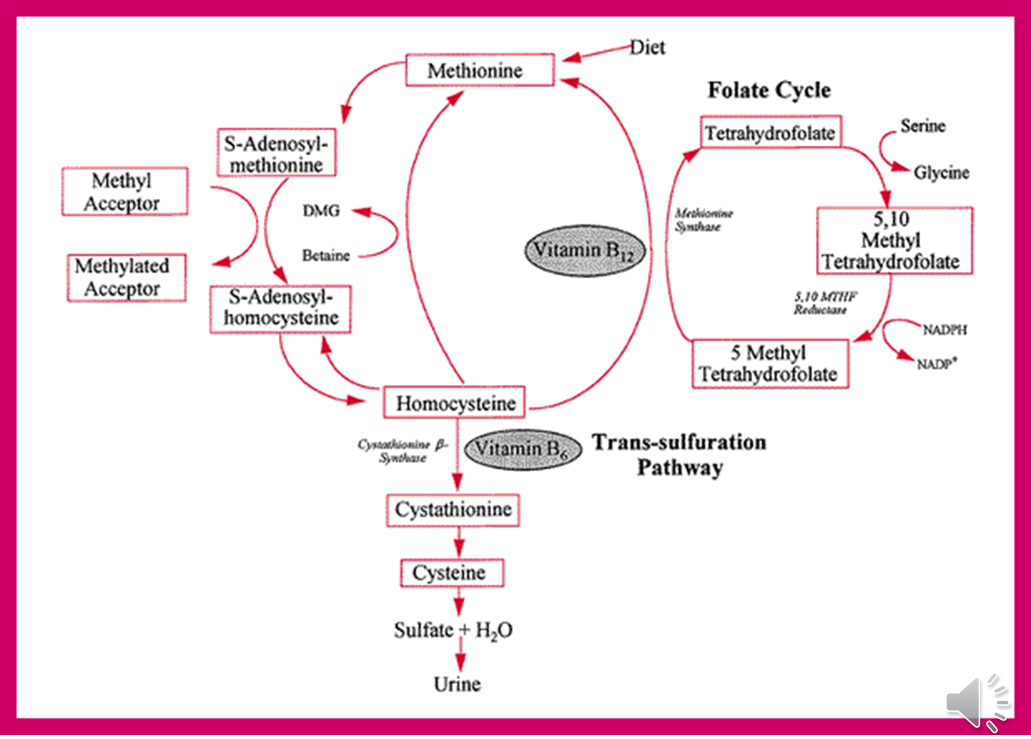
Homocystinuria Metabolic Pathway
Methionine (taken in via diet) → Homocysteine via multiple steps
Homocysteine → Cysteine via Cystathionine Beta-Synthase (CBS) + B6 (coenzyme)
Block of CBS = Elevated Homocysteine and decreased Cysteine levels
Homocysteine converted back to Methionine (Methionine synthase + B12 coenzyme)
Untreated Homocystinuria
Marfanoid Skeletal changes
Tall and thin, long arms legs fingers
Chest pectus
High-arched palate
Crowded teeth
Ectopia Lentis (dislocation of lens)
Can be first clinical manifestation (typically 3-5 years): different displacement DOWN (vs UP in Marfan’s)
Neuropsycatric Disease
Spontaneous Veinous/Arterial Thromboembolism
Up to 25% by age 15: casue stroke, death pulmonary embolism
Diagnosing Homocystinuria
Newborn screening: via tandem Mass Spect.
Elevated Methionine levels
false negative: accumulation of MET not high in the first few days
false positives: multiple conditions cause elevated MET: liver disease, hypermethioninemia (MET elevated, but not homocysteine)
Biochemical Confirmation
Plasma AA analysis
elevated homocysteine + MET
low/absent Cysteine
Urine: elevated homocysteine (NOT AS SPECIFIC as plasma AA)
CBS enzyme activity: Skin fibroblasts and hepatocytes
Genetic Confirmation
CBS gene sequencing and dup/del analysis
Many mutant alleles, mostly compound heterozygotes
Homocystinuria Treatment
Diet
Restrict methionine and protein
MET-free metabolic formula
Medication
Vitamin B6 (also called Pyridoxine)
BBS cofactor challenge (50% B6-responive)
Supplement folate and B12 (allow for conversion of homocysteine back to methionine)
Betaine
offers alternative route for homocysteine remethylation: a methyl group donor which lowers homocysteine levels by remethylating homocysteine to methionine
used in patients not dramatically B6-repsonive
Outcome
near normal IQ if treated early and effectively
Marfan-like complicaitons significantly reduced
Homocystinuria Carrier Detection
Carrier Testing
If mutation found in affect Individuals
Linkage analysis if mutation not found on affected individuals
Prenatal/Preimplantation Diagnosis
CBS enzyme activity assay in amniocytes (not possible on CVS)
Amniotic fluid homocysteine levels
Mitochondria
Site of oxidation phosphorylation: via the electron transport chain embedded in the inner mito membrane
Produce ATP
The other biochemical processes occur in the Mito:
Pyruvate oxidation
Krebs Cycle
Fatty Acid Beta-Oxidation
Oxidative Phosphorylation
The electron transport chain
Inner membrane has 5 distinct protein complexes embedded: some encoded by nuclear DNA, some encoded by Mito DNA
Use NADH + FADH coming form Krebs cycle break down of Actyl-CoA
Fatty Acids (generated via F.A. B-Oxidation)
Pyruvate (generated via glycolysis)
Electron transport down the chain of complexes: creates gradient by pumping IN H+ ions
Complex V uses gradient to Generate ATP as H+ ions move OUT
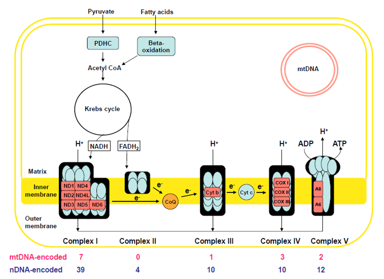
Why does Mitoondiral diase present with LACTIC ACID and MITO PROLIFERATION?
Lactic Acid
Glycolysis (break down of 1 glucose molecule) produces: 2 ATP + 2 Pyruvate + NADH
NADH: shuttles (H+) into the ETC → LOTS OF ATP MADE → NAD- is produced, ready to ‘shuttle’ a new (H+)
Pyruvate: gets converted to Acetyl-CoA → A-CoA gets used in the KREBS CYCLE to make more NADH
ATP: just a small amount
When ETC is not working right due to MITO DIEASE: NADH cannot release it’s (H+), no NAD- is ‘renewed’
Pyruvate must react with NADH instead → NAD- + Lactate is produced
NAD can be used in glycolysis to make small amount of ATP
Lactate builds up → LACTIC ACIDOSIS
Mitochondrial Disorders
Oxidative Phosphorylation/Electron Transport chain dysfunction
Two varieties:
Secondary Mitochondrial dysfunction: Non-genetic conditions
Hypoxemia (inadequate Oxygen for Oxidative Phosphorylation)
Medication: valproic acid, HIV meds
Toxins: cyanide, rotenone
Primary Mitochondrial Disease
mitochondrial DNA itself or nuclear DNA mutations
General Features of Mitochondrial diease
Mitochondrial disease preferentially affects tissues with the HIGHEST energy demands →
BRAIN: seizures, ataxia, stroke-like episodes
MUSCLE: myopathy, hypotonia
HEART: cardiomyopathy, conduction defects
EYES: ***PTOSIS*** eye muscle weakness, optic atrophy
EARS: Hearing loss
Mitochondrial Genome
The Mitochondrial Chromosome: encodes 37 genes
only 3% of Mito. proteins are encoded by mito DNA
97% are encoded by nuclear DNA and imported into mitochondria
Complex 1
46 total proteins
MtDNA encoded: 7
nuDNA: 39
Leigh Syndrome
Leukodystrophy
Complex 2
4 proteins: ALL nuDNA ENCDOED
Leigh Syndrome
Paraganglioma
Pheochromocytoma
Complex 3
11 proteins
MtDNA: 1
nuDNA: 10
Leigh syndrome
GRACILE syndrome
Complex 4
mtDNa: 3
nuDNA: 10
Leigh Syndrome
Hepatopathy
Cardioencephalomyopathy
Leukodystrophy/tubulopathy
Complex V
mtDNA: 2
nuDNA: 14
Maternal Inheritance
mtDNA mutations can only be inherited through the mother
all mito provided by the ovum
no mito contriubted by the sperm
Heteroplasmy
Mito genomes can differ between mitochondria in a given cell and % of mutant mtDNA can vary in an individual from cell-to-cell and tissue-to-tissue
Each cell has up to 1000 mitochondria, each with their own copy of the mito genome
mtDNA mutation rate is 10-20x nuclear DNA mutation rate
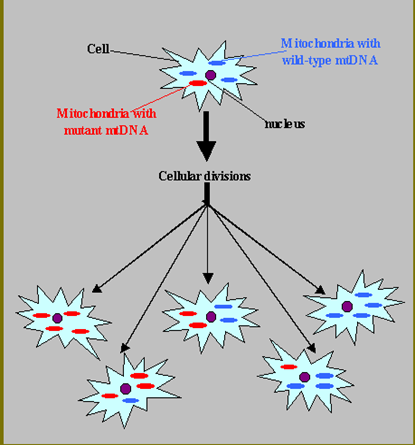
Threshold Effect.
energy requirements vary between tissues
mtDNA mutation burden varies tissue to tissue (heteroplasmy)
Tissue specific % mutant mtDNA threshold for disease
Phenotypic variability results
Example: Brain and Muscle have a lower threshold than Skin and Kidney
Mitochondrial Disorders: Presentation
Can present in almost any way and vary from person to person, but 3 general categories
“Classic” Mitochondrial diseases: reproducible, multi-organ pattern
Unexplained multi-organ dysfunction:
Hearing loss short stature
Diabetes + hypertrophic cardio myopathy
ophthalmoplegia +ptosis
Unexplained single organ syndrome: just hearing loss, epilepsy, GI
Often elevated Lactic acid in Blood or CNA and Mitochondrial proliferation in muscle
Mitochondrial Encephalomyopathy, Lactic Acidosis and Stroke like episodes (MELAS)
Age of Onset: before 40yo (average 5-15)
Clinical
Stroke like episodes + Epilepsy, Dementia
Muscle weakness (myopathy), Cardiomyopathy, Lactic Acidosis
Hearing-Loss, Retinopathy, Diabetes
CT/MRI: Infarcts→ but not seen in vasuclar regions: infarct occurs due to region engery insufficney from Mitocondrial
Etiology: heterogeneous mtDNA mutations (Often mt-t RNA) → VERY dependent on Heteroplasmy with individual
Myoclonic Epilepsy with Ragged Red Fibers (MERRF)
Adolescent onset
Clinical manifestations
Epilepsy (myoclonic)
Muscle weakness (myopathy), Lactic acidosis, Ataxia
Encephalopathy, Hearing Loss
EMG
EEG:
Muscle Biopsy: (if done on affected muscle) will show ‘ragged red fibers’ caused by mitochondria proliferation
Etiology: Single mtDNA-tRNA mutation 80 to 90%
Leber’s Hereditary Optic Neuropathy (LHON)
Age of onset 20-24 yo
Clinical:
Acute or sub-acute bilateral central vision loss→ Rapid progression to blindness (usually confined to optic nerve)
Rarely: heart block, dystonia, MS-like symptoms
Fundoscopy: early tortuous retinal arteries, followed by optic atrophy
Etiology: 95% mtDNA “ND” (electron transport subunit) gene mutations MATERNAL INHERITANCE
****4:1 M:F ration → X-linked modifier genes that make females less affected****
Chronic Progressive Ophthalmoplegia (CPEO)
Chronic Progressive Ophthalmoplegia (CPEO)
External ophthalmoplegia (eye weakness) → can’t look in certain directions
bilateral ptosis (eyelid drooping)
mild myopathy (limb weakness)
Onset ***AFTER*** 20yo (slowly progressive)
Etiology:
Mainly mtDNA deletions→ can be smaller or larger chunks of mtDNA (smaller =CPEO, larger=KSS)
Majority are SPONTEOUS
Kearns Sayer Syndrome
<20 years + ophthalmoplegia (ptosis w/ eye-muscle paralysis) + pigmentary retinopathy + HEART BLOCK + Ataxia
mtDNA Deletions
small deletion = CPEO
LARGE deletion = KSS
Earlier onset than CPEO (before 20yo) and EXTRA systemic mito. disease features (ataxia)
*****CARDIAC CONDUCTION DEFECTS → HUGE FEATURE****
INHERITANCE
Single large-scale mtDNA deletion disorders are usually sporadic/de novo rather than showing the classic maternal transmission you associate with many mtDNA point variants.
KSS does not usually produce a pedigree full of maternally affected relatives.
Sengers Syndrome
Gene: AGK (dysfunction impairs mitochondrial oxidative phosphorylation and energy production)
MITOCONDRIAL DIEASE
Inheritance
Autosomal Recessive
Clinical Features
Eyes
***CONGENTIAL CATARACTS***
Cardiac
**** Hypertrophic Cardiomyopathy****
Metabolic
Lactic Acidosis
Muscle
Skeletal Muscle Myopathy
[ **Congenital CATARACTS** + **HYPERTROPHIC CARDIOMYOPATHY** + LACTIC ACIDOSIS + skeletal MYOPATHY ]
Subacute Necrotizing Encephalopathy (Leigh Syndrome)
Large spectrum of 75 genes (mito and nuc. but mostly nuc.) that cause an energy failure in the brain
*****USUALLY AUTOSOMAL RECESSIVE****
6-12 months onset - death by 3-5 years (25% have later onset or slower forms)
Clinical: (often abrupt decompensations/regression with infection/fever)
Developmental ***REGRESSION***
Seizures, Ataxia, Hypotonia, spasticity
Ophthalmoplegia, Nystagmus, Optic atrophy
Diagnostic Testing
MRI: ***SYMETRIC LESIONS OF BASAL GANGLIA***
Elevated Lactic Acid in blood or Cerebral spinal fluid
10% mtDNA mutation
90% nDNA mutation
Lower % of mitochondria with the mutant mtDNA→ have NARP instead of Leigh (HETEROPLASMY)
Leigh Etiology
Genetic Heterogeneity
10-30% mitochondrial DNA mutations → maternal inheritance
90-70% nuclear DNA mutations → Classic Mendelian
HETEROPLASMY AFFECT: If a lower # of mito. in a cell have these mutations = Later onset Neuropathy, Ataxia, Retinitis Pigmentosa (NARP)
Pyruvate Dehydrogenase Complex (PDHC) Deficiency: Clinical + Testing
Gene: PDH1
Failure to convert Pyruvate to Actyl-CoA (via PDH)
Lactic Acid levels elevated (***PDHC most common cause of Lactic Acidosis***)
Inheritance
**** X-LINKED****
Point mutation in NUCLEAR DNA
Clinical Features
Progressive intermittent neurologic deterioration
hypotonia, seizures, ataxia, ophthalmoplegia, dystonia
Presents similar to mitochondrial dysfunction
Suggestive Abnormal Tests
Plasma: increased Lactic Acid + Pyruvate, but normal ratio of Lactic Acid: Pyruvate
Distinguished from other Mitochondrial Disease: Pyruvate levels are NOT elevated
Cerebral Spinal Fluid: increased Lactic Acid
Pyruvate Dehydrogenase Complex (PDHC) Deficiency: Metabolism +Etiology
Failure to convert Pyruvate to Actyl-CoA (via PDH)
Lactic Acid levels elevated (PDHC most common cause of Lactic Acidosis)
Etiology: PDHC is a multisubunit complex
Catalytic components: E1, E2, E3
Regulatory component: PDH Phosphatase
Confirmation:
PDHC enzyme activity assay
Sequencing of
E1 → PDHA1 : MOST COMMON , X-Linked (males only)
E2 → DLAT, Recessive
Mitodoncrial Diease: Work Up
Serum levels: increased anion gap + metabolic acidosis
Lactic Acid: Pyruvate ratios (>30 Mito. Dis ; <10 PDHC Def.)
Imaging: brain MRI, Spectroscopy ( LA peaks over brain regions( BasalGang)
Basal Ganglia hypodensities: generalized atrphy
Hypoplastic corpus callosum if fetal lactic acidosis
Muscle Biopsy
Genetic Testing
Mitochondrial Disease: Muscle Biopsy
Allows for:
Detecting ragged red fibers (mito. proliferation)
Abnormal mitochondria proliferation
Detecting enzyme activity of the chain-genes
Mutational analysis of mitoDNA
Pitfalls:
need 1 gram of flesh (large amount)
biopsy of moderately affected muscle
may not distinguish exact genetic mechanisms
Genetic Testing for Mitochondrial Conditions
mtDNA:
Leigh Syndrome
LHON
MERRF (blood/muscle)
MELAS (blood/muscle)
NARP (blood/muscle)
KSS/CPEO (muscle)
nDNA (all in blood)
Leigh syndrome
MNGIE
Mohr-Tranebjaerg
Friedreich’s Ataxia
AR spastic paraparesis
AD PEO
Mitochondrial Disorders: Treatments
Less evidence for specific treatments that actually improve outcomes
Trials with Vitamins that optimize Electron Transport chain function:
Carnitine
Biotin
thiamine
Riboflavin
High Fat/ Low Carb diet: low carb→ less glycolysis→ less LA
Avoid Mito toxic meds
Reduce LA, control acidosis (dialysis/vent)
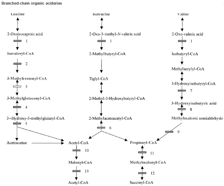
Organic Acidemias Background
Primarily disorders of Amino Acid Catabolism: Mainly
Branch Chain Amino Acids (BCAA)
Lysine
Toxicity comes from accumulation of ORGNIC ACIDS not from an A.A. acid accumulating
Causes metabolic acidosis with increased “Anion Gap”: Decrease in main anion Bicarbonate (HCO3-)
Secondary toxic effects of acidosis
Mitochondria→ Lactic acidemia
Urea Cycle → Hyperammonemia
Bone marrow→ Bone marrow suppression
CNS function→ Encephalopathy/Mental retardation
Major Presentations: Neonatal encephalopathic acidosis, late chronic/intermediate
All autosomal recessive
Metabolic Acidosis
Blood pH low due to excess acid (H+) vs Base (HCO3-)
normal range pH 7.3-7.45 (measure via Atrial Blood Gas)
Normal HCo3- level: 22-26 mEq/L
Mutiple etiologies for Metabolic Acidosis
Lowered HCo3- : loss through GI (diahrria), Renal tubule acidosis, Medications
Elevated H+: creation of abnormal acids in blood due to starvation, diabetes; Lactic acidosis due to mitochondrial dysfunction, Organic Acidosis
Clinical Consequences
Neonatal: non-specfic, similar to UCDs presenations,
Lethargy, vomting, Tachypena, Hypotonia, Seizures, Coma, Death
Adult: Devleopmental Delay, Ataxia, Neurological Deficits, (then the neonatal presenations)
Organic Acidemias
Newborn Screening detects many Organic Acidemias
Organic Acidemias Treatment
Restrict Dietary Protein disease specific amino acid free formulas
Prevent Catabolism provide sufficient protein free calories
Reverse Acidosis ± Hyperammonemia
Hemodialysis
Ammonia and lactic acid scavengers
Sodium bicarbonate, sodium benzoate, phenylbutyrate
Cofactor therapy for specific Disorders
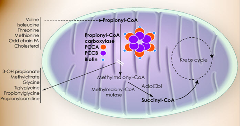
Propionic Acidemia Metabolism
Failure of Propionyl-CoA carboxylase
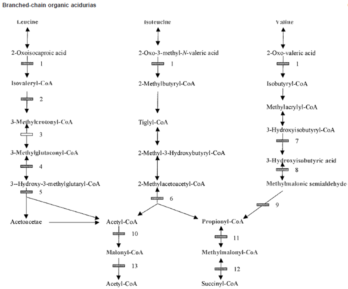
Step 11 of Isoleucine and Valine metabolism:
Propionyl-CoA → Methylmalonyl-CoA via Propionyl-CoA carboxylase activity
Propinoyl-CoA: the activated mitochondrial form
Propionic Acid: free organic acid (interferes with NH3 removal, other stuff)
Propinolycarnatine (C3 - what NBS measures): the ‘detox’ / transport form that excess P-CoA gets converted to so it DOESN'T become Propionic Acid
Propionic Acidemia
Also known as Ketonic hyperglycemia: high level of glycine and ketone bodies
Autosomal Recessive
Incidence 1:100,00 (higher in Saudia Arabia and Inuit)
Genetic Defect
Propinyl-CoA Carboxylase (PCC) alpha or beta subunit genes
some genotype/phenotype correlation (null alleles/deletions more severe)
BIOTIN cofactor for PCC
PA accumulation due to PA production from
MET/THR/VAL/ISO catabolism,
gut bacteria,
odd chain FAs
Untreated Propionic Acidemia
Classical Neonatal Encephalopathic Form
Normal at birth
Within a few days
Poor feeding, lethargy, vomiting hypotonia →encephalopathy, seizures, coma, death
Late-Onset Form
Developmental delays/regression
cyclic vomiting
protein intolerance
growth impairment
hypotonia
metabolic basal ganglia stroke
cardiomyopathy
Acute episode of toxic encephalopathy
Rare Cardiac Subtype isolated cardiomyopathy
Diagnosing Propionic Acidosis (PA)
Newborn Screening
Elevated Propionyl Acylcarnitine (C3) and ratio to other carnitine species (Propionyl-CoA gets combined with Carnatine to try buffer high Prop-CoA levels)
other etiologies: Methylmalonic Acidemia, Cobalamin Defects, Maternal B12 Deficiency, False +
Confirmatory Testing
Atrial Blood Gas: Elevated ammonia, low glucose, high acidosis, increased anion gap
Complete blood count: suppression of bone marrow→ less blood cells
Urine Organic Acid Analysis: High 3-OH-proprionate, mthylcitrate, tigly/proprionylglycine but NOT MMA
Plasma Amino Acid profile:
elevated glycine + glutamine, not homocysteine (seen with Cobalamin defects)
Acyl-Carnitine Profile: Elevated C3 acylcarnitine, not C4-DC unless SUCLA2 deficiency
PCC enzyme activity: can measure PC enzyme in leukocytes or fibroblasts
PCC Genotyping
Gene sequencing w del/dup analysis (99% detection rate)
Treating Propionic Acidosis
Acute Acidotic Encephalopathy
Remove acids and ammonia hemodialysis
severe hyperammonia: ammonia scavengers
Reduce PA production Protein restriction 24-25hr
Prevent catabolism: glucose and lipids IV
Enhance PA excretion: IV Carnitine
Decreased PA production in Gut: Antibiotics (Metronidzole
Biotin:
Chronic Treatment
protein restriction and MTVI-free metabolic formula
Oral Carantine, Biotin, and Antibiotics
Avoid decompensation
unresponsive to Tx → liver transplantation
Propionic Acidemia Deficiency Outcome
Treamtnet improves surivial, but invariable there is an affect to some degree
Neurodevleopmatl disabilty
metabolic basal ganglia stroke
seiures
pancreatisis
cardiomyopathy
gorwth impairment
nuetorpnia, AA defience
renal failure
premature ovarian fialure
hearing and vidual defecits (optic nerve atrphy)
Propionic Acidemia Deficiency Screening
Carrier Screening:
PRenatal Diaongis:
amontic fluid orgnaic acid measurment possible (some false negatives)
Methylmalonic Acidemia Pathway
Isoleucine and Valine
Methlymalonyl-CoA → Succinyl CoA via Methylmalonic-CoA mutase activity
Methlymalonyl-CoA accumulates
Methylmalonic Acidemia
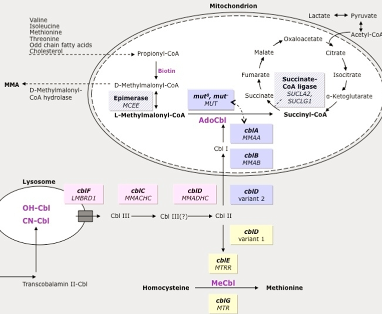
Increased Methylmalonic Acid but not homocysteine (other forms of MMA have elevated homocysteine→ not primary MMA, but related to Adenosyl Cobalamin - A )
Genetic Defect: mutation of multiple genes cause similar phenotype
60% Methlymalonyl-Co mutase gene mutation (MUT)
37% Cobalamin A,B,D2 (MMAA, MMAB, MMADHC)→ the upstream vitamins that will be converted into Adenosyl Cobalamin→ leads to dysfunctional MM-Co mutase
Untreated Methylmolaynic Acdiemia
Infantile Subtype: Most common mut0, cblB mutations
Normal at Birth
Within days to weeks: poor feeding, lethargy, vomiting, hypotonia, encephalopathy→ progress to seizures, coma, death
Intermediate phenotype: mut-, cblA, cblD2
Normal for month to years: fialure to thrive, devleopmental delay, hypotonia, poriten aversion→ risk of carastrophic decompensations
Benign Adult form: typically asymptomatic, can decomapnste
Diagnosing Methylmolaynic Acdiemia
Newborn screening: Elevated Propinoyl Acylcarnitine (and ratios) → but non specific
Confirmatory testing
Atrial Blood Gas, Ammonia Levels, Completel blood ocunt:
Hi AG metabolic acidsosi
Elevated ammonia
Low gluclose
pancytopenia
Urine Organic Acid: High MMA
Plasma Amino Acid profile: high glycine + glutamine, no Homocystine (Hcy)
CblC/D/F - Hcf + MMA high ;
cblD2/E/G - Just hcf High
Enzyme activity: fibroblasts
Genotyping on genes = 95%
Treating Methylmolonic Acidemia
Treat acute acidotic encephalopathy
Remove acids+amonia: hemodialysis
Reduce MMA production: protein restriction
Prevent catabolism: IV glucose and lipids
Severe hyperammonemia: Amonia scavengers
Decrease gut bacteria: Antibotics
HYDOXYCOBALAMIN (B12) injects: cofactor
Chronic Treatment
protien restriciton and MTVI-free meatolibc fomumal
L-Carnitine + OH-B12
Avoid decompensation
Methylmalonic Acidemia Treatment outcome
Most patient will have some degree of mental impairment, long term affects
Methylmalonic Acidemia Diagnosis
Prenatal/Preimplantation:
Ammonitic organic acid fluid analysis possible
Enzyme activity of CVS and amniocentesis
Iso-valeric Acidemia
issues with the Isovaleryl-CoA dehygroenase enzyme
LEUCINE PATHWAY ONLY
Build up of Isovalryl-CoA (Isovaleric Acid)
Disorder of Leucine metabolism only (unlike PA or MMA)
Genetic Defect
IsoValeryl-CoA Dehydrogenase (IVD) Gene Mutation
results in increased Isovaleric Acid
Sweaty feet odoer is prominent
Untreated Isovaleric Acidemia
Severe Neonatal Onset form
Normal at birth
Within day: poor feeding, lethargy, hypotonia, Sweaty feet order → encephalopathy, seizure, coma, death
Mid/Late Onset Form
unexplained failure to thrive and developmental delay
Benign Adult Form: Typically, asymptomatic but can mildly decompensate
Diagnosing Isovaleric Acidemia
Newborn screening: elevated Isovalerylcarnitine (C5 acylcarnitine) - used to ‘buffer’ Isovaleric Acid that builds up when Acylvaleryl-CoA builds up
Confirmatory testing
Blood tests:
High ammonia
Low glucose
High metabolic acidosis
Urine organic acid
High IVA
High isovaleryl glycine
Plasma AA levels:
High glycine
High glutamine
Enzyme activity: Fibroblast
Genotyping: exact genes unknown
Treating Isovaleric Acidemia
Treat Acute Acidotic encephalopathy
Remove acids and ammonia: hemodialysis
Reduce IVA production: protein restriction 24-26hrs
Prevent Catabolism: IV glucose and lipids
Enhance IVA excretion: IV carnitine
If hyper ammonia: ammonia scavengers
GLYCINE SUPPLMENTAITON-BINDS IVA
Chronic Treatment
Protein rection and LEUCINE-free metabolic formula
Oral L-Carnitine and L-Glycine
Avoid decompensation
Isovaleric Acdiemia Otucome
Outlook with Treatment is one of the best if treatment done early and effecetively enough
can be comepltely asymptomatic as long condition is monitored
Leucine tolerance gets better with age
Even if diaognsis is after neonatal period, and evne with major encaplapthic event in neonatal period—> longer term out look is vairable : CAN BE OK
Isovaleric Acidemia Prenatal Diagnosis
Ammniotic fluid can be checked for organic acids
Biotinidase Deficiency Pathway
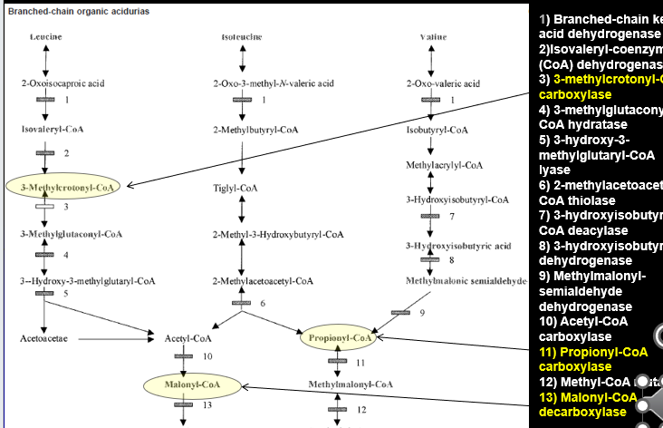
Biotin is a vital cofactor for of number of different enzymes:
ALL ARE CARBOXYLASES
3-Methylcrontoyl-CoA carboxylase (Leucine)
Propinoyl-CoA Carboxylase (Isoleucine and Valine)
Malonyl-CoA decarboxylase
When there are mutations in the BIOTINADASE gene: Biotin is not properly recycled → these blocks develop
Biotinidase Deficiency (BTD)
Late multiple Carboxylase Deficiency
Slightly increased incidence in Hispanic and Middle Easter
Gene Defect: Biotinidase (BTD) gene
failure to recycle biotin = biotin deficiney
Biotin co-factor for the carboxylases: cannot combine and make function enzyme
Untreated Biotinidase Deficiency
Affect depends on the residual enzymatic activity when biotin absent
Profound Deficiency (<10% enzyme)
Clinical Features
No acute acidodic encepahlopathy decompensatio events : GRADUAL
Normal at birth
Symptoms develop after few months”
Developmental delay, seizures, hypotonia, ataxia
hearing loss, visual problems,
***alopecia***, ***eczema*** (unique to BTD)
Partial Deficiency (10-30% enzyme)
intermittent symptoms with stress
Symptoms can be irreversible once present
Diagnosing Biotinadase
Newborn Screening: Elevated C5-OH Acylcarnitine, but not specfic to BTD
Confirmatory Testing
Blood:
High ammonia
High acidosis
Low gluclose
Urine Organic Acids: multiple organic acids b/c Bitonaisde affects multiple enzymes → referred to as ‘Multiple Carboxylase Defeicieny’ (MCD) on uOA
( )
Elevated Hydroxy-Isovalyrl-carnatine (C5-OH)
Enzyme activity: IMPORTANT STEP: blood sample
If Biotinadase activity is normal→ then issue is probably Holocarboxylase Deficiency (presents the same way but is earlier)
Genotyping: sequencing 99% detection
Treating Biotinadase Deficeincy
Rarely severyl acidotic or hyperammonemic
may occasionally need sodium bi-cabonate (adress acidty)
may occasionally need amonia scavnerge (adress amonia levels)
Insitute Biotin therapy immediately
Chronic treatment
Biotin
No protien restction
Avoid raw egg whites (has protein that binds Biotin)
Biotinadase Deficiency outcome
Extremely great outlook for patients (one of the best for Organic Acidemias)
As long as treatment is implemented BEFORE the development of severe symptoms
If detected after symptoms, some are irreversible: optic atrophy, hearing loss, developmental delay can presist
Biotinadase Deficiency Prenatal diagonsis
Biotinadase enzyme activity can also be measuredin the amniocytes and the amniotic fluid
Glutaric Acidemia Type 1 (GA1) Metabolism
NOT a Branch Chain Amino Acid metabolism disorder
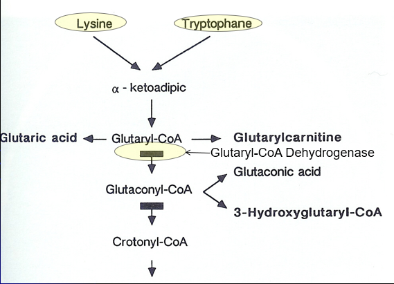
Breakdown of LYSINE and TRYPTOPHANE
Lysine + Tryptophane → Alpha ketoadipic→ Glutyrl-Coa
Glutyrl-Coa→ Glutaconyl-CoA (shunt to Glutaontic Acid) via Glutaryl-CoA Dehydrogenase activity
Glutaric Acidemia Type 1 (GA1)
“Cerebral” Organic Acidemia: Often normal
Genetic Defect: Glutaryl-CoA Dehydrogenase (GCDH) gene mutation causing defective Lysine + Tyrptohan metabolism
Glutaric Acidemia Type 1 (GA1) Symptoms
Often normal at birth or only macrocephalic
symptoms often begin prior to 2 years of age
May start with a Sudden neurologic decompensation: 75% by 14months → fever, illness, metabolic stress
Primary symptoms
Progressive Macrocephaly
Acute encephalopathic crisis
Hypotonia,
Loss of motor skills,
Feeding difficulty,
SOMETIMES Seizure
***Movement Disorder****
speciosity
dystonia (involuntary movements)
Glutaric Acidemia Type 1 (GA1) Diagnosis
Newboarn screening: Elevated C5-DC (glutaryl) Acylcarnitine
many False negatives
Confirmatory Testing
Blood: elvated ammonia, low gluclose, Aciditiy
Plasma + Urine: C5-DC glutaryl acylcarnitine + glutaric acid
Enzyme activity: fibroblast
CT/MRI: Cerberallar atrophy, basal ganlia infact and hemorrhage
Genotyping
Glutaric Acidemia Type 1 (GA1) Treatment
Reverse/Prevent Catabolism When sick: protien free calroeis during metaoblic stress
Dietary Mdofication
Low LYSINE and TRYOPTHAN
MEdicaitons
B2 (Riboflavin) is a COFATOR
Carntine: Binds Glutaric acid and remvoes it
Avoid Valproate (Bind Carnitine)
The Urea Cycle
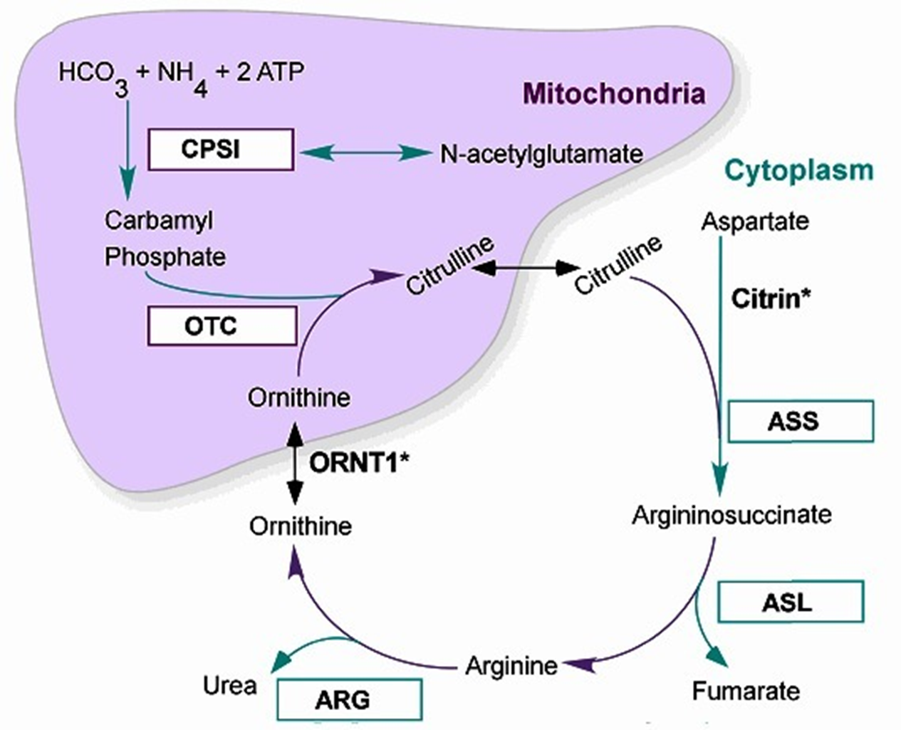
6 Major Enzymatic reactions that occur in the liver within the Hepatocytes: the mitochondria + cytoplasm
2 Major functions
removal of nitrogenous waste (produced mainly from protein catabolism) → Ammonia incorporated into Urea for Excretion
Synthesis of amino acids: Arginine, Ornithine and Citrulline (become ESSENTIAL A.A. in deficiencies of the UREA CYCLE ENZYMES)
Major presentations of Urea Cycle Disorders:
Severe Neonatal Hyperammonemic encephalopathy (exception: Argine deficiency + late/mild onset variants)
All Autosomal Recessive except for Ornithine transcarboxylase deficiency (OTC):
OTC is X-Linked Recessive → only affects Males
The 6 Urea Cycle Disrders
Correspond to the 6 steps of the Urea Metabolic cycle: taken individual relatively uncommon, but all together 1:8-35,000
N-Acetyl Glutamate Synthetase Deficiency
Carbamoyl Phosphate Synthetase (CPS1) Deficiency
****Ornithine Transcarbamylase (OTC) Deficiency****—> *****MOST COMMON*****
Arginosuccinic Acid Synthetase I (ASS1) Deficiency → also called Citrullinemia I
Arginosuccinic Acid Lyase (ASL) Deficiency
Arginase (ARG) Deficiency
Ammonia (NH3)
Ammonia is the product of the metabolism/catabolism of protiens/amino acids
Normal serum ammonia levels
Adults <35 mmol/L
Neonates <100 mmol/L (immature liver cells + increased tissue catabolism surrounding delivery)
Hyperammonemia: happen with great degree with IEMs of Urea Cycle But also seen with other metabolic disorders like Organic Acidemias (excess acid decreased Urea Cycle activity, lesser extent that UCDs)
Causes Neuronal excitotoxin increased extracellular glutamate +overexcitation of NMDA receptors→ Cell death and Cerebral Edema
Clinical Consequence
Acute severe elevation: seizures, coma, death
Mild chronic elevations: Brain atrophy, cognitive impairment
Classic UCD Presentation: Early
Neonatal Hyperammonemia Encephalopathy
In utero: protected my maternal urea Cyle activity of liver cells
At Birth in first 48hours: Ammonia levels rise quickly
Decreased feeding w/ vomiting
Lethargy
Tachypnea (rapid breathing)
Seizure activity
Followed by Rapid
Encephalopathy/Coma
Respiratory Failure
Cerebral Edema and Death
Late-Onset UCD Presentations
Variable age of onset and severity of Chronic and/or recurrence/Fluctuating symptoms:
Headache, Vomiting Ataxia and incoordination
Psychiatric/Behavioral disturbance: Delirium, ASD, ADD/ADHD, Manic episodes
Cognitive impairment: DD/MR, executive processing defects, early dementia
Often exacerbated/precipitated by:
Fever, Illness, fasting, post-partum, protein load (self restrict protein)
STILL AT RISK FORM HYPERAMMONEMC ENCEPHALOPATHY:
even if previously asymptomatic, can still be fatal
Mutiple Etiologies for Hyperammonemia
Things besides UCDs (MOST COMMON CAUSE) that can also cause the accumulation of excess ammonia:
Generalized Liver Deiasie (Acute or Chronic)
Non-genetic Causes: infections, Toxins, Trauma, Ischemia etc.
Genetic Causes
Non-IEM: Alpha-1 Antitrypsin (accumulation in liver=chrossis), Alagille syndrome etc
Non-UCD IEM
Aminoacidopathies: Tyrosinemia Type I
Organic Acidemia: elevated Lactic acid which inhibit NAGS
Primary Mitochondrial disorders
Fatty Acid oxidation Defects: Reye-Like syndrome
Carbohydrate Metabolic defects: Galactosemia, Fructosemia
Metal Processing defects WILSONS DIEASE, hemochromatosis
Primary UCDs