neuroscience exam 2
1/37
There's no tags or description
Looks like no tags are added yet.
Name | Mastery | Learn | Test | Matching | Spaced |
|---|
No study sessions yet.
38 Terms
describe the experiment that provided the first direct evidence for the existence of a chemical transmitter
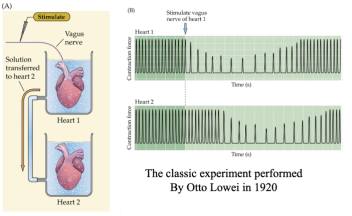
Otto Loewi experiment:
provided the first direct evidence for chemical neurotransmitter
Loewi placed 2 beating frog hearts in their own perfusion chamber
after stimulating the vagus nerve of the first frog heart, the heart beat more slowly
Loewi took the fluid from chamber of the first heart and applied it to the second heart
after that, the second heart slowed down as well
he named the molecule causing the second heart to slow down “vagusstoff” aka acetylcholine and proved that neurons communicate through chemical transmission
had the idea in a dream and woke up in the middle of the night to do the experiment which won him a Nobel Prize
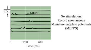
explain the term miniature end plate potential (MEPP) and what is the basis of MEPP
miniature end plate potential (MEPP): a small depolarization of the postsynaptic terminal that is caused by a single vesicle
when the nerve is NOT stimulated, the mean amplitude for a MEPP is 0.4mV (in a distribution, most MEPPs are 0.4mV)
there are still spontaneous fusions of single vesicles and those lead to small MEPPs
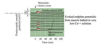
what does the term quantal hypothesis mean? what is the evidence that supported this hypothesis?
quantal hypothesis: neurotransmitters are packed together in “quanta” that are released probabilistically
though each vesicle only changes the membrane potential by about 0.4mV, the total EPP is due to the release of many of these vesicles, and their combined effect is enough to produce a large change in membrane potential
evidence:
experiments conducted by Bernard Katz
Katz observed that the membrane potential would change spontaneously without stimulation
those changes would be shaped like EPPs but had a much smaller amplitude
this led to the discovery of MEPPs
Katz then stimulated the frog neuromuscular junction with very low concentrations of extracellular calcium (Ca2+)
Katz was able to find a low enough Ca2+ that produced some EPP response that was equal to the size of 1 MEPP
other responses had no EPP observed
other responses less frequent EPP responses that equaled multiples of single MEPPs
what is the effect of reducing extracellular calcium concentration on synaptic transmission?
decrease in extracellular Ca2+ concentration results in a smaller end-plate potential
this is due to Ca2+ causing the release of neurotransmitters from vesicles
if less Ca2+ enters the presynaptic terminal from the extracellular space, the probability of vesicle fusion will decrease and thus result in a smaller response in the postsynaptic neuron
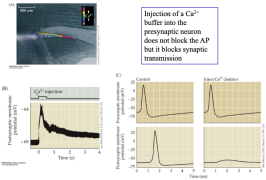
how would injecting a calcium chelator/buffer into the presynaptic site affect synaptic transmission?
all presynaptic events responsible for synaptic transmission are initiated by the influx of Ca2+ (ex. the release of reserved vesicles from the actin, the release of transmitter from docked vesicles, etc.)
injecting a Ca2+ chelator (thus inactivating Ca2+) would stop synaptic transmission
what are the 2 major roles that calcium plays in transmitter release? how does calcium exert these effects?
1) Ca2+ releases vesicles from the reserve pool
Ca2+ releases vesicles by activating calmodulin which activates protein kinases which phosphorylate the synapsin cap on the vesicles causing it to release the vesicle from the actin thus the vesicle diffuses away and floats down to the presynaptic membrane dock
2) Ca2+ initiates the fusion of docked vesicles in order to release their neurotransmitter
Ca2+ activates synaptotagmin protein on docked vesicles
synaptotagmin inserts itself into the presynaptic membrane and pulls the vesicle down, fusing the vesicle with the membrane and creating a pore that releases the neurotransmitter
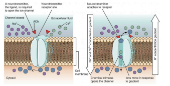
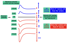
what is the definition of a reversal potential? what is the reversal potential for the nicotinic ACh (nACh) receptor at the neuromuscular junction and how is it computed? how can you measure the reversal potential of a channel experimentally?
reversal potential: the potential at which the net flow of current of a given channel starts to reverse or switch directions
ex: equilibrium potential for Na+ is +67mV
Na+ ions move through ion channels based on voltage difference
if membrane potential relative to the inside of the cell is more negative than ENa, Na+ ions flow into the cell
if membrane potential relative to the inside of the cell is more positive than ENa, Na+ ions flow out of the cell
Thus ENa is called the reversal potential
in real cells multiple ions are involved
in nACh receptors of the neuromuscular junction, the reversal potential would be the potential at which the influx of Na+ in and the efflux of K+ out would be equal
This is around -10mV and is computed as the average of Na+ and K+ equilibrium potentials
you can measure reversal potential of a channel experimentally by voltage clamping at multiple increasing/decreasing membrane potentials until the net current measured becomes zero
how would the reversal potential of the nACh receptor change if the extracellular Na+ concentration is increased? how would it change if extracellular concentration of K+ is increased?
increased Na+ concentration
neuron has greater driving force pushing Na+ in compared to a normal neuron
you would have to voltage clamp at potentials more positive than normal to counter the increased Na+ influx (a higher reversal potential)
increased K+ concentration
neuron has reduced driving force pushing K+ out compared to a normal neuron
you would have to voltage clamp at potentials more positive than normal to push out the same amount of K+ (a higher reversal potential)
you could verify this by looking at the Nernst equation and recognizing that increased extracellular concentrations always increase the equilibrium potential of a positively charge ion which would raise the reversal potential
a chemical neurotransmitter is found to depolarize one neuron but to hyperpolarize another. explain how this is possible
the identify of the neurotransmitter does not necessarily determine whether it depolarizes or hyperpolarizes neurons
if a neurotransmitter depolarizes neurons, then it is the receptor that causes an influx of Na+ when the NT binds
if a neurotransmitter hyperpolarizes neurons, then it is the receptor that causes an efflux or K+ or influx of Cl- when the NT binds
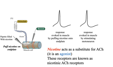
explain the terms agonist and antagonist. what are agonists and antagonists of the nicotinic ACh (nACh) receptors and muscarinic Ach (mACh) receptors?
agonist: a chemical that binds to receptors and mimics the action of a chemical that usually binds to the receptor
nACh: nicotine
mACh: muscarine
antagonist: a chemical that blocks a receptor and prevents the effect that binding to the receptor would usually cause
nACh: curare
mACh: atropine
what are the 3 criteria for establishing that a chemical is a neurotransmitter?
1) stimulating the neuron must cause the release of the chemical from that neuron
2) the effects of stimulation can be mimicked by applying the chemical directly to the postsynaptic cell
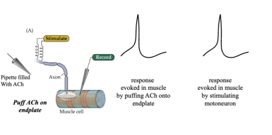
3) the response must have the same pharmacology — the responses to the external application of the chemical must respond to various drugs in the same way neuron stimulation would in response to the same drugs
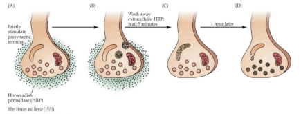
describe the experiment that provides direct evidence for recycling of synaptic vesicles
horseradish peroxidase (HRP) is introduced to the synaptic cleft of the frog neuromuscular junction
after stimulating the presynaptic terminal with a train of action potentials, the location of HRP can be visualized through electron microscopy
thus HRP can be used to track the location of elements that were originally in the synaptic cleft over time
after washing extracellular HRP, intracellular HRP can be found immediately after the stimulation within coated vesicles
after a few minutes, the coated vesicles disappear and the HRP can be found within the endosome
after an hour after stimulation the HRP can be seen within synaptic vesicles revealing the recycling process
which 3 proteins play a key role in exocytosis?
synapsin: attached to synaptic vesicles and dock vesicles to the intracellular actin cytoskeleton at resting state
once phosphorylated by Ca2+ bound calmodulin, synapsin dissociates from the vesicle and allows for vesicle migration to the active site of the synapse
SNARE: vSNAREs on the vesicle and membrane SNAREs on the cell membrane side are attracted to each other
once the vesicle is freed from the actin cytoskeleton, the two types of SNAREs attach to one another and dock the vesicle against the cell membrane
synaptotagmin: merge the vesicle with the membrane
when the presynaptic terminal is stimulated by an action potential, voltage-gated calcium channels open, allowing for an influx of Ca2+ ions which will bind to the synaptotagmin located at the docking site between vesicles and the membrane which will then facilitate the merging thus releasing the contained neurotransmitter
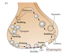
which 2 proteins play a key role in endocytosis?
clathrin: clathrin triskelions coat the membrane from the interior of the presynaptic terminal, shaping the coated pits into spherical vesicles
eventually released from around the vesicle, leaving behind the naked, complete vesicle
dynamin: binds along the base of the vesicle budding from the cell membrane
cuts away the newly created vesicle from the membrane
what are the 2 major advantages of metabotropic receptors over ionotropic receptors?
signal amplification
much longer effect after activation
what is one major advantage of ionotropic receptors over metabotropic receptors?
receptors open much quicker
describe the steps in a typical metabotropic receptor mediated response. which steps involve amplification?
Activation:
1) a transmitter binds to the receptor changing the receptor conformation to allow for binding of G-proteins.
2) G-protein binds to the receptor, this then changes the conformation of the G-protein leading to GDP being replaced by GTP on the G-protein
signal amplification occurs here as many G-proteins are activated.
3) GTP binding then catalyzes a reaction where the beta-gamma complex of the G-protein dissociates from the alpha subunit on the G-protein.
4) the GTP bearing alpha subunit of the G-protein goes on to activate adenylate cyclase which will convert ATP to cAMP and phosphate
the signal is amplified again as each adenylate cyclase produces many cAMP molecules.
5) cAMP will go on to bind and activate protein kinase A which phosphorylates and activates targets proteins
amplification happens once more as each protein kinase phosphorylates many ion channels.
Termination:
1) the neurotransmitter unbinds from the receptor meaning that the receptor no longer activates G-proteins.
2) internal GTP-ase activity of the alpha subunit converts GTP to GDP and the alpha subunit fall off of the adenylate cyclase rendering it inactive.
3) phosphodiesterase converts cAMP to AMP and PKA is no longer active without cAMP.
4) the phosphate is removed from the target protein via phosphatase.
where in the brain are the 2 major targets of dopaminergic projections and what is their behavioral role?
striatum (motor control)
forebrain (reward)
definition of spatial summation
spatial summation: occurs when the neuron receives many inputs from different presynaptic neurons simultaneously
these excitatory and inhibitory postsynaptic potentials will then be summed in the dendrite and propagate passively to the cell body where they will affect the probability of the cell firing an action potential
definition of temporal summation
temporal summation: the summation of multiple signals from a single presynaptic neuron when the incoming signals are close enough in succession
rapid succession of signals cause part of the postsynaptic potential to the first input spike to still be present by the time the next input spike is received and thus will sum together
what are the main differences between AMPA and NMDA glutamate receptors? what are the requirements for opening NMDA receptors? what ions are AMPA and NMDA glutamate receptors permeable to?
main differences
NMDA receptors are permeable to Ca2+, Na+, and K+ while AMPA receptors are only permeable to Na+ and K+
NMDA receptors are blocked by Mg2+ while AMPA receptors are not
requirements for opening NMDA receptors
require both a depolarization and the binding of glutamate to open
this combination expels the Mg2+ from blocking the pore allowing an influx of ions
what is short term synaptic facilitation and what causes it?
when the presynaptic neuron is given two pulses in a fairly short period, the postsynaptic cell experiences two postsynaptic potentials–the second one is larger than the first
thus, changes in the presynaptic site can mediate changes in the postsynaptic site
this interval is VERY short–around 10 ms!
this occurs because the Ca2+takes time to leave the presynaptic cell, and if the second pulse happens before the Ca2+leaves the cell, the Ca2+can accumulate and result in more neurotransmitter release and therefore larger postsynaptic potential
what is short term synaptic depression and what causes it?
when the presynaptic site is stimulated at a regular interval, the magnitude of the postsynaptic potential will slowly decrease
if the presynaptic site is stimulated at a very high rate for a short period of time, the magnitude of the postsynaptic potential dramatically decreases
this is because stimulating the presynaptic site too quickly depletes the synaptic vesicles and, in turn, releases neurotransmitters at a faster rate than they can be replenished.
explain the term behavioral sensitization and how can it be studied in Aplysia?
Behavioral sensitization: the increase in the response to a stimulus after a novel aversive stimulus
Aplysia study
touching the siphon and when it is touched, the Aplysia quickly contracts their gills
if the siphon is repeatedly touched, the gill eventually contracts less and less because it gets used to the sensation (i.e. habituation)
when the tail is electrically stimulated (causing a painful sensation) at the time of touching the siphon (a non-painful sensation), the gill recovers the original size of the effect, as if the siphon was being touched for the first time.
how does the modulatory interneuron in the Aplysia mediate the enhancement underlying behavioral sensitization?
when the Aplysia tail is shocked, sensory neurons innervating the tail are activated
these sensory neurons innervate modulatory interneurons, exciting them
these modulatory interneurons make connections with the presynaptic terminals on the sensory neurons of the siphon
here, the interneurons release serotonin, which binds to G-protein coupled serotonin receptors in the sensory neuron
the activated G-protein dissociates from the receptor and binds to adenylyl cyclase which stimulates production of cAMP to then bind to PKA
PKA then phosphorylates potassium channels, causing fewer channels to be open
when the sensory neuron is then stimulated and calcium flows into the cell, the reduced efflux of K+ causes a prolonged depolarization which enhances the calcium influx
more calcium will result in more neurotransmitter release, causing a stronger reaction in the motor neuron.
definition of long-term plasticity (LTP)
Long-term potentiation: a process in which frequent stimulation of synaptic connections causes strengthening of these synapses
deal with synaptic plasticity
definition of long-term depression (LTD)
LTD: a low frequency of stimulation of synaptic connections causes the connection to be weakened
deal with synaptic plasticity
Hebb’s rule
Hebb’s rule: when the presynaptic and postsynaptic elements at a synapse are active together, the connection will be strengthened.
“cells that fire together, wire together.”
for example: if two pre-synaptic inputs arrive at the postsynaptic neuron at the same time and are sufficient to cause the postsynaptic neuron to fire an action potential, the synapses from both of the neurons will be strengthened, as opposed to if they arrived separately and are below the threshold for evoking a spike in the postsynaptic neuron.
what are 2 ways to induce LTP in the synapses between Schaffer collaterals and CA1 pyramidal cells in the hippocampus?
tetanic stimulation of the Schaffer collaterals
simultaneously stimulate Schaffer collaterals at a low rate and strongly depolarize CA1 cells
what do the terms “specificity” and “associativity” mean in the context of LTP in the hippocampus?
specificity: LTP will occur specifically in the pathways that are tetanically stimulated and not in inactive pathways
associativity: if a pathway is weakly stimulated at the same time, an adjacent pathway is strongly stimulated, then LTP can occur in the weakly stimulated pathway (i.e. they become associated with each other).
what is the mechanism of LTP expression (i.e. how does LTP affect the synapse)?
LTP occurs when a strong enough depolarization unplugs Mg2+from many NMDA receptors, given that this depolarization coincides with the release of glutamate from the presynaptic neuron
this leads to Ca2+ entry into the terminal of the postsynaptic neuron, which leads to activation of protein kinases such as Cam Kinase II, which phosphorylates proteins that facilitate trafficking of additional AMPA channels from storage in the recycling endosome to the synaptic membrane
now, the postsynaptic neuron can more easily be stimulated
what is the mechanism of LTD expression?
LTD occurs when Ca2+in the postsynaptic terminal is low and changes slowly
this leads to stronger activation of phosphatases, which dephosphorylate key trafficking proteins
this facilitates removal of AMPA channels from the synaptic membrane to storage in the recycling endosome
now the postsynaptic neuron is no longer as easily stimulated
what triggers LTP?
tetanic stimulation and/or simultaneous weak stimulation and postsynaptic depolarization → high and fast influx of Ca2+through NMDA receptors → more active protein kinase (CaMKII) → LTP
what triggers LTD?
weak, less frequent stimulation that does not coincide with postsynaptic repolarization → low and slow influx of Ca2+ → more active phosphatase → LTD
explain the term silent synapse and how are silent synapses converted to active excitatory synapses?
silent synapse: a synapse where there is no localized excitatory postsynaptic potential due to the fact that the postsynaptic membrane contains only NMDA receptors and no AMPA receptors
silent synapses still release glutamate, which binds to NMDA receptors
the binding of glutamate alone does not open the NMDA channels
when glutamate is released in the synapse and binds to NMDA receptors while the postsynaptic cell is strongly depolarized, the magnesium blockage of NMDA receptors clears and allows positive ions into the postsynaptic cell, including Ca2+
Ca2+will bind to calmodulin, which eventually leads to the phosphorylation of key trafficking proteins, which lead to the insertion of AMPA receptors which are stored in the recycling endosome into the postsynaptic membrane, “activating” the silent synapse
what mechanisms contribute to the consolidation of LTP into a long-lasting change in synaptic strength?
the late, long-lasting phase of LTP first consists of changes in protein synthesis triggered by protein kinases activated in the early stage of LTP
these long-term changes are further maintained and reinforced by changes in gene expression and transcription factor activity within the cell
specifically, an active protein kinase travels into the nucleus and phosphorylates CREB, a transcription factor
phosphorylated CREB binds to CRE, which begins a cascade resulting in changes in protein synthesis which are then trafficked to the synapse and can lead to structural changes
explain the term Spike timing-dependent plasticity. which conditions facilitate LTP and which LTD?
order matters; a presynaptic event must activate a postsynaptic event
LTP only occurs if the presynaptic cell fires shortly before the postsynaptic cell
LTD only occurs if the postsynaptic cell fires shortly before the presynaptic cell
in both cases, the two sets of spikes must happen within a very short interval of less than 40 ms
describe key differences and similarities between the long-term synaptic plasticity mechanisms mediating sensitization in Aplysia and LTP in the mammalian Hippocampus.
differences:
LTP in the hippocampus is postsynaptic, involves Ca2+ as a second messenger, and is mediated by insertion of additional AMPA receptors into the synaptic membrane
sensitization in the Aplysia is presynaptic, involves serotonin and a G protein coupled signaling cascade, and is mediated by increase in the probability of release of neurotransmitter
similarities:
both involve an early transient phase and a late consolidation phase which requires changes in gene expression and protein synthesis
both involve coincidence detection
both involve second messengers, intracellular effectors and protein phosphorylation