Pathophysiology Exam 1
 Call Kai
 Learn
Learn Practice Test
 Spaced Repetition
 Match
Match Flashcards
 Knowt Play
1/263

There's no tags or description
Looks like no tags are added yet.
Name | Mastery | Learn | Test | Matching | Spaced | Call with Kai | Chat |
|---|
No analytics yet
Send a link to your students to track their progress
264 Terms
Pathology
Study of Disease
Pathologist
One who studies disease
Pathogens
Microorganisms or agents that cause disease
Pathologic
Caused by a disease process
Pathogenesis
Description of how a particular disease progresses
Pathogenesis: Acute Disease
Short-term disease with sudden onset
Pathogenesis: Chronic Disease
Long-term disease or slow healing process
Homeostasis
State of normalcy in the body
Disease
Change from normal where symptoms occur with a pathologic state present
Disorder
Abnormality of cellular or organ function
Syndrome
Group of symptoms caused by a specific disease
Etiology
The cause of a disease
Etiology: Idiopathic
Cause of disease is unknown
Etiology: Iatrogenic
Problem related to medical treatment (e.g., chemotherapy N/V)
ex: no thyroid → new problems
Etiology: Nosocomial
Disease acquired in a hospital setting
Top 2 Nosocomial Infections
Urinary Tract Infection (UTI) and Pneumonia (2nd most common but most deadly)
Predisposing Factors
Age, sex, environment, lifestyle, and hereditary factors that increase disease risk
Subjective Data
Patient-reported information (Chief Complaint, history of present illness (HPI), medical/social history)
Objective Data
Measurable healthcare findings (Physical Exam, Vitals, Labs, Diagnostic Tests)
Differential Diagnosis (DDx)
Systematic diagnostic method used to identify a disease when multiple alternatives exist. We use the DDx as guidelines to run tests to rule out or confirm the actual diagnosis.
Ex: pt complains of chest pain
DDx: Angina, MI, costochondritis, anxiety, PE, etc.
Nucleus Function
Cellular site of DNA synthesis and DNA transcription into RNA
Ribosome Function
Cellular organelles (free or attached to ER) that translate RNA into proteins
Mitochondria Function
Cellular site of aerobic metabolism producing ATP
Aerobic Respiration
Consumes Glucose and O2 in mitochondria to yield 32-36 ATP
Anaerobic Respiration
Consumes Glucose without O2 in cytoplasm, producing 2 ATP and Lactic Acid (produced in cytoplasm)
Anabolic Reactions
Energy-consuming chemical reactions ('building' or 'synthesis')
Catabolic Reactions
Energy-releasing chemical reactions ('breakdown' or 'digestion')
Potassium Gradient ([K+])
[K+]ECF < [K+]ICF; Potassium diffuses out of the cell
![<p>[K+]ECF < [K+]ICF; Potassium diffuses out of the cell</p>](https://assets.knowt.com/user-attachments/249f3d01-c8a0-4e0f-bb73-39172a0dd3c8.jpg)
Sodium Gradient ([Na+])
[Na+]ECF > [Na+]ICF; Sodium diffuses into the cell
![<p>[Na+]ECF > [Na+]ICF; Sodium diffuses into the cell</p>](https://assets.knowt.com/user-attachments/0feecb91-6384-4a48-9af3-3b880f01ea2b.jpg)
Calcium Gradient ([Ca2+])
[Ca2+]ECF > [Ca2+]ICF; Calcium is stored in ER and mitochondria with low cytosolic levels
![<p>[Ca2+]ECF > [Ca2+]ICF; Calcium is stored in ER and mitochondria with low cytosolic levels</p>](https://assets.knowt.com/user-attachments/f1843a64-943e-4778-a4b7-3a444e28df1a.jpg)
Cell Stress Outcomes (not testable?)
Cellular adaptation, reversible cell injury, irreversible cell injury, apoptosis, or necrosis
Causes of Cell Injury
Hypoxic injury (lack of oxyxgen) → MCC, unintentional/intentional physical trauma (many types), free radicals/ROS, radiation, nutritional imbalances (kwashiokor, vitamin A toxicity), chemical injury, infections, immunologic injury, and temperature extremes
Saturated Fat Excess
Risk factor for Atherosclerosis (decreases blood flow, most common cause of death and heart disease)
Sugar Excess
Risk factor for Type II Diabetes Mellitus
Anemia Deficiencies
Deficiency in Iron, Vitamin B9 (Folate), or Vitamin B12
Scurvy Cause
Deficiency of Vitamin C (affects collagen)
Pellagra Cause & Symptoms
Deficiency of Vitamin B3 (Niacin); characterized by 3 D's: Dementia, Dermatitis, Diarrhea
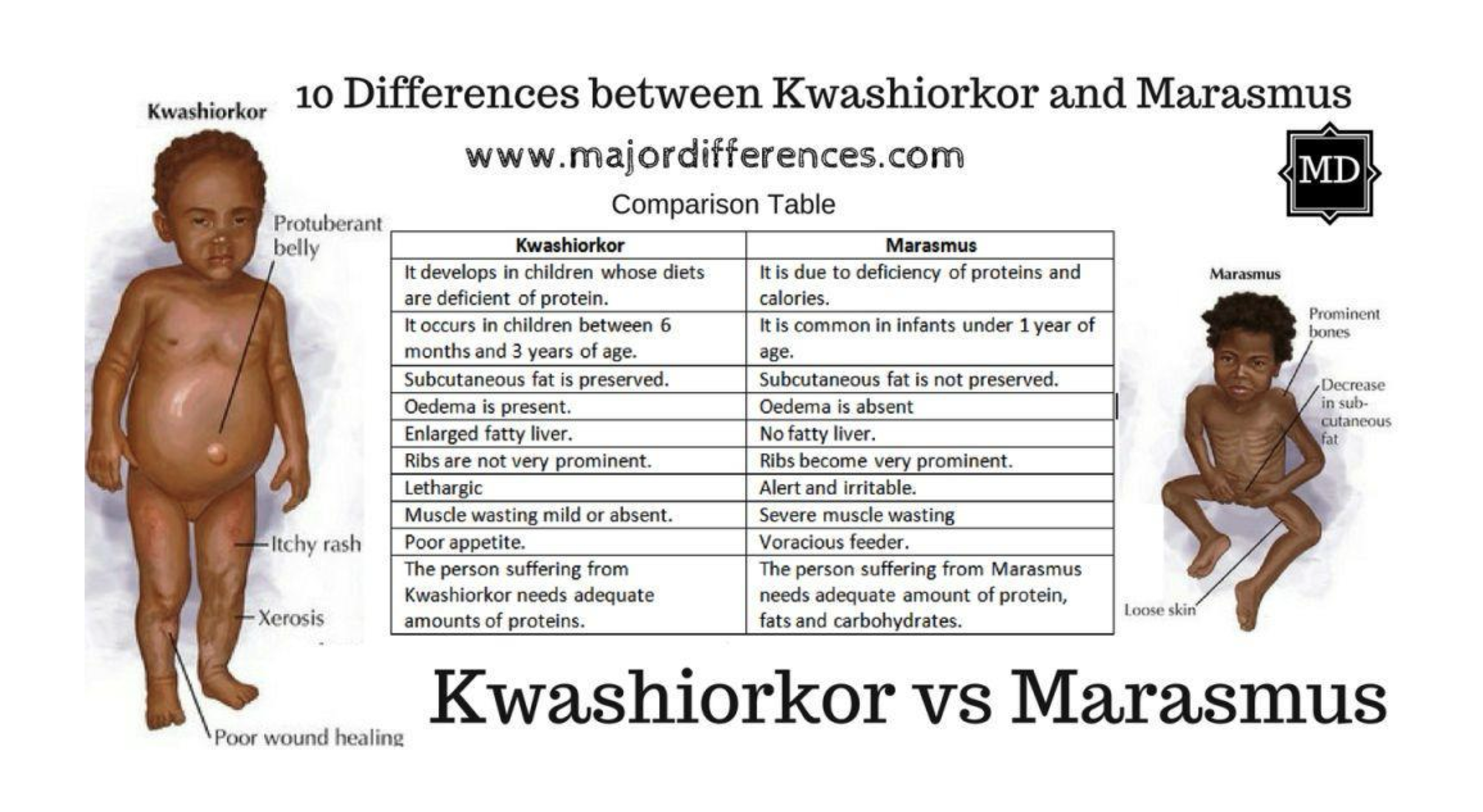
Kwashiorkor Cause
Severe protein deficiency
Distended abdomen
Albumin → has to do with absorption; lots of fluid pools in abdomen

Marasmus Cause
Severe total calorie deficiency (proteins, sugars, and fats)
Free Radicals
Electrically uncharged atoms or atom groups with an unpaired, unstable electron
Reactive Oxygen Species (ROS)
Physiologic free radicals used in the electron transport chain and released during inflammation
Types of free radicals found in the mitochondria and neutrophils
Antioxidants > free radicals
Oxidative Stress
Occurs when Free Radicals > Antioxidant system capacity
Can cause a chain reaction of damage:
lipid peroxidation → destruction of phospholipid bilayer
alteration of proteins → misfolding
alteration of DNA → mutations
Lipid Peroxidation
destruction of unsaturated fatty acids generates ROS → chain reaction → destruction of phospholipid bilayer (organelles and cell) → ‘leaky cell’
Cellular adaptation: Atrophy
Decrease in cell size

Cellular adaptation: Hypertrophy
Increase in cell size due to increased work demand or hormones (e.g., Cardiomegaly from HTN)

Cellular adaptation: Hyperplasia
Increase in cell number due to increased rate of cellular division (e.g., BPH, callus)
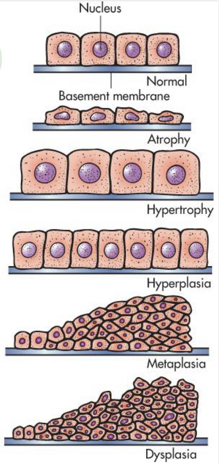
Cellular adaptation: Metaplasia
Reversible replacement of one mature cell type by another less mature cell type
Ex: Bronchial metaplasia
Ex: Barrett’s Esophagus

Barrett's Esophagus
Esophageal metaplasia where normal stratified squamous epithelium is replaced by simple columnar epithelium with goblet cells (usually found in lower GI)
Cellular adaptation: Dysplasia
Deranged cellular growth ('abnormal cell'); Not a true cellular adaptation but rather an atypical hyperplasia. Abnormal changes in the size, shape, and organization of mature cells.
Clinical: The amt of dysplasia determines the tumor grade
Ex: cervical dysplasia seen on a pap smear

Bronchial Metaplasia
Reversible replacement of bronchial ciliated columnar cells with stratified squamous epithelial cells due to smoking
Loss of cilia and mucus producing glands
Bronchial metaplasia can be reversed if the inducing stimulus (cigarette smoking) is removed

Hypoxic injury: Hypoxia
Lack of sufficient oxygen to cells; Most Common Cause (MCC) of cellular injury
Hypoxic injury: Ischemia (reduced blood supply)
Lack of blood flow into vessels that supply the cell with oxygen and nutrients
Ex: Atherosclerosis, thrombus, high altitude, anemia
Hypoxic injury: Anoxia
Total lack of oxygen supply (e.g., embolus causing MI, CVA, PE)
Effects of Hypoxic Cell Injury: 1. ATP depletion
Anaerobic metabolism - until glycogen stores are depleted
Na+/k+ ATPase & Ca2+ ATPase are affected
Increased intracellular Na+ & Ca2+
H2O follows Na+ → cell swelling
RER swelling → ribosomes detach → decrease in protein synthesis
Effects of Hypoxic Cell Injury: 2. Intracellular Calcium Effects
Ca2+ activates intracellular catabolic enzymes
Decrease in ATP, membrane damage, DNA damage
Increase in mitochondrial permeability → increase in ROS (reactive oxygen species)
Effects of Hypoxic Cell Injury: 3. Oxygen-derived free radicals
Oxygen-derived free radicals = Reactive oxygen species (ROS)
Destruction of cell membrane and nucleus
Effects of Hypoxic Cell Injury: 4. Defects in membrane permeability
Leakage of intracellular enzymes, can be detected in the blood
How do you diagnose an MI? Liver failure?
Cellular response to stress: Irreversible Injury (Cell death): Apoptosis
Apoptosis
Programmed cell death ('cell suicide') characterized by cell shrinkage without inflammation
Cellular response to stress: Irreversible Injury (Cell death): Necrosis
Unprogrammed cell death characterized by cell swelling and inflammatory changes
Necrosis: Coagulative Necrosis
MC in kidney, heart, skin
Cause: hypoxia/ischemia/anoxia → infarction
Tissue appears pale
Necrosis: Liquefactive
Typically affects neurons/glial cells in the brain, GI
Cells ‘digested’ by their own hydrolases - tissue becomes soft
Walled off from healthy tissue, forming cysts
Necrosis: Caseous (cheese like)
MC w/ infection of mycobacterium tuberculosis → lungs
Combo of coagulative and liquefactive
Cells disintegrate/denature but debris is walled off
Necrosis: Fat
MC in breast, pancreas, liver
Saponification (soap)
Gangrenous Necrosis: Dry Gangrene
Gangrene from coagulative necrosis; skin is dry, dark brown/black, and shrunken
Gangrenous Necrosis: Wet Gangrene
Gangrene from liquefactive necrosis of internal organs; swollen, cold, black, almost always infected → foul odor
Gangrenous Necrosis: Gas Gangrene
Wet gangrene caused by Clostridium infection, bacteria forma gas bubbles in tissue
5 Cardinal Signs of Inflammation
Calor (heat), Rubor (redness), Tumor (swelling), Dolor (pain), Functio Laesa (loss of function)
Pain Definition
Unpleasant sensory & emotional experience associated with actual or potential tissue damage

Referred Pain
Pain felt in an area distant or removed from its original point of origin

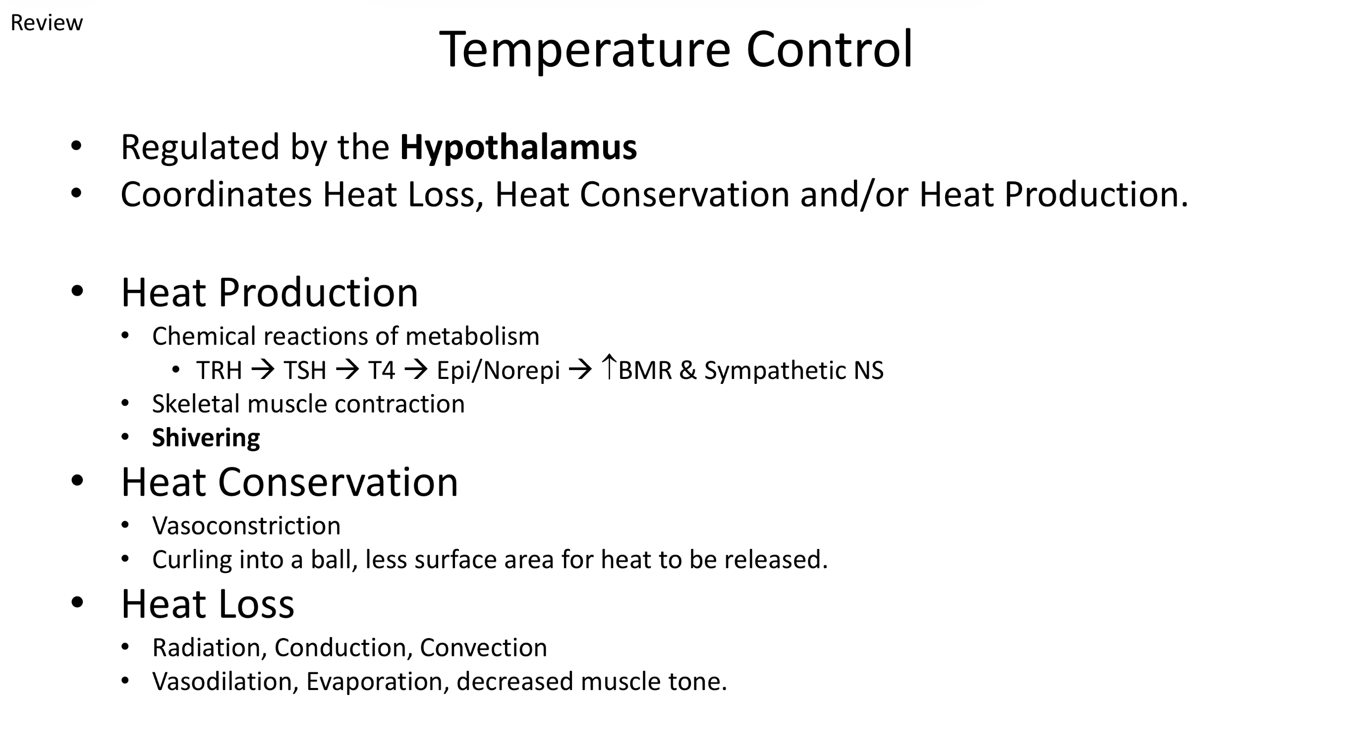
Hypothalamus Temperature Control
Regulates heat production (shivering, BMR), heat conservation (vasoconstriction), and heat loss (vasodilation, sweating)
Fever Functions
Stimulate the immune system
Create an inhospitable environment for invading pathogens
Benefits of Fever
Kills many microorganisms
Decreases serum levels of iron, zinc, and copper - needed for bacterial replication
Promotes lysosomal breakdown and autodestruction of cells
Increases lymphocytic transformation and phagocyte motility
Augments antiviral interferon production and phagocytosis
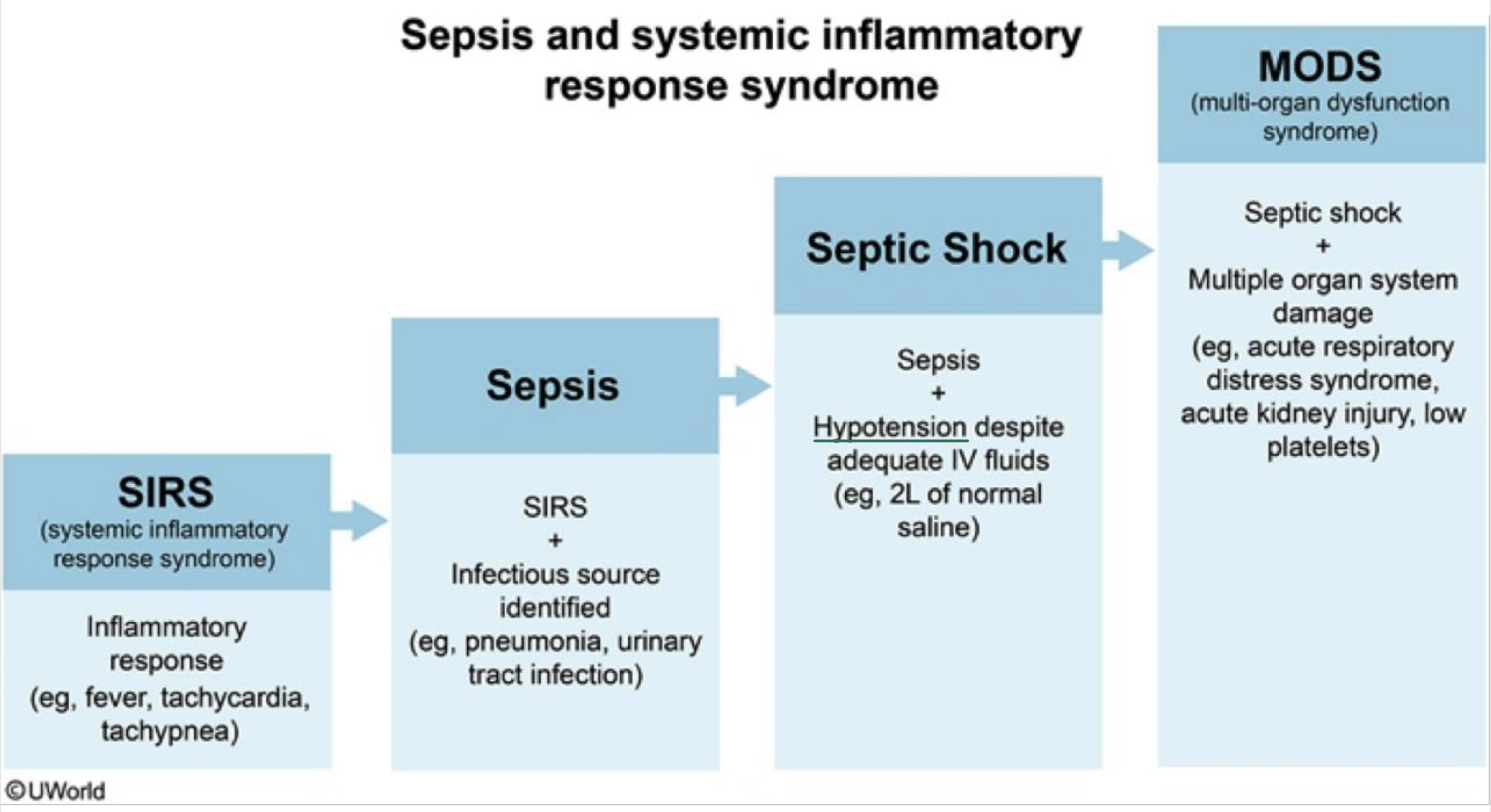
Sepsis
Massive systemic inflammatory response to an infection
Lungs (pneumonia is the MCC)
Urinary tract (UTI is a very common cause of sepsis in elderly)
Other locations: skin, abdominal organs
1/3 of the time, no identifiable source
Septic Shock
Sepsis accompanied by severe, refractory hypotension
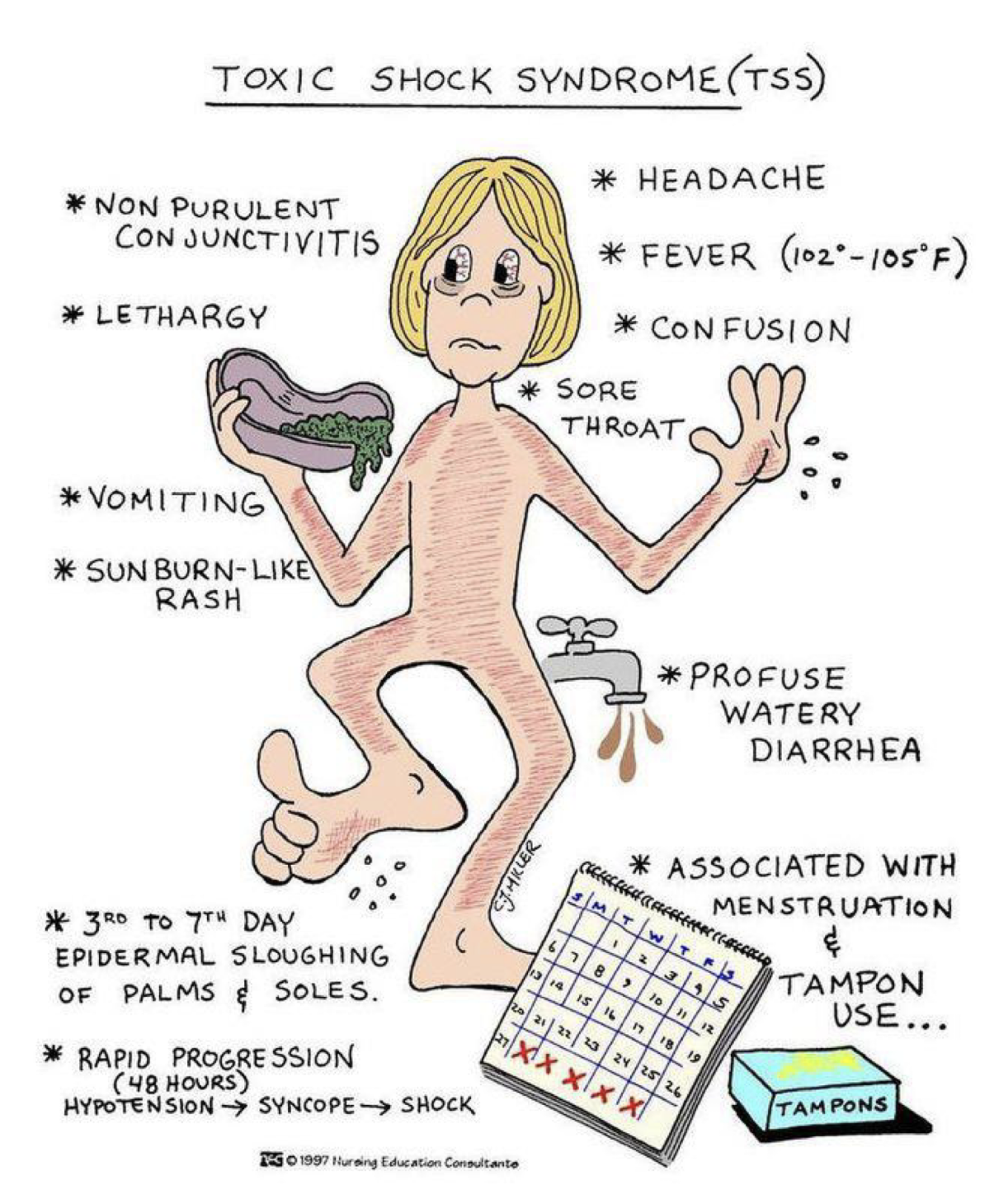
Toxic Shock Syndrome (TSS)
Life-threatening bacterial infection causing high fever, hypotension, V/D, headache, myaglia, AMS, and sunburn-like rash
Predisposing factors: Age
Alzheimer’s, CVD
Predisposing factors: Sex
Hypothyroidism F>M
BPH, PCOS
Predisposing factors: Environment
Pollution, radiation
Predisposing factors: Lifestyle
ETOH, tobacco, drugs, unprotected sex, multiple sex partners, stress, lack of exercise, poor diet
To make a diagnosis: Medical history, physical exam, diagnostic labs and tests
Medical history
Chief complaint
“what brought you in today”
HPI
many questions about the CC
Physical exam
Inspection
Auscultation
Palpation
Percussion
Diagnostic Labs & Tests
Medical History: SOAP
Subjective, Objective, Assessment (Diagnosis), Plan
Mitochondira contains many . . .
Free radicals (O-), isolated from the rest of the cell
Ions are always moving from . . .
High to low concentrations trying to reach their electrochemical equilibrium
ATP is required to move . . .
an electrolyte against its concentration gradient (active transport)
What would happen to intracellular concentrations if there were no ATP available?

Cell stressors: Trauma
Injured tissue can disrupt blood flow
Cell stressors slide: Pathologic
Ionizing radiation (UV rays, X rays)
Metabolism of certain chemicals (CCl4)
Increased mitochondrial membrane permeability due to hypoxic cell injury
Cellular adaptation
The cell’s response to protect itself from injury
Toxic shock syndrome causes
Staph aureus
Group A beta hemolytic strep
Tampons, contraceptive sponges/diaphragm
Wounds, post surgical

Toxic shock syndrome testing
CBC, Blood culture
Slide: Sepsis and systemic inflammatory response syndrome
Slide: Sepsis: SIRS Criteria

Sepsis Summary Slide: Fever
IL-1, IL-6, TNF
Sepsis Summary Slide: Vasolidation
Hypotension
Impaired O2 delivery leads to tissue hypoxia
Sepsis Summary Slide: Treatment
Antibiotics, IV fluids, ventilation
Vasopressin (ADH) → causes vasoconstriction
Sepsis: Causes
MCC: Bacterial infections (staph, strep, e. coli)
Very common in indwelling central line catheters
Other causes: Fungal, viral, parasites
Bacterial exotoxins that act as superantigens
Sepsis: Signs and Symptoms
Signs and symptoms
Fever, tachycardia (increase in HR), tachypnea (increase in RR)
Pallor, altered mental status (AMS)
Bacteremia, leukocytosis
If hypotensive → septic shock
Fever: Pathogenesis
Exogenous pyrogens (endotoxins from pathogens) stimulate phagocytes to release endogenous pyrogens (IL-1, IL-6, TNF-a)
IL-1, 1L-6, & TNF-a stimulate the hypothalamus to make more PGE2 → raises the set point for the body temperature
Body temp is now lower than the set point
Heat generation and heat conservation occur
Slide: Inflammatory Modulators

Gangrenous necrosis
Clinical term, death of tissue resulting from hypoxic injury