pos
 Call Kai
 Learn
Learn Practice Test
 Spaced Repetition
 Match
Match Flashcards
 Knowt Play
1/741

There's no tags or description
Looks like no tags are added yet.
Name | Mastery | Learn | Test | Matching | Spaced | Call with Kai | Chat |
|---|
No analytics yet
Send a link to your students to track their progress
742 Terms
basic structure of chromosomes-
centromere at centre
telomeres at ends- protective, repetitive sequence of DNA
there are replication origins
genes spread around
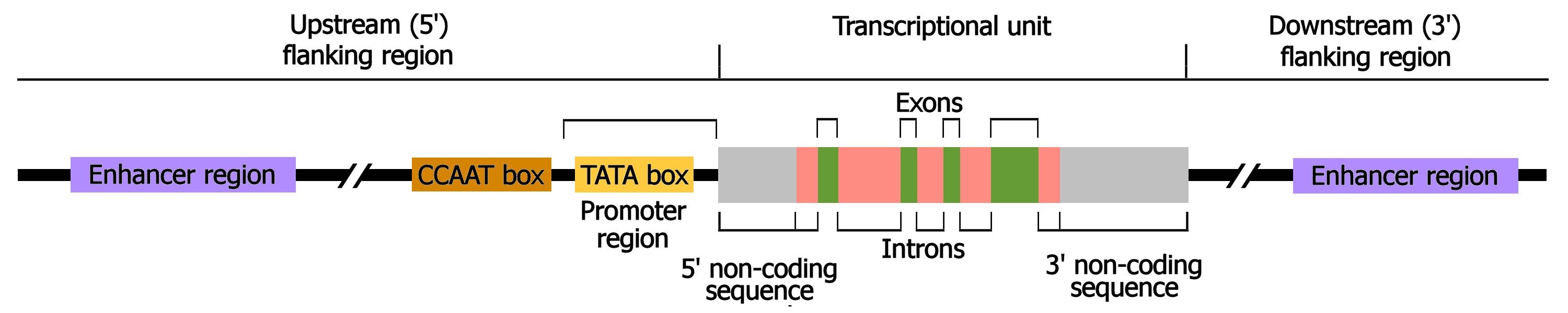
structure of a eukaryotic gene
promotor region
exons and introns
TATA box in promotor- initiation of transcription
transcription initiation site
translation initiaton site in exon 1 (ATG)
translation termination site
transcription termination site
what is the central dogma
DNA to RNA to protein
(transcription, splicing, export, translation, folding)
what are RNA polymerases and what are the 3 types
protein complexes made up of 8-14 subunits
Pol 1 - ribosomal RNA
Pol 2- mRNA for translation into protein
Pol 3- small RNA (tRNA)
transcription initiation
transcription factor binds to eukaryotic promotor
formation of preinitation complex
RNA pOL 2 begins elongation
what do transcription factors bind to
gene specific transcription factors bind to regulatory sequences of the gene
gene definition
A locus (or region) of DNA, which is made up of nucleotides and is the molecular unit of heredity
chromosome definition
A thread-like structure of nucleic acids and protein found in the nucleus of most living cells, carrying genetic information in the form of genes.
genome definition
The complete set of genes or genetic material present in a cell or organism
initiation
general transcription factors bind , eg TBP to TATA box
pre initiation complex formed
rna polymerase holoenzyme binds to the promotor region
where do transcription factors bind to?
regulatory sequences
what is a transcription factor homodimer
2 protein monomers that bind together to regulate gene expression
trans activating domain
regions of transcription factors which bind to coactivator complexes to activate transcription
helix turn helix motif
two alpha helices joined by a short strand of amino acid
recognition and binding of DNA is done by the 2 helices, one at n terminus and one at c terminus
c terminal binds to major groove, n terminal helps to position the complex
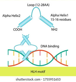
other dna binding motifs
zinc finger
leucine zipper
helix loop helix
elongation
what does rna polymerase do
what helps maintain the correct length
what does the zipper do
RNA polymerase travels along from the 5’ to 3’ end (upstream to downstream)
rudder in the upstream end of the RNA-DNA hybrid helps maintain the correct length, etc
the zipper is another protein loop within RNA polymerase and maintains boundaries of the transcription bubble especially at the downstream end
what are topoisomerases
regulate dna overwinding or underwinding
5’ cap addition
7 methyl guanosine triphosphate cap
this is a modified guanine cap to the 5’ end of mRNA
protection from degradation
initiation of protein synthesis
3’ polyadenylation
example of the sequence
what recognises it
what happens then to the mRNA
what then happens at the 3 end
what binds
why does this happen
sequences of the pre mRNA such as the AAUAAA sequence are recognised by the cleavage and polyadenylation specifity factor (CPSF)
then an endonuclease cleaves mRNA at specific site
enzyme poly-a-polymerase joins multiple adenine nucleotides to the 3’ end
poly a binding proteins bind to the newly formed tail
provides stability and facilitates exit of mRNA from the nucleus
xm2
nuclease that degrades remaining nacsent transcript and leads to termination of transcription
splicing
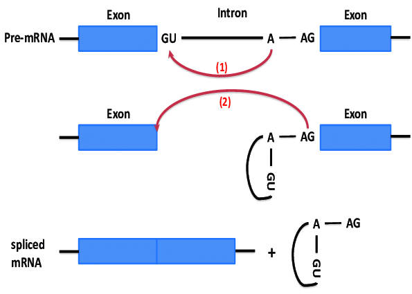
introns always begin with the residues GU and end with AG and an A somewhere towards the middle
phosphodiester bond is attacked between GU and A
3 prime end attacks phosphodiester bond between A and AG joining exons and removing the introns
how can proteins be altered for a specific function?
alternative splicing
what is the genetic code
20 amino acids
4 diff nucleotides
if code is x nucleotides , number of possiblity is 4 to the power of x
codons
charging of tRNAs with amino acids
what does amino acyl tRNA synthetase do
what does it then attach and what is released as a result
what is created
what does the enzyme bind this to
what then happens and what kind of bond is formed
what molecule is released
amino acyl tRNA synthetase binds an amino acid and ATP
the enzyme catalyses a reaction that attaches AMP to amino acid releasing pyrophosphate
this creates a high energy aminoacyl-adenylate intermediate
The same enzyme, aminoacyl-tRNA synthetase, binds the activated aminoacyl-adenylate and its corresponding tRNA molecule.
amino acid is transferred from AMP to the 3 end of tRNA which forms an ester bond
AMP is released, as the amino acid is now covalently linked to the tRNA, creating a "charged" aminoacyl-tRNA.
what are the site of the ribosome
p site holds trna with growing polypeptide chain attached
e site holdsthe trna which will exit
a site holds aminoacyl trna
elongation cycle
what is it
what is delivered to the a site and what escorts it to where and where does it bind to
what does this consume
what catalyses formation of a peptide bond
what happens and what 2 things does this need
process of adding amino acids to a polypeptide chain
aminoacyl trna is delivered to the A site. elongation factor escorts the trna and binds it to the ribosome consuming GTP (to GDP)
peptidyl transferase catalyses formation of peptide bond
ribosome shifts one codon along in the 3 direction. facilitated by elongation factor 2, this requires GTP.This action moves the tRNAs: the now uncharged tRNA from the P-site moves to the E-site, and the tRNA carrying the polypeptide chain moves from the A-site to the P-site, freeing up the A-site for the next cycle.
translation initiation
what do the eukaryotic initation factors bind to
what does initiator tRNA carry and what does this do
what does the small ribosomal subunit do
what happens to the GTP on the tRNA and what does this cause and allow
what sits on the p site
eukaryotic initiation factors bound on the 5’ end join to the (poly a binding protein) bound on the poly a tail of the 3’ end
initiator tRNA carries methionine, GTP and initiation factor 2, and the 40s ribosome both recognise the initiation factors bound to the 5 and 3 ends. They all interact
small ribosomal subunit finds the start codon, using protein initiation factors. but some are easier to find than others (purine 3 residues upstream of A -Kozak sequence) 40s subunits slides down
GTPase activity converts the GTP on the tRNA to GDP
causes conformational change, and GDP is release
this allows the large ribosomal subunit to bind
methionine sites on the p site of the ribosomal complex
ready to begin elongation
termination
stop codon do not have a corresponding trna to recognise
instead when a ribosome reaches the stop codon the a site accepts a protein called release factor instead of tnra
release factor hydrolyses the bond between trna in the p site and the amino acid in the polypeptide chain, freeing it
two ribosomal subunits and other structures dissociate
antibiotics targeting translation
chloramphenicol
erythryomycin
tetracycline
streptomycin
chloramphenicol binds to 50s rrna inhibiting formation of a peptide bond
erythromycin binds to 50s of rrna and prevents movement along mrna
tetracycline inhibits trna anticodon reading of mrna codon
streptomycin changes shape of 30s rrna and causes mrna to be read incorrectly
functions of chromatin
package and condense dna
prevent dna damage
control dna expression
what are nucelosomes
how many base pairs
what is the core comprised of
what are the cores separated by
what brings nucleosomes together and what does this do
what do multiple stacked rings form
what is formed when they are coiled further
‘beads on a string’
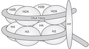
around 146 base pairs of dna wrapped around 1.7 times around a core
core is comprised of an octamer of histone proteins
nucleosome core proteins are separated by linker proteins
histone H1 brings nucelosomes together to condense chromatin further
multiple stacked rings form the solenoid fibre/30nm fibres
these are coiled further to form chromosomes
pcr
heat to 94 to denature (separate) dna strands
cool to 50-60 to allow primers to anneal
72 to allow elongation of new strands using taq polymerase
so at the end of the first cycle 2 new strands
begin cycle two by denaturing and annealing again
elongation generates strands of different sizes depending on the template strands
sanger sequencing
mix small amount dideoxy with deoxy nucleotides
cycle through multiple rounds of pcr
each ddNTP contains fluorescent tags
separate by gel electrophoresis
detect by laser
(chain synthesis terminates when ddNTP is incorporated)
technical applications of PCR that could be used to an advantage in veterinary medicine
VNTR
RAPD
AFLP
they are all used to determine breed, pedigree and/or parentage
what components are essential for basic PCR?
DNA polymerase
all 4 deoxyribonucleotides
dsDNA template
a pair of primers which bind to opposite polynucleotide strands and have their 3 ends pointing towards each other
what is PGE characterised by
diarrhoea/ weight loss
poor weight gain
hypoalbuminaemia
examples of parasitic nematode worms casusing bovine pge
ostertagia (abomasum)
cooperia and neatodrus (small intestine)
life cycle of ostertagia
adult ostertaga in abomasum
egg containing L1 larva passed in faeces
eggs hatch on pasture
develops to L3 larva, migrates from faeces to grass
L3 larva ingested by cow
this L1 to L3 development is temperature dependent. it takes up to 2 weeks or can be even slower
the prepatent period is 3 weeks, or up to 6 months if the development is arrested
typical strongyle eggs in faeces: oval, thin clear wall, bundle of cells. 80um
3rd stage larva is 750 um.
adult around 1cm long, brown. they emerge from the gastric glands and live/mate on abomasal mucosal surface
pathogenesis
where do larvae migrate to on ingestion
when do adult worms emerge
what happens if a large number of worms emerge at the same time
on ingestion larvae migrate to gastric glands of abomasum to continue development
adult worms emerge from glands around 18 days later
if a large number of worms emerge at the same time
pH increases from 2-7 as less acid produced as gastric glands are affected
pepsinogen cannot be activated to pepsin
abomasal epithelium becomes leaky
plasma proteins lost into gut lumen causing hypoalbuminaemia, weight loss and diarrhoea
what can happen to gastric mucosa
thickened, hyperpastic
raised nodules
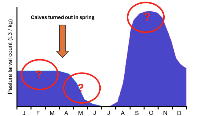
some of the L3 larvae survive on pasture overwinter
the new eggs deposited in early spring develop slowly to L3 as it is too cold
as the weather warms some existing L3 die as drier conditions and trapped in cow pats. but rate of L1 to L3 dev increases.
Then L3 increase furthe as more calves are infected and shed and eggs in faeces
what does rate of infection depend on?
host appetite
number of infective larvae (L3) on pasture
so disease is most common in calves where they are grazing permanent pasture and kept at a high stocking density
what happens to L3 is ingested in late autumn or early winter?
L4 arrest in gastric glands
then L4 resume development and emerge from glands in waves (type 2 disease)
immunity to ostertagia
slow to develop - takes the whole grazig season
may dip over winter ad re established upon turnot (2nd grazing season)
adult cattle solidly immune
epidemiology in beef herds- spring calving
calves at foot with cows
spring mortality of L3 occurs before calves eat much grass
so immun cows eat most of grass and pass few eggs
so very low disease risk
epidemiology in autumn calving
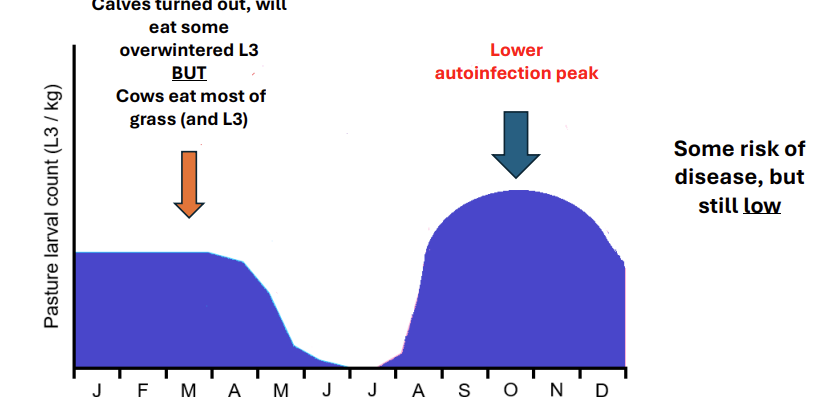
calves turned out at start of year and will eat some outwintered L3 BUT cows eat most
then lower autoinfection peak
some risk but still low
type i disease
typically calves in first grazing season
mid july onwards
morbidity high, mortality low
diarhoea common (larval damage to gastric glands)
weight loss
type ii disease
typically yearlings
late winter/ spring following first grazing seasn
prevalence low, only some affected
mortality more likey
± diarrhoea with anorexia and thirst
hypoalbuminea more marked, weight loss
control of type i disease
use clean pasture
delay turnout untul after spring mortality in L3
strategic anthelmintic use
anthelmintic use
macrocyclic lactone wormers last 5 weeks eg doramectin
prepatent period for ostetagia s 3 weeks
so dosing needed every 8 weeks
options
dose and move to fresh pasture in july before autoinfection peak
but can still get disease if L3 high, increased risk of spreading resstant worms to fresh pastur
dose every 8 weeks from july and keep on same pasture
dose at spring turnout and 8 weeks later reducing autoinfection peak
give an intraruminal device before turnout which reduces autoinfection peak
control of type ii
cattle exposed to low challenge at pasture in late autumn
unlikely to require worming treatment at housing
cattle exposed to medium/ high challenge at pasture in late autumn or cattle of unknown origin
likely to require worming treatment at housing
what is meant by a protein structure hierarchy
-primary, secondary, tertiary, quaternary(eg 2 beta and 2 alpha globin polypeptides with a heme group)
what type of amino acids are mammalian proteins
how many amino acids
what is the peptide bond classified by
how many amino acids makes a protein
shape of peptide bond
mammalian proteins are alpha amino acids
20 amino acids
carboxyl group reacts with amino group to form a peptide bond and lose a water molecule
catalysed by peptidyl transferase (28s ribozyme-ribonucleic acid enzymes)
within a polypeptide chain, individual amino acids = residues
when >50 amino acids are in a polypeptide is a protein
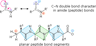
peptide bond is planar (partial double bond between carbonyl O and N)
bonding determining protein structure
covalent primary
hydrogen secondary
hydrophobic+VDW tertiary
electrostatic within tertiary helps maintain
VDW/ electrostatic quaternary
alpha helix
what runs parallel to the helix axis
what winds around the axis
what does htis lead to
how many amino acid residues per complete turn
beta sheets
what orientation do H bonds run to the chain direction
alpha helix- H bonds run parallel to helix axis. alpha carbon backbone winds around an axis so that each carbonyl O atom is H bonded to each amino N of the amino acid located 4 residues closer to C terminus
The standard alpha helix has approximately 3.6 amino acid residues per complete turn
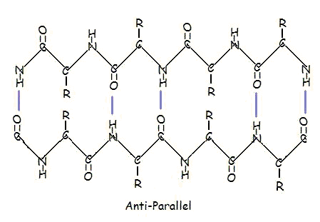
beta sheets- H bonds run perpendicular to chain direction
parallel and antiparallel
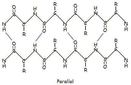
types of r groups
nonpolar, polar, electrically charged
cysteine
can form disulphide bonds as it has an SH group
examples of tertiary proteins
-catalase
-triose phosphate isomerase
-actin
tertiary domain
the smallest stable unit of a tertiary structure. a domain is defined as that region of a polypeptide chain that can fold into an autonomous stable tertiary structure
domain shuffling- when domains have been switched around between proteins through evolution
quaternary
haemoglobin- needs to deliver oxygen tissues. needs high enough affinity to pick up oxygen but low enough to release it.
t state- oxygen unbound
r state- oxygen bound. so that the 3 other units have higher affinity
small changes in oxygen concentration can dramatically affect binding to haemoglobin giving a sigmoidal curve of oxygen binding
collagen
what type of protein
what is the general structure
extracellular fibrous protein but not an alpha helix
3 helical chains that wind around a central axis
general structure is Gly-X-Y where X can be any amino acid especially proline lysine or hydroxyproline
Glycine is small so glycines from each chain fit at the centre of each helix
H bonds between the chains
skin and problems
thick layer of cells and sebaceous gland secretions
wounds
vector borne pathogens
mucous membraness
mucociliary escalator, peristalsis
coughing and sneezing, vomiting and diarrhoea
secretions- physical and anti microbial properties
commensal microflora
what does the innate immune sstem use for detection
uses pattern recognition receptors to detect microbial components that are itrinsically foregin
protein associated molecular patterns eg
lipopolysaccharide in gram negative
peptidoglycan in gram positive
mannose sugar in prokaryotic carbohydrate molecules
bacterial PAMPs
pathogen associated molecular patterns
pattern recognition receptor
where can they be found
what type in each
can be found in the cytoplasm, cell membrane, inside vessicles or as soluble molecules int he tissue fluid/plasma to detect PAMPS
cytoplasm: NOD receptors
membrane bound: TLRs
soluble: complement c3 protein, mannose binding lectin, c reactive protein
toll like receptors- what do these recognise
2
4
3 and 7
what are they predominantly expressed by
tlr2 recognises peptidoglycan- gram positive
tlr 4 recognises LPS - gram negative
tlr3 and 7 recognise viral nucleic acid
predominantly expressed by neutrophils and macrophages
how does innate to viral work
how are they detected
how do cells respond and what effect does this have
do not have strutural pamps
are detected by presence of double stranded rna produced during replication (not present in mammalian cells) or DNA in the cytoplasm
cells respond by producing type 1 interferon
this includes interferon alpha, beta, omega
interferons have a paracrine effect
function of type 1 interferon
resistance to viral replication
act on neighbouring
increased deradeation of viral mrna
inhibition of viral protein sythesis
increased antigen presentation of viral antigens
what is the paracrine effect
innate immunity to viruse
viruses can infect any nucleated cells
all nucleated cells can respond to viral infection by producing type 1 interferon
interferon omega can be used to treat persistent viral ifnection of cat eg FeLV/FIV
natural killer cell- large granular lymphocyte
what does it recognise
what does it release
recognise decreased levels of MHC molecules on host cells
there is decreased production during viral protein synthesis
some virus block transport to cell surface to prevent expression
this decrease is a symptom of viral infection.
the NK cell releases toxic granules killing the cell before the viral replication is complete
innate- cellular mechanisms
what is the response
how is the micro organism killed
what helps and enhances
recognition of pathogen (membrane, vesicular and cytoplasmic PRR)
response is phagocytosis and inflammation
phagocytic organisms attach to the organism and use pseudopodia to capture it forming a phagosome
then it stimulates a respiratory burst and toxic metabolites (eg oxygen free radials, hydrogen peroxide) are pumped into the vesicles to kill the micro organism
subsequent fusion of the lysosomes with the phagosome (phagolysosome) releases proteolytic enzymes and anti microbial mediators (defensins and lactoferrin)
increasingly acidic pH results in digestion of the microorganism
enhanced phagocytosis (opsonisation) can be achieved with the help of the antibody IgG and or complement CC3b
destruction of endocytosed organisms can be enhanced by stimulation of cytokines released by T helper cells
innate- humoral mechanisms
recognition of pathogen- soluble PRR
response: killing of foreign organism, enhacned phagocytosis and inflammation
tlr
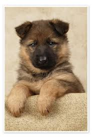
what could NOD2 receptor defect cause
crohns in man
ibd or anal furunculosis in german shepherd
what does recogn tiion of bacteria by macrophage tlr lead to
phagocytosis and inflammatory resposne
respiratory burst
enhanced cellular aeorbic metabolism
reactive oxygen intermediates are formed
superoxide anion
hydroxyl radicals
hydrogen peroxide
this isthe oxygen dependednt mechanism of bacteiral killing
lysosomes
3 types
defensins
cationic anti microial peptides that damage bacterial cell wall
lactoferrin
binds and chelates free iron, which is required for bacteiral growth
acid proteases
digestive enzymes active at low pH
inflammatory mediators-histamine
what is it released by
produced by mast cell degranulation in tissues
anti histamine
pro inflammatory cytokines
examples
what are they synthesised by
tumour necrosis factor a
synthesised predominantly by wbc and macrophages
corticosteroids
lipid mediators of inflammation
prostaglandin and leukotrienes
derived from arachidonic acid by action of cycloocygenase and lypoxygenase enzyme
nsaid
localised effect
inflammation
systemic
hypothalamus
fever
liver
acute phase response
bone marrow
neutrophil and monocyte mobilisation
speed up production
acute phase response
where are the proteins produced by in response to what
examples
what do they do
are they specific or non
acute phase proteins are produced by the liver in response to pro inflammatory cytokines
serum amyloid protein, C reactive protein and mannose binding lectin stick to bacterial cell walls
act as opsonins to enhance phagocytosis and stimulating complement activation
but are non specific
complement
where are complement proteins found
what steps do they form
what is the end product
complement prtoens are found in the blood
series of enzyme activation steps forming an aomplification cascade
small amount of activation is amplified to generate a large response
similar in nature to clotting cascade but with a diff trigger and outcom
the end product of the cascade is the polymerisation of C9 monomers to form a C9 polymer forming a membrane attack complex
a tube like structure that create holes in the cell walls of bacteria causing them to lyse
inactive C3 in the blood dissociates into C3a and b in the presence of bactiera- it is a pro enzyme
C3b is deposited onto the surface of the microbe and acts as an enyzyme to catalyse the formation of the MAC and also act as an opsonin (phagocytic cells express c3b receptor)
C3a binds to receptors on local tissue mast cells triggering degranulation and stimuating a inflammatory response
what are 3 examples of physical barriers
thick stratified squamous epithelium (skin and lower urinary tract)
mucociliary escalator (resp tract)
peristalsis, vomiting and diarrhoea when necessary (alimentary)
example of biochemical barriers
lactic and fatty acis in sebum from sebaceous glands of skin
enzyes
acid in stomach
antibacterial peptides eg defensin
how do commensal organisms provide protection
compete with organisms for space
provide natural antibiotic
what do gamma delta t cells do
react to stress proteins that are upregulated on the surface of infected mucosal epithelial cells
overall oucome of complement activation
lysis of the bacteria by the MAC
enhacend phagoytosis of bacteria coated in complement proteins C3b
inflammation at the site of complement activation C3a
where are these prrs located
TLR 2
TLR 4
TLR 5
TLR 9
NOD2
peptidoglycan
lipolysaccharide
flagellin
prokaryotic DNA
muramyl dipeptide