W2L3 cerebral cortex
1/59
There's no tags or description
Looks like no tags are added yet.
Name | Mastery | Learn | Test | Matching | Spaced |
|---|
No study sessions yet.
60 Terms
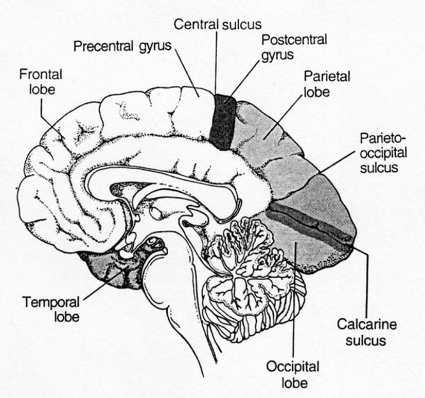
name 3 specialized functions of the specific lobes in the cerebral cortex, and examples for each
–Special senses (e.g. somatic sensation or vision or hearing)
–Motor control (e.g. initiation of voluntary movements)
–Cognitive (e.g. different language or memory functions)
how are the primary areas orientated in the cerebral cortex
paired/matching areas in each lobe & hemisphere
couples are found as one in the lobe and another in the hemisphere
what is functional contra-laterality
(meaning opposite side)
involved with events occurring on the opposite side of the body or of external space
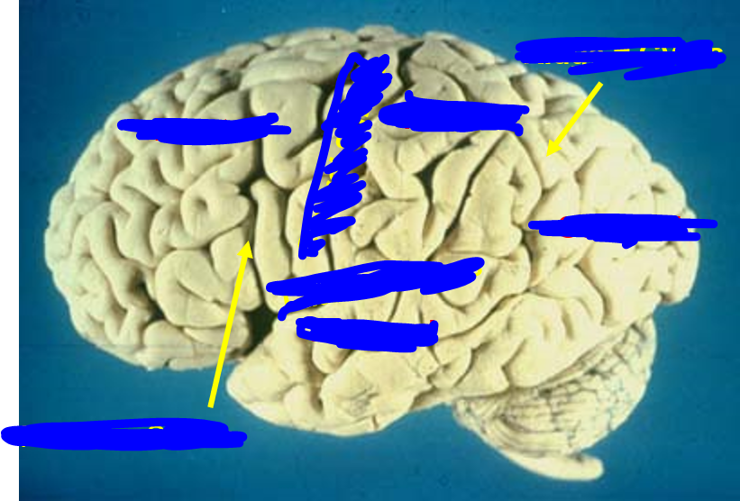
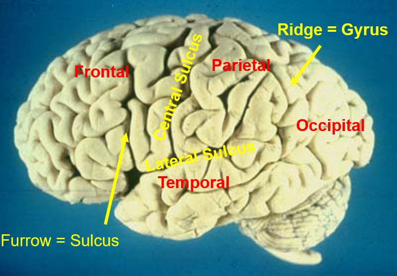
what does topography reveal
•contain MAPS of the body or of external space
function of left and right hemispheres of cerebral cortex
•Left = language functions; Right = spatial awareness/attention
what is the C19 phrenology THEORY
35 personality traits ascribed to different cortical regions- enlarged gyri (felt as ridges on overlying skull) show inc. development, while depressions mean underdevelopment
what are primary cortical areas
A small part of each lobe in each hemisphere, concerned with the most basic or lowest levels of sensory or motor function
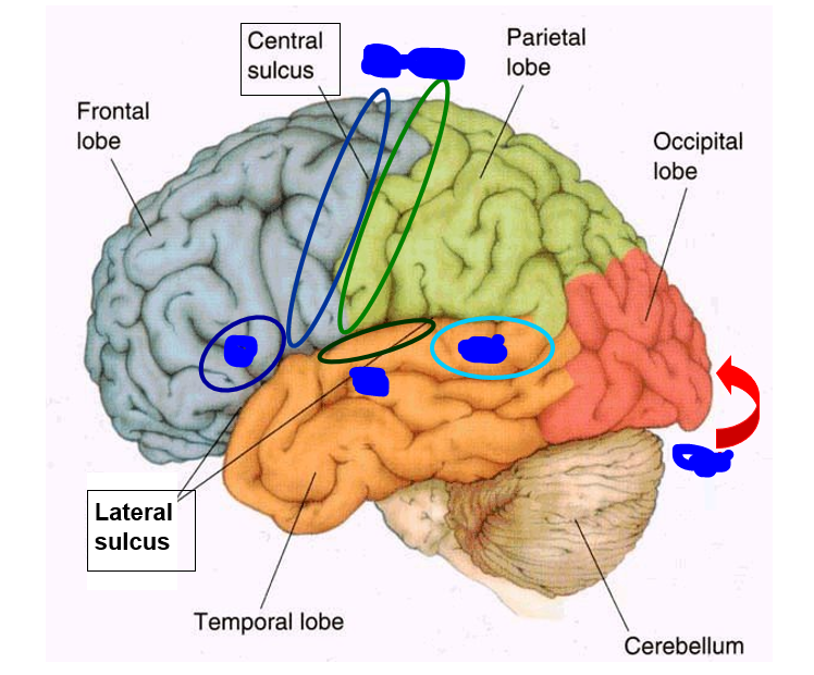
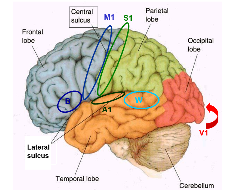
name the 4 primary cortical area, and what lobes they are found at
•Primary Somatosensory (S1) Cortex (L & R parietal lobes)
•Primary Motor (M1) Cortex (L & R frontal lobes)
•Primary Auditory (A1) Cortex (L & R temporal lobes)
•Primary Visual (V1) Cortex (L & R occipital lobes)
what is the postcentral gyrus, what can be seen there
long continuous gyrus, starting from bottom, continuing into the medial cortex
can find the s1 cortex here
what happens if there is unilateral damage to the parietal lobe, post-central gyrus
•Unilateral damage results in hemi-anaesthesia (loss of tactile, thermal, pain & joint sensation, opposite side of body)
Topographic Representation or Map of the parietal lobe, post-central gyrus
•Sensory Homonculus
what does the sensory map of the (left) post-central gyrus look like?
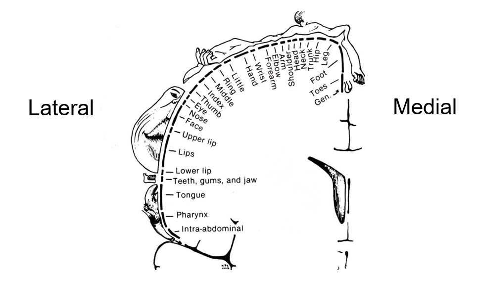
why is the map of the body considered to be upside down
the lowest part of the body is displayed on the highest part of the brain
looks like body is hanging from a pole
what is the function of the frontal lobe, pre-central gyrus
•Micro-stimulation (Wilder Penfield) results in movements of opposite body parts
what does unilateral damage to the frontal lobe, pre-central gyrus cause
•Unilateral damage causes hemi-plegia (paralysis, opposite side of body)
Topographic Representation or Map of the frontal lobe, pre-central gyrus
Motor Homonculus
How do different Primary Areas acquire their different functions?
via long-range axon pathways that relay in the THALAMUS
Why do Primary Areas deal with only the opposite side of the body/space?
because axons cross the midline (a.k.a. ‘decussate’) once, somewhere along their pathway in the CNS
Are there anatomical differences between different Primary Areas that relate to their Functional Specialization?
subtle differences in their cellular structure (‘cyto-architecture’/ micro anatomy)
what are the 2 things that pathways inside CNS consist of
separate nuclei in the grey matter
long range axon pathways in the white matter
what do the separate nuclei in the grey matter do
•Operate as ‘relay’ stations, which process information before passing it on via the white matter axons
what do the long range axon pathways in the white matter do
•Transfer this information to neurons at the next relay
where is the thalamus found, how is it paired and what is it made up of
•Central brain location
•Paired: left & right sides separated by narrow 3rd Ventricle
•Comprises many nuclei
what does the thalamus do (2 points)
•Receive input from specific sensory or motor pathways
•‘Relay’ to specific Areas of Cerebral Cortex, via axons in the Internal Capsule (which is a large white matter pathway)
what are the 4 ‘primary’ relay nuclei, what does each do
VL- ventral lateral, relays motor signals to primary motor cortex
VP- ventral posterior nucleus, relays somatosensory info to lateral cortex
MG- medial genicular, relays auditory info to auditory primary cortex
LG- lateral genicular, eye up to primary visual cortex
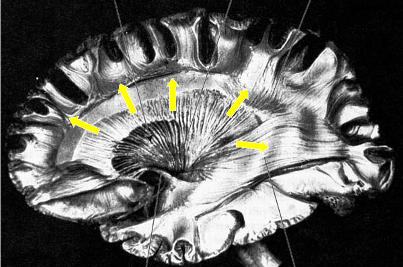
what are internal capsules
Thalamic axons ‘radiating’ towards the Cortical Grey Matter
what is corona radiate
radiating crown of axons travelling from internal capsule to different areas of the cerebral cortex (seen with yellow arrows here)
what is the cortical cytoarchitecture made up of, what can be found in it
6 Layers of the Grey Matter: neuron cell bodies, different sizes & densities
why is layer 4 darker, and what is it made up of
darker as it has loads of neuron cell bodies (stellate neurons, which are only found in layer 4)
layer 4 is mainly stellate/granule cells, receive thalamic input
what does layer 4 do
acts as local circuit neurons, excite the cells above and below
what is found in the other layers
mainly pyramidal cells, long-range projections
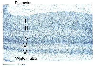
what are the blue cells here
all the blue dots are cell bodies of corticol neurons
note the darker 4th layer, and layer 1 containing almost no neurons at all, as it only has synaptic connections
what are the 4 types of cortical connections, name the layers they are found in
•Thalamic Input (layer 4)
•Inter-Cortical ‘Association’ projections (layers 2 & 3)
•Inter-hemispheric projections via corpus callosum (layers 2 & 3)
•Subcortical descending projections (layers 5 & 6)
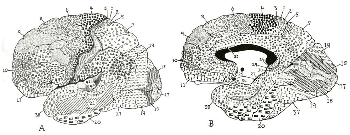
what does brodmann’s area describe
Korbinian Brodmann (~1900), identified 52 discrete areas of human cerebral cortex, based on differences in layer thickness, cell size & distribution (i.e., cytoarchitecture)
what is expressive aphasia, what causes it
inability to express language, can be caused by focal lesion in the inferior frontal lobe
what is receptive aphasia, what causes it
inability to understand spoken language, caused by focal lesion in left superior temporal lobe
2 type of memory
Procedural: Motor Skill Acquisition
Declarative: Facts & Episodes
3 points about procedural memory
•No conscious access
•Acquisition difficult (practice), long-term retention
•Cerebellum & Premotor Cortex (e.g., Broca’s Area)
4 points about declarative memory
•Conscious recollection
•Acquisition easy (single events), retention variable
•Short-term, ‘Working’: Pre-Frontal Cortex
•Long-term ‘Autobiographical’: Limbic System
what are 5 things (3 with working memory, 2 with personality) changes that damage to the pre-frontal cortex can do
Working Memory Loss
Difficulty organizing moment-by-moment behaviour
Numerous plans, instantly abandoned
Distractible
+ Personality changes
Impatient of restraint
Licentious & Profane
what can bilateral hippocampus removal cause
Anterograde Amnesia
•unable to form new long-term personal memory of any autobiographical life events
which MAPs is inverted
the somatosensory map in the post-central gyrus is inverted
what does it mean when we say inverted MAPs
sensory info coming from the top part of the body (eg, fac) is processed by neurons in the lower part of the gyrus, with sensory info coming from lowest part of body processed by neurons at upper part of gyrus
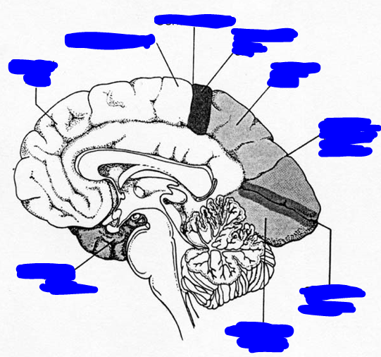
what does the visual hemifield MAPs in the banks of the calcarine sulcus look like
is distorted, many neurons in the posterior half of the cortex process sensory info coming from a small part of the central retina (fovea, macula), with relatively few neuros at the anterior end of the cortex, devoted to processing info coming from a bigger region of the peripheral retina
name 3 white matter pathways
optic nerve, internal capsule, corpus callosum
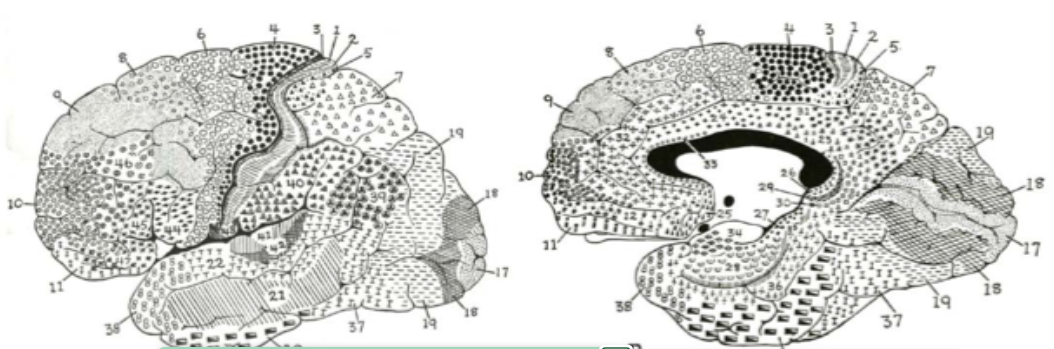
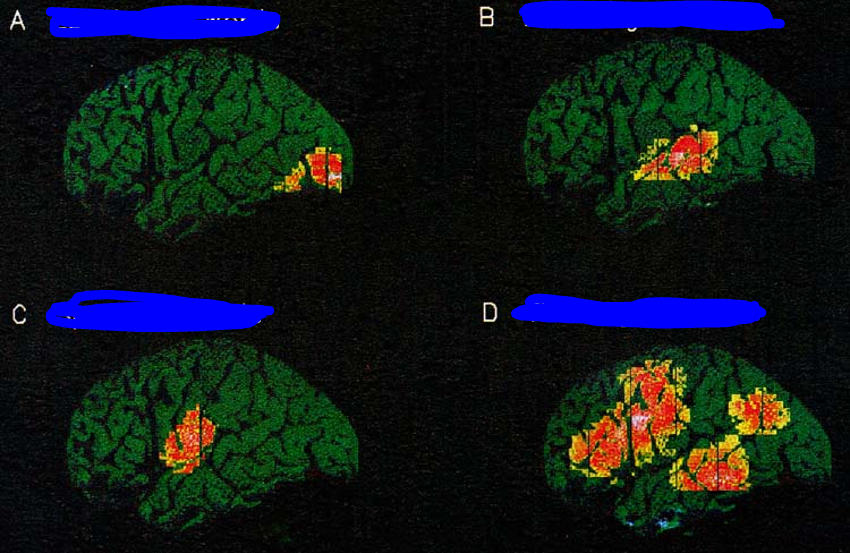
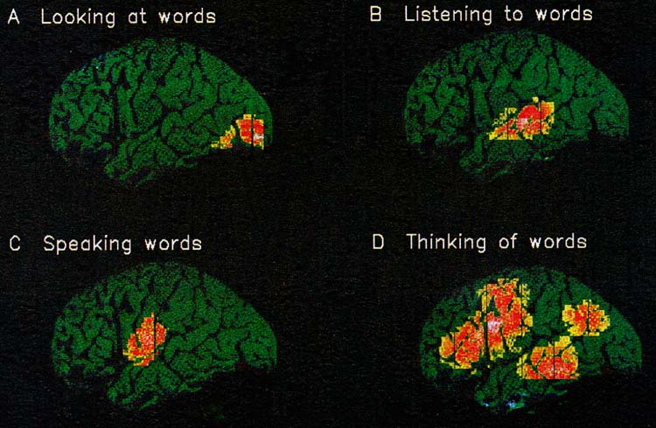
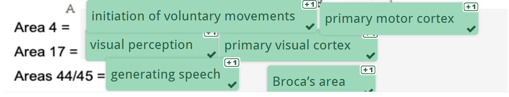
name the functions of areas 4, 17 and 44/45
Four main lobes of the cerebral cortex and their primary functions
Frontal lobe: Motor control and voluntary movements
Parietal lobe: Somatic sensation
Temporal lobe: Hearing and auditory functions
Occipital lobe: Visual functions
What is meant by "functional contralaterality" in brain function?
Functional contra laterality is the phenomenon where each hemisphere of the brain controls and processes sensory input from the opposite side of the body
What is the primary somatosensory cortex (S1) responsible for?
The primary somatosensory cortex (S1) processes sensory input from the body, such as touch, pain, and temperature, and contains a map of the opposite body parts known as the sensory homunculus
What is the role of the primary motor cortex (M1)?
The primary motor cortex (M1) controls voluntary movements of muscles, and like the somatosensory cortex, it has a distorted map of opposite body muscles called the motor homunculus
What is the motor homunculus, and where is it found?
The motor homunculus is a topographic map of muscles in the primary motor cortex (M1), showing areas responsible for different muscle movements
What is Broca's area and its associated function?
Broca's area, located in the left inferior frontal gyrus, is involved in speech production. Damage to this area leads to expressive aphasia, where patients have difficulty speaking but can understand language
What is Wernicke's area responsible for?
Wernicke's area, located in the superior temporal gyrus of the left hemisphere, is essential for understanding spoken language. Damage here results in receptive aphasia, impairing comprehension but leaving speech production intact
What role does the corpus callosum play in the brain?
The corpus callosum connects the left and right cerebral hemispheres, allowing information to be shared between them
How is the visual map in the primary visual cortex (V1) organized?
The visual map in V1 is inverted and distorted, with large areas devoted to central vision and smaller areas for peripheral vision
What is the function of pyramidal cells in the cerebral cortex
Pyramidal cells are long-range projection neurons that transmit signals from one region of the brain to another, primarily found in layers 2-3 and 5-6 of the cortex