MRI and US
 Call Kai
 Learn
Learn Practice Test
 Spaced Repetition
 Match
Match Flashcards
 Knowt Play
1/83

There's no tags or description
Looks like no tags are added yet.
Name | Mastery | Learn | Test | Matching | Spaced | Call with Kai | Chat |
|---|
No analytics yet
Send a link to your students to track their progress
84 Terms
What are major laboratory tests/ signs that there is something wrong with the pancreas
Serum amylase: May be indicative of acute pancreatitis if raised
Lipase: Raised in acute pancreatitis and cancer of the pancreases
Glucose: Increased in diabetes
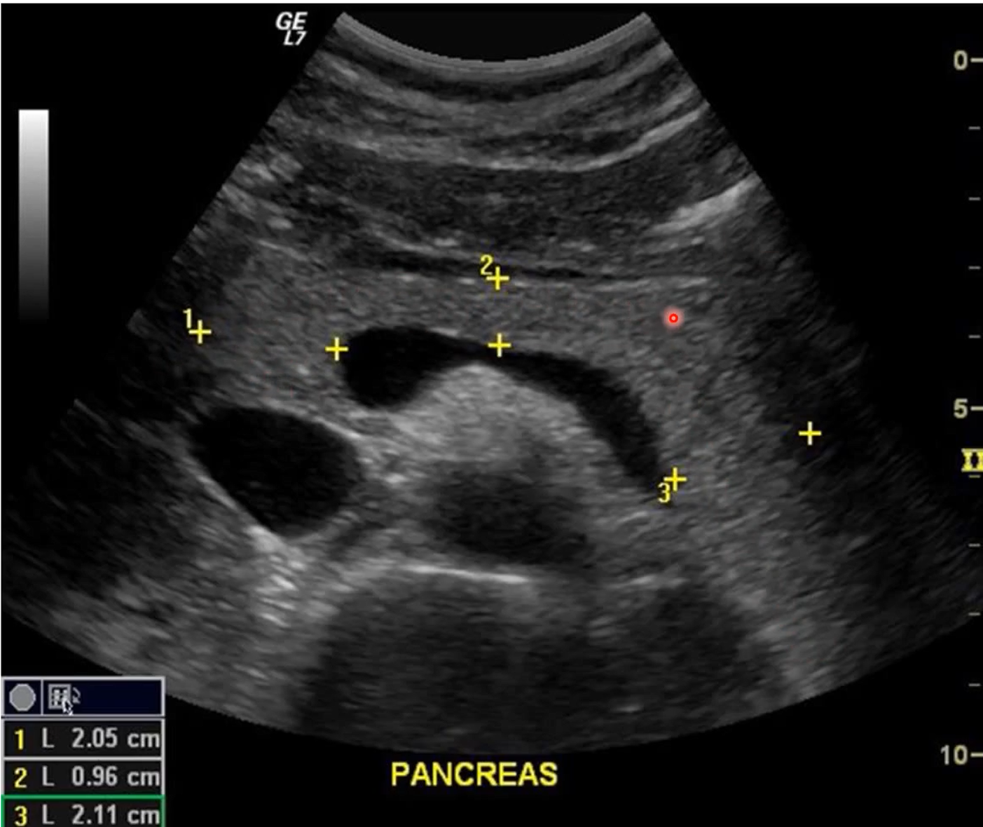
Which areas of the pancreas are which number
1: head
between 1 and 2: neck
2: body
3: tail
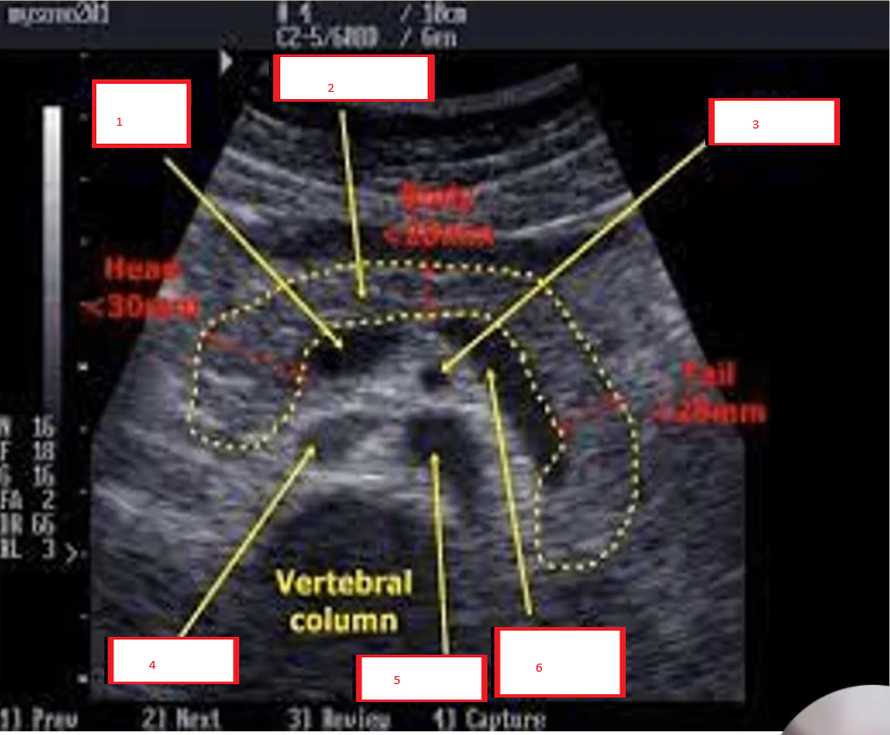
Portal vein
Pancreas
superior mesenteric artery (SMa)
IVC
Aorta
Splenetic vein
What is the patient preparation for standard abdominal ultrasound
Minimum 6 hour fast. sips of water is okay, can take medication, diabetics may eat small meals but no fats (fatty food)
Preferably a morning appointment
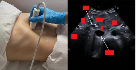
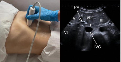
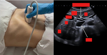
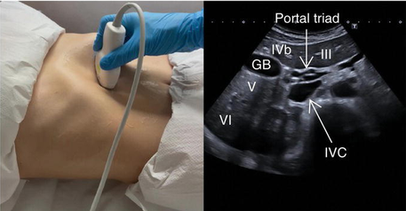
get a picture of pancreas long and label it
Pancreas scan technique
Many different techniques may be needed:
Varied breathing patterns
varied patient positions
patient in a erect position
water-filled stomach
Vascular landmarks are important
How to scan Pancreas
Pt supine
start in transverse scan plane in upper epigastrium region
scan inferiority using left lobe of liver as a window
identify vascular landmarks: aorta/IVC, celiac axis, SMA, splenic vein/ portal confluence, SMV
Volume scan thoroughly in 2 scan planes
Look for pancreatic duct and CBD
What is pt preparation for renal scan
Fast for 4-6 hours
A distended urinary bladder is required for a renal examination
Ask the pt to finish drinking 600-1000ml one hour prior appointment time
What are indications for renal US
Haematuria
Abnormal renal function tests
Abnormal PSA levels
Back or pelvic pain
Recurrent UTIs
Known congenital abnormalities
High blood pressure
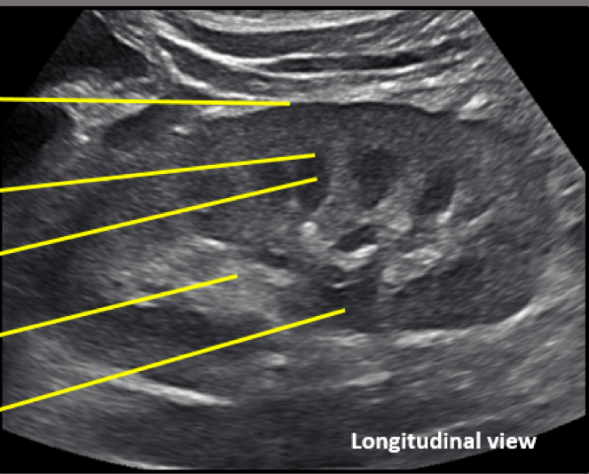
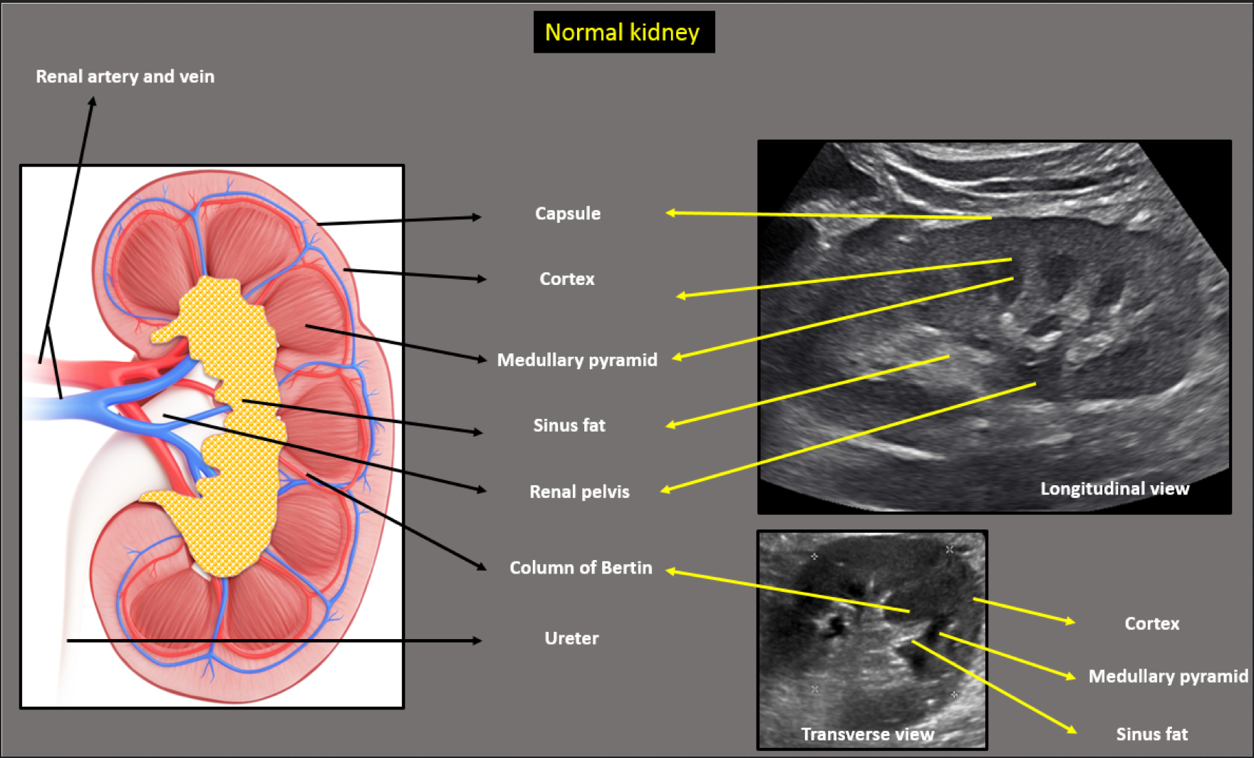
How does the kidney appear on US
Renal capsule: echogenic line
Renal cortex: Outer most tissue, hypoechoic
Renal medulla: Consists of the medullary pyramids, less echogenic than cortex
Renal parenchyma: Consists of cortex and medullary pyramids
Renal sinus: Central, hyperechoic region that contains renal pelvis, calyces, blood vessels and variable amount of fat
What is the scan technique for renal US
Check probe orientation
Coronal and sagittal volume scans
Long measurement (bipolar) (normal measurement is around 9-12 cm)
Transverse axis volume scan
Add CD to ensure that there is perfusion to the cortex of the kidney. Power Doppler is also useful.
How do you demonstrate pathology in renal US
Must be imaged and measured in two perpendicular planes
What are common pathology in renal US
Renal cysts
Calculi
Obstructive uropathy
Solid renal masses
inflammatory disease
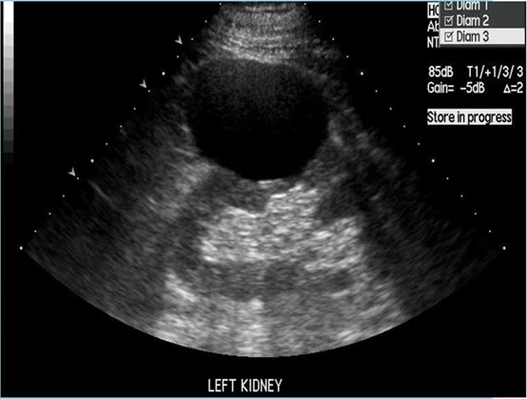
What pathology is shown here
Renal cortical cysts
benign and fluid filled
Anechoic
well-defined thin walls
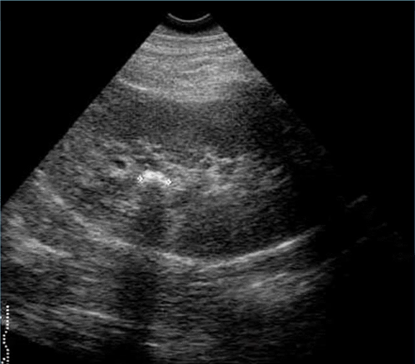
what pathology is this
Renal calculi
Larger calculi = echongenic foci + shadowing
Small calculi = echogenic foci - shadowing
What is the spleen US technique
Pt supine
Transducer in coronal plane
Inspiration, expiration or gentle respiration
Abnormality?
Oblique plane
Anterior scan?
Cant see spleen in supine position?
Which transducer?
What is the spleens appreacne in US
shape = inverted comma shape
normal surrounding structures =
echogenicity = haemogonious
Size = depends on age and gender (12 cm for female, 13cm for males)
Elastography role =
What is ultrasound frequency define as and what frequency does medical ultrasound use
Ultrasound frequency is defined as the number of ultrasound waves per second
Medical ultrasound machines use waves with a frequency ranging between 2 and 15 MHz.
What does frequency mean in US
Frequency refers to the number of cycles per second emitted by the ultrasound probe over one second and is expressed in hertz (Hz).
What does Amplitude mean in US
Amplitude is the height, or strength, of a wave defined by the distance between the peak and the average of the wave's highest and lowest points. Power in ultrasound refers to the square of wave amplitude, or difference between the maximum and average values, of propagated waves.
What are the 4 ways US waves interact with human tissue
absorption of ultrasound by tissue
reflection from interfaces (this includes diffuse and specular reflection);
scattering of the ultrasound beam due to diffraction effects (this includes Rayleigh scattering from blood cells and often from tissue parenchyma)
refraction, or the change in direction of the ultrasound beam when moving from one medium into another
What are the different US imaging modes and describe them
A-Mode (Amplitude mode): The transducer sends a single pulse of ultrasound into the medium
B-Mode (Brightness mode): B-mode is a 2d image of the area that is simultaneously scanned by a linear array of 100-300 piezoelectric elements rather than a single one as in A-mode.
M-mode (Motion mode): A single beam is an US scan can be used to produce a picture with a motion signal, where movement of a structure such as a heart valve can be depicted in a wave-like manner.
What are the 5 MRI-specific safety precautions
Magnetic field
Crogens
Gradients
RF power deposition
Contrast agents
How does MRI safety labeling work
red = MR unsafe: items should not enter the MRI scanner room
Yellow: MR conditional: items may safely enter the MRI scanner room only under very specific condtions
Green: MR safe: items pose no safely hazards in the MR enviroment
How many MRI zones are there an how do they work
Zone 1: general public/ MRI access area (negligble MRI hazard)
Zone 2: unscreended MRI patients/ MRI pt screening and preperation
Zone 3: Screened MRI patients and personal having access to MRI room
Zone 4: Screened MRI patient under direct supervision of MRI personal
What is the Gauss line
The 5 Gauss line refers to the boundary around the scanner where the magnetic filed strength falls to 5 gauss (0.5 millitesla). Beyond the the 5 Gauss line the magnetic field is considered sufficiently weak not to interfere with external devices or pose risks to general public health.
What is the Faraday cage
The Faraday cage is built around the MRI unit in zone 4. It is made of copper lining that has induced eddy currents that oppose the main magnetic field
What casues acoustic noise in MRI and how is it mitigated
The primary source of acoustic noise in the vibration of gradient coils due to rapidly switched electrical currents interacting with the strong static magnetic field. This is mitigated through the of ear protection such as earplugs and earmuffs
What is MRI quenching
Quenching refers to using a cryogen usually helium to cool the magnet in the MR scanner
What is MRI slew rate
The speed at which a gradient magnetic filed can be turned on and off during a n MRI scan. It measures how quickly the gradient reaches its maximum amplitude.
What is MRI SAR
SAR is specific absorption rate, it measures the rate at which energy is absorbed by the body when exposed to radio frequency electoprgenatic filed during an MRI scan. Units watts per kilogram (W/kg), is used. Factors affecting it are tissue conductivity, RF frequency, flip angle, duct cycle and pt size and shape.
What is gadolinium used for in MRI
Gadolinium is a positive contrast agent (T1 agent) that is primarily used to shorten T1 relaxation times leading to increased signal intensity
What must you do to MRI a pregent pt
gain written contesnt
scan using normal mode (SAR)
Avoid IV GAD
Monitor SAR levels and scan patient rolled 15 degrees to the left to reduce compressing the IVC
What are relaxation times in MRI
T1 (longitudinal relaxation time) represents the time it takes for the longitudinal magnetisation (aligned with the external magnetic field) to recover after perturbation (e.g., radiofrequency pulse). Gadolinium shortens T1 by enhancing the longitudinal magnetisation recovery.
T2 (transverse relaxation time) is the time it takes for the transverse magnetisation (perpendicular to the external field) to decay due to interactions between neighboring protons. Gadolinium has a minor impact on T2 relaxation.
What is GBCAs and what are its types
Extracellular Agents: These small molecules distribute nonspecifically in blood and extracellular spaces. They are commonly used for imaging tumors, inflammation, and magnetic resonance angiography (MRA)
Blood Pool Agents: Used mainly for MRA, these agents have longer intravascular half-lives, allowing extended imaging beyond the arterial first-pass phase.
Hepatobiliary Agents: Designed for liver lesion diagnosis, including gadobenate dimeglumine (MultiHance) and gadoxetic acid (Eovist, Primovist).
What are the different MRI coils and what do they do
Main filed coils: These are principal magnet windings and superconducting shim and shield coils
Shim coils: used to improve homogeneity
Gradient coils: essential for imaging and include active sheilds
RF body coils: transmits the B1 field
Surface coils: primarily detect MR signals
What is the aliasing (wrap) artifact in MRI
Aliasing refers to when the filed of view (FOV) is smaller than the body part being imaged
Resolved by:
enlarging FOV
Anti-aliasing software
switch phase and frequency directions
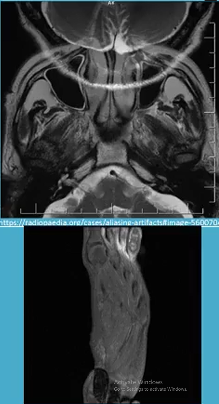
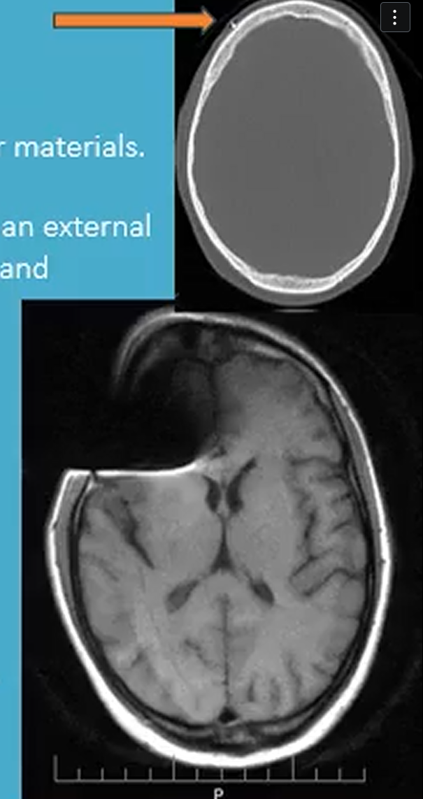
What is the susceptibility artifact in MRI
Susceptibility artifact occurs due to differences in magnetic susceptibility between tissues or materials
What is the RF noise artifact in MRI
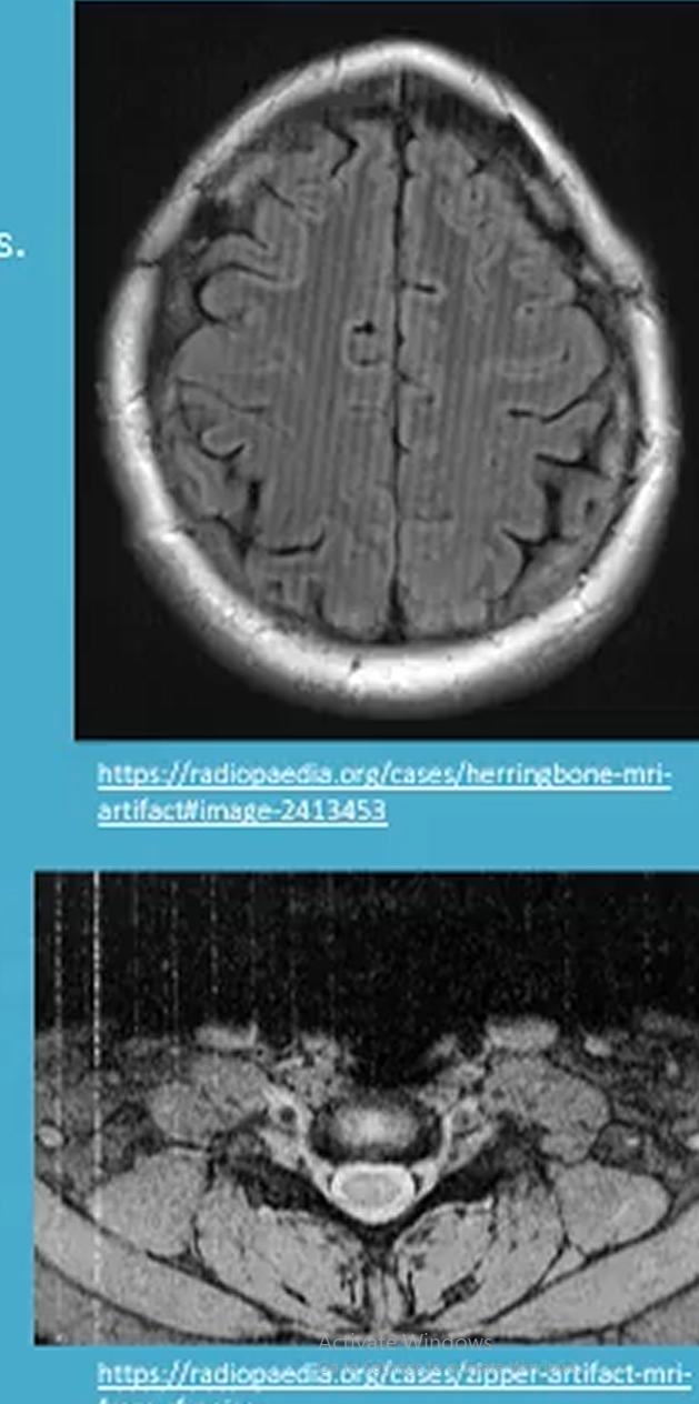
RF noise artifact appears as static on the image. Occurs die to electromagnetic interference from external sources such as nearby electrical equipment. A specific RF noise is herringbone artifact.
Resolved by:
Shielding external sources of EMI
keeping scanner room door closed
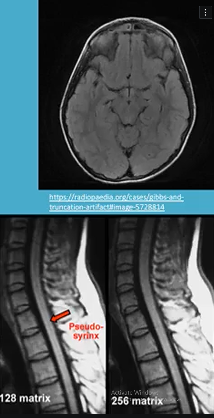
What is the Gibbs artifact in MRI
Gibbs artifact looks as a series of oscillations or ripples near sharp edges in the image. Caused by finite sampling of the signal during image reconstrucation
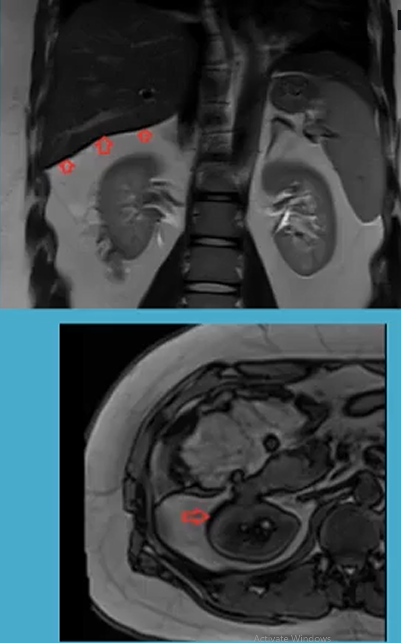
What is the Chemical shift artifact in MRI
The chemical shift artifact occurs due to spatial misregistration of fat and water molecules
What is the knee MRI protocol
Indications:
ACL, mensical or ligament etars
tumor
infection
Sequences:
3 plane localizer
Axial PD FS
Coronal OD
Sagittal PD
Sagittal PD FS
Coil:
knee coil or flexible coil
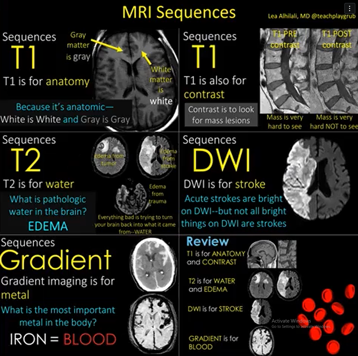
What is the Brain MRI protocol
Indications:
Headssaches
Tumour
Infection
Stroke
Sequences:
3 plane localizer
Sagittal T1
Axial T2
Axial FLAIR
Axial DWI
Axial Gradient
Coil:
head coil
What is the Spine MRI protocol
Indications:
Tumor
MS
Nifection
Trauma
Sequences:
3 plane localizer
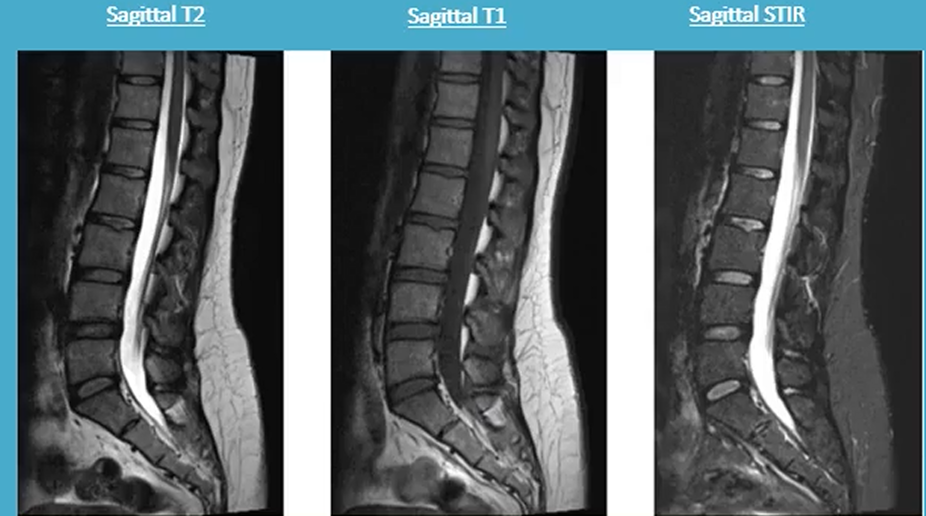
Sagittal T2
Sagiottal T1
Sagittal STIR
Axial T2
Coil:
C-spine: posterior element of head coil on table
T-spine: posterior element of head coil on table
LS spine: Spine array coil on table
Whole spine: Spine array coil on table
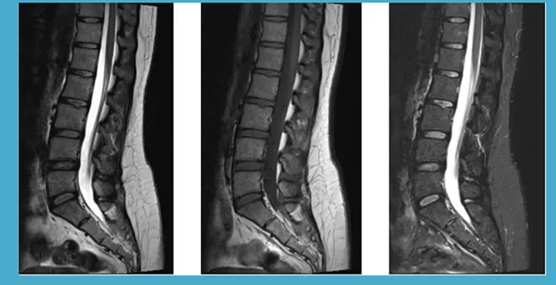
What are these images
L-spine
How does an MRI work
Each proton spins in a random way in the body creating a magnetic field. This filed is manipulated by MRI to create a signal to make an image
The magnetic filed causes by the protons charge has a direction and magnitude which is given a vector symbol and called the magnetic dipole moment. These moments are random when not under the effect of a magnet. The net magnetic vector (NMV) is almost zero.
When the patient is in the main magnetic filed (Bo) which is along the Z axis some charges align with some align against it (align with = low energy state, align against = high energy state. the difference between the low and high states is the NMV (same direction as Bo
Spin of proton under the influence of Bo is called Larmor frequency
Applying a radio frequency (RF) causes the protons axis to spin on the magnetic dipole moment, this is called resonace
When the RF pulse is turned off the dipole moment of the protons can go back to align to Bo. This gives of energy which the MRI detector reads to create an image
The time it takes for the magnetic vector to reach 63% of its original state is called the T1 time of relaxation for the tissue the proton is in
T1 images are carted by taking the signal early TE (short time to echo) and T2 is taken from long TE
How are T1 MRI images done
Protons spins are random and NMW is almost zero. patient is then placed in the magnetic filed of the machine
RF pulse is is applied flipping magnetic moments of protons 90 degrees
RF pulse is now turned off and the magnetic moment of each proton starts to spin back to align with Bo. This gives of energy the MRI detector reads to create an image
The information to create the image is when the magnetic vectors reach 63% of its original state this is called the T1 time of relaxation
How are T2 MRI images done
Protons spins are random and NMW is almost zero. patient is then placed in the magnetic filed of the machine
RF pulse is is applied flipping magnetic moments of protons 90 degrees
RF pulse is now turned off and the magnetic moment of each proton starts to spin back to align with Bo. This gives of energy the MRI detector reads to create an image
Compared to T1 imaging T2 allows protons to de phase by using a longer TE and TR
This longer TE and TR phase gives the T2 image
What are the difference between T1 and T2 MRi images
T1:
has a short time to echo (TE) and repetition time (TR)
CSF = dark
muscle = gray
spinal cord = gray
fat = bright
disk = gray
air = very dark
inflammation = dark
T2:
has a long TE and TR
CSF = bright
muscle = dark gray
spinal cord = light gray
fat = light
disk = bright
air = very dark
inflmation = bright
What is an ultrasound
A very high frequency sound that travels at 1540 meters/second in soft tissue
It causes pressure oscillations in the medium through which it is traveling
Explain diagnostic US
A short pulse of Ultrasound is transmitted into the body by the trandcuer
Structures in the tissues cause reflection and scattering of the ultrasound as it travels through the tissues
Some of these reflected and scattered energy travels back to the transducer converting it into electrical signal (echoes)
These signals are sued to create the image
What is Doppler US
Used to obtain information on blood-flow
Doppler effect: the change in perceived wave frequency when there is a relative motion between wave source and observer
What do the following mean
Echogenicity:
Anechoic:
Hyopechoic
Hyperechoic:
Isoechoic:
Echotexture:
Homogeneous:
Heterogeneous:
Echogenicity: describes the strength (brightness) of the echoes dispaled
Anechoic: no internal echoes
Hypoechoic: less echogehnic than adjacent tissue (dark)
Hyperechoic: More echogenic (bright)
Isoechoic: Same echogenicity as adjacent tissue
Echotexture: the echo consistency of the organ or structure
Homogeneous: smooth and uniform echotexture
Heterogeneous: coarse or irregular echotexture
What are the different imaging artifacts in US
Resolution artifacts: artifacts which diminish resolution within an image by giving incorrect representation of the shape and size of objects
Axial and lateral resolution artifacts: closely separated objects may appear as one object in the image, due to inadequate axial and lateral resolution
Acoustic speckle: The net echo signal received back at the transducer face from a small group of scatterers will be large or small depending on how the echoes from individual scatterers have interfered with each other.
Slice-thickness artifact: due to the finite thickness of the ultrasound beam in the elevational direction (perpendicular to the scan plane)
Attenuation artifacts: variation in the degree of attenuation of the US beam
Acoustic enhancement: Increased amplitude of displayed echo signals from regions posterior to low attenuation structures such as fluid.
Acoustic shadowing: decrease in amplitude of displayed signals from the region along the beam direction, posterior to solid or echogenic masses.
Propagation artifact: structures which incorrectly appear in the display as a result of non-convetional beam paths
Miscellaneous artifacts:
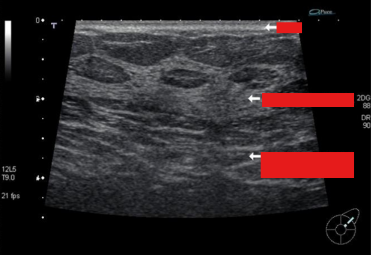
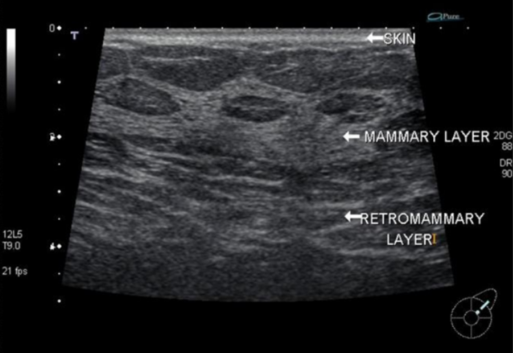
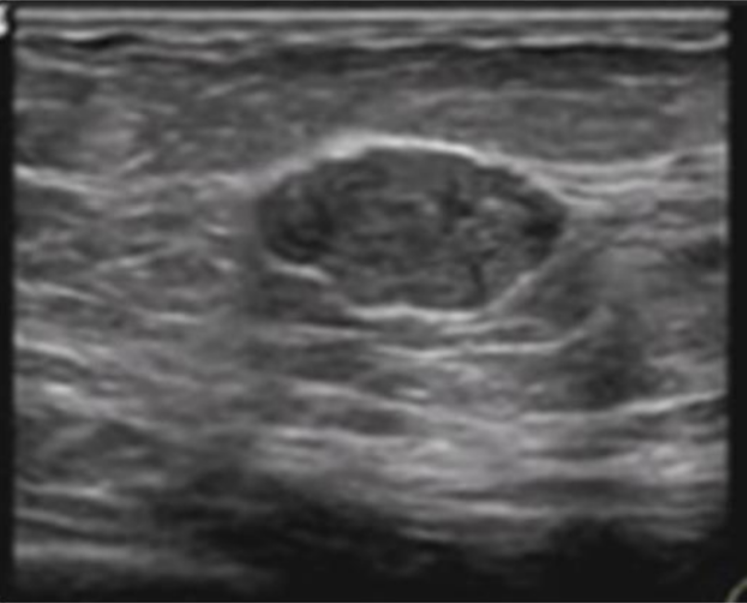
What is the pathology on this breast US
Well circumscribed hypoechoic fibroadenoma
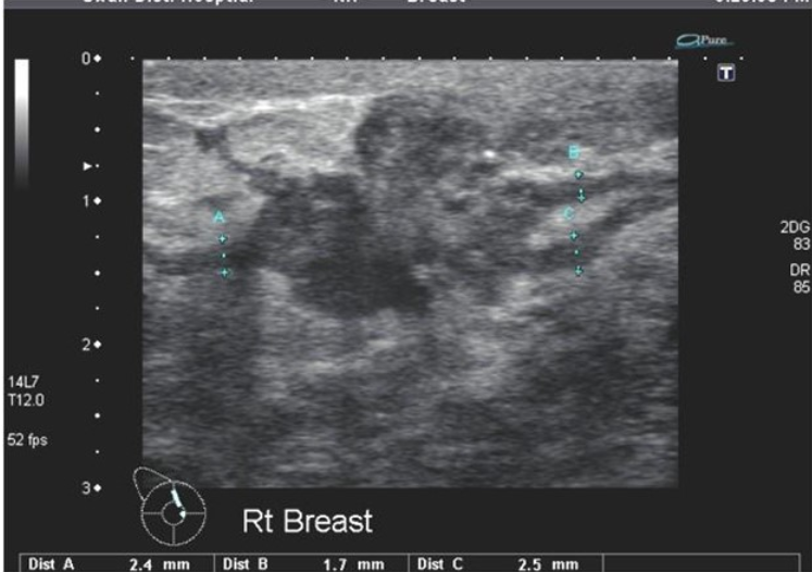
What is the pathology on this breast US
Ductal carcinoma with extension seen invading surrounding parenchyma
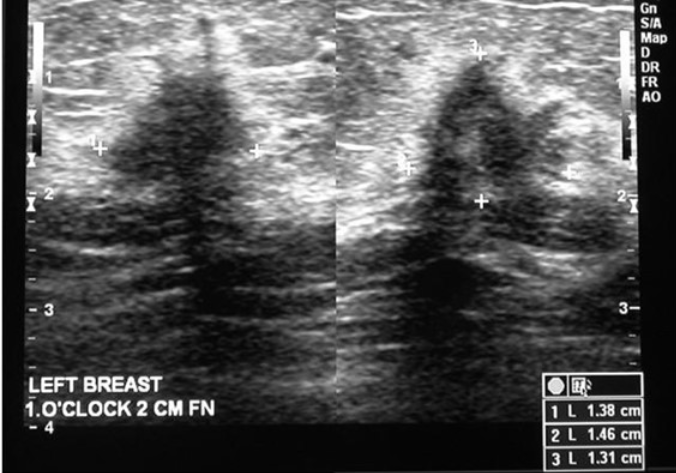
What is the pathology on this breast US
Longitudinal and transverse images of a ‘typical’ ductal carcinoma
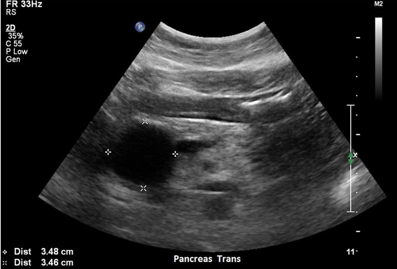
What pathology is this
An enlarged heterogeneous pancreas consistent with acute pancreatitis.
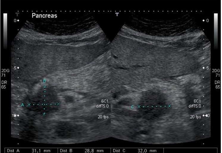
What pathology is this
Longitudinal and transverse views through the pancreatic head/uncinate process demonstrating a pancreatic carcinoma.
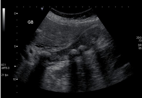
What pathology is this
Cholecystitis with bile stasis and obstructive calculus in neck.
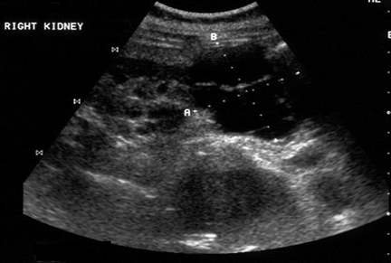
What pathology is this
Septated right kidney lower pole cyst
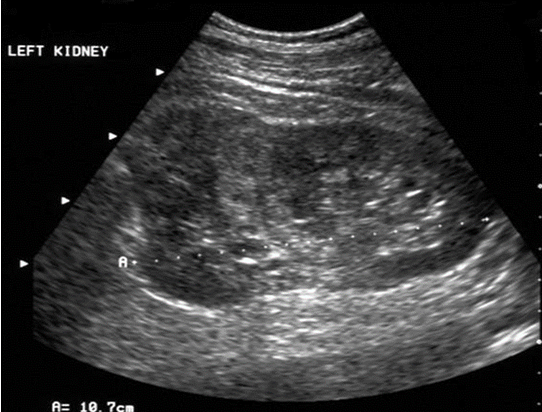
What pathology is this
Renal cell carcinoma (RCC) of the upper pole of the left kidney seen as an isoechoic cortical mass.
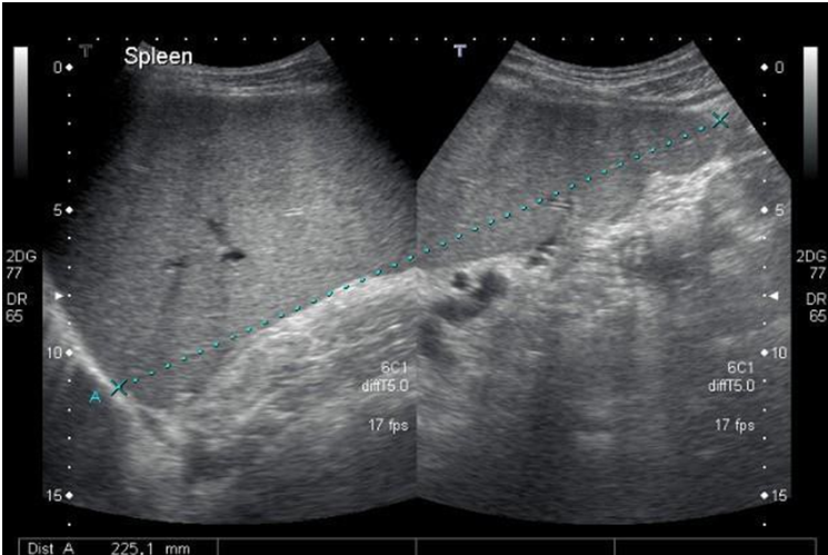
What pathology is this
splenomegaly. It is described as splenomegaly if the longest axis measuring greater than 12-14 cm.
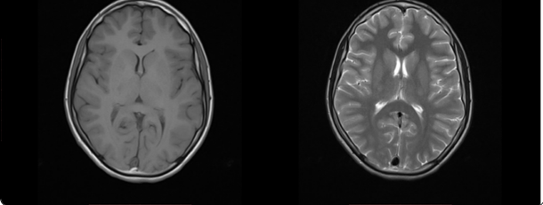
Which is which and why
T1 as only 1 tissue is bright which is the fat
T2 as 2 tissues are bright the fat and water
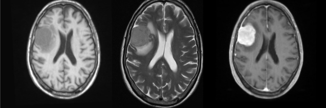
Which is which
T1
T2
T1 + contrast
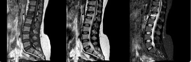
Which is which
T1
T2
STIR (T2 image that is manipulated so that fat is nullified
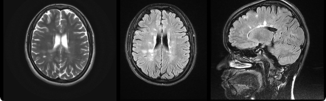
Which is which
T2
Flair (T2 which has CSF nullified)
Flair
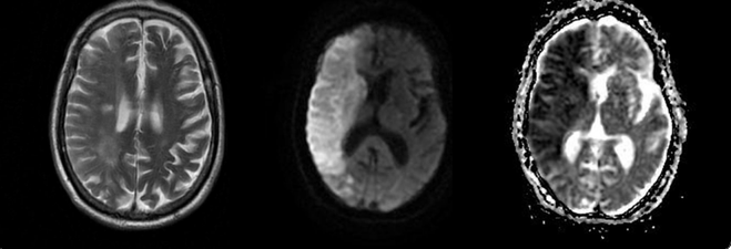
Which is which
T2
DWI
ADC
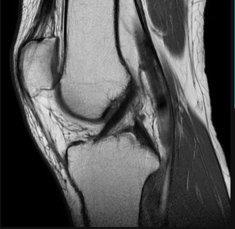
Normal?
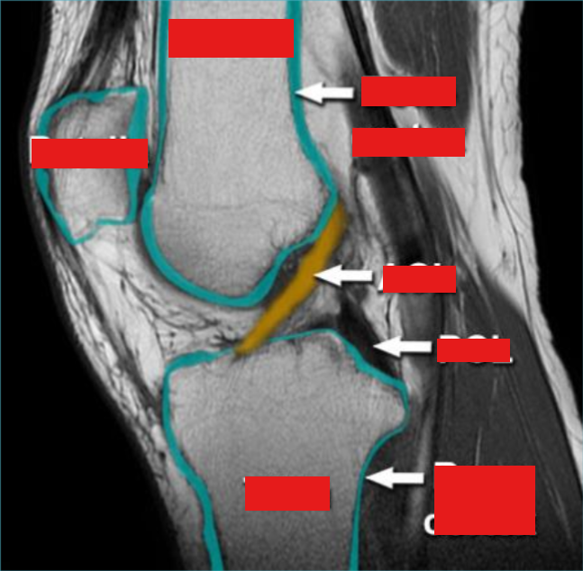
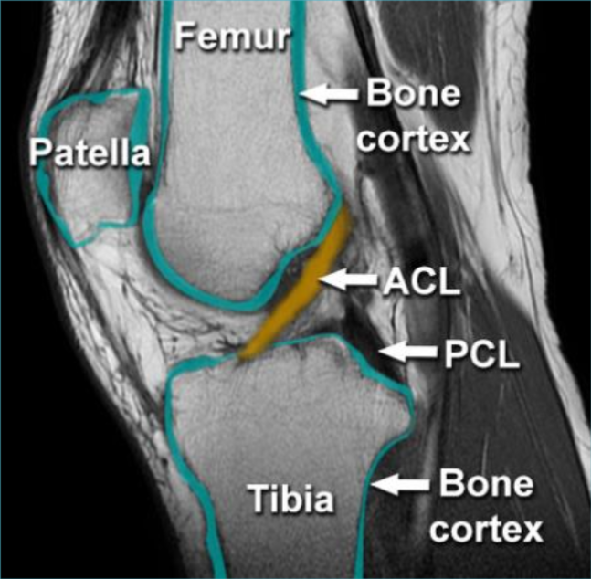
Normal knee MRI
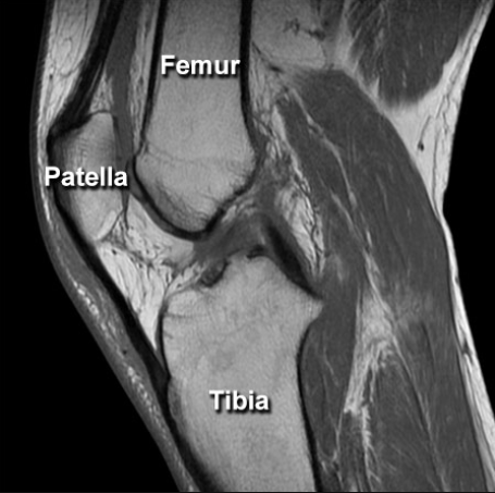
Normal?
ACL tear
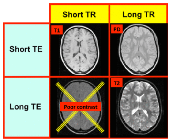
What do the following =
Short TR/Short TE →
Long TR/Short TE →
Long TR/Long TE →
Short TR/Short TE → T1-Weighted image
Long TR/Short TE → PD-Weighted image
Long TR/Long TE → T2-Weighted image
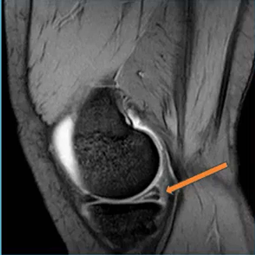
What is this
Meniscus tear
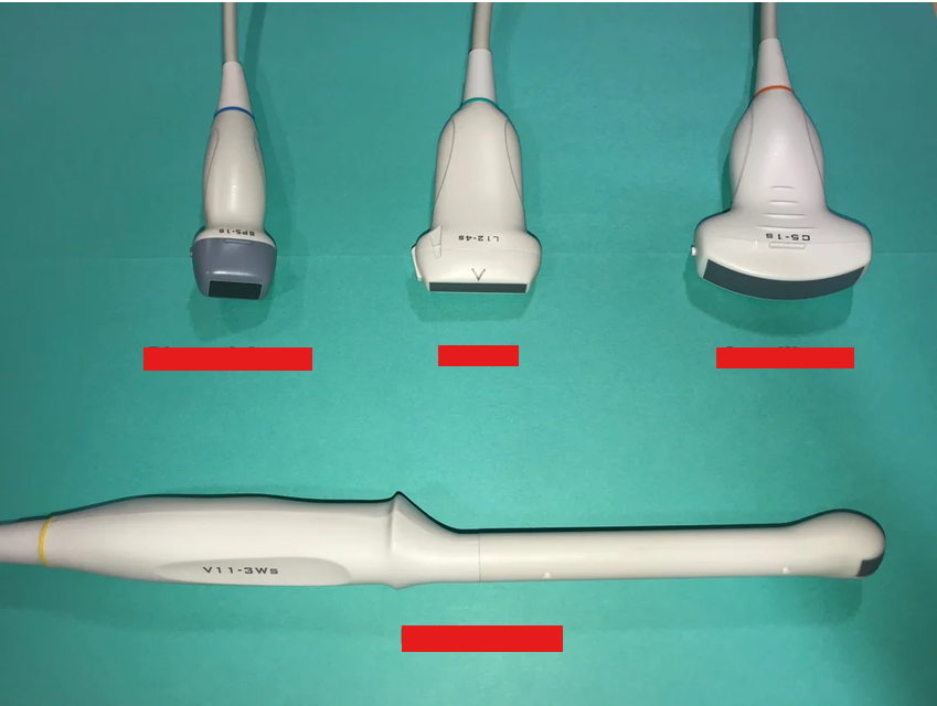
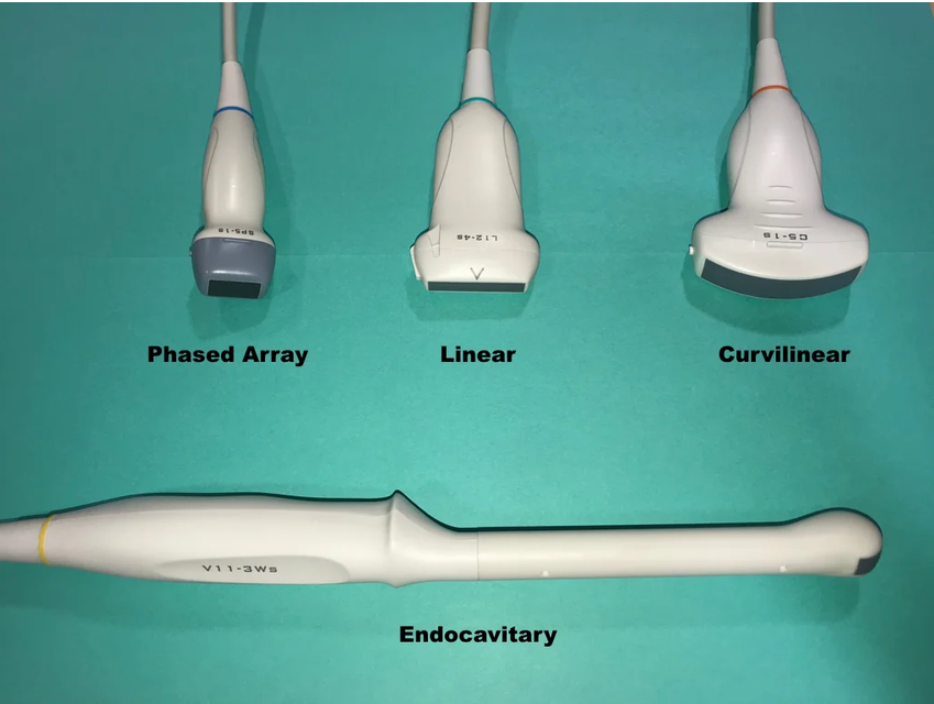
When are the folwing usually perfomed:
nuchal translucency (NT)
morphology
growth assessment
nuchal translucency (NT): first trimester (11-14 weeks)
morphology: second trimester (18-22 weeks)
growth assessment: third trimester (32-36 weeks)
Explain the bio effects of thermal and non-thermal interactions in US
Thermal interactions: heat production by the ultrasound wave, the higher the frequency the greater the absorption and degree of heating obtained
Non-thermal interactions:
Cavitation: Sound waves traveling through liquid contain tiny gas bubbles they cause then to grow and then collapse causing high temperature and pressure that can damage surrounding tissue
Radiation force: Tiny pushes causes by ultrasound waves
Acoustic torque: Sounds waves twisting objects they encounter
Acoustic streaming: High-intensity sound waves can cause a flowing motion within a liquid, like tiny whirlpools.
How much heat is too much in US
Safe limit: When scanning fetus below 1.5 degrees celcius to avoid potential harm
What real timer labels can you use to monitor ultrasound safely
Thermal Index (TI): This measures how much a tissue's temperature might increase due to the ultrasound's power. A higher TI indicates a greater potential for heating.
Mechanical Index (MI): This measures the likelihood of transient cavitation occurring during the exam. Values below 0.7 are considered safe