Skin & Skeletal Practical Exam (BIO 121, Parkland College)
 Call Kai
 Learn
Learn Practice Test
 Spaced Repetition
 Match
Match Flashcards
 Knowt Play
1/163

There's no tags or description
Looks like no tags are added yet.
Name | Mastery | Learn | Test | Matching | Spaced | Call with Kai | Chat |
|---|
No analytics yet
Send a link to your students to track their progress
164 Terms
Epidermis
Outer most layer of cells/stratified squamous epithelium
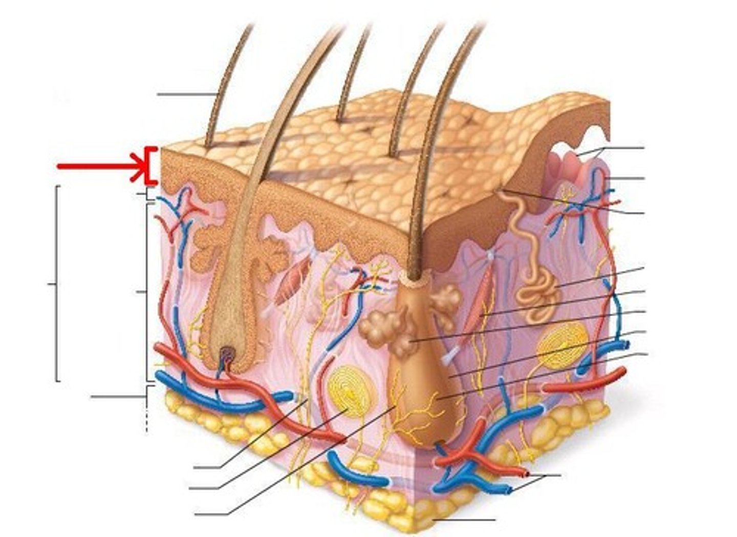
Stratum corneum
outermost protective layer of epidermis, 5 - 30 layers keratinized cells [FIRST LAYER/CORNEA]
![<p>outermost protective layer of epidermis, 5 - 30 layers keratinized cells [FIRST LAYER/CORNEA]</p>](https://assets.knowt.com/user-attachments/65a4758c-c815-4e80-abe0-c7e9b1734d8e.jpg)
Stratum basale
Basal (deepest) layer of epidermis, most cells are basal cells [BASE]
![<p>Basal (deepest) layer of epidermis, most cells are basal cells [BASE]</p>](https://assets.knowt.com/user-attachments/93099abf-9d74-492d-a398-9ad28d257355.jpg)
Dermis
Located between epidermis and hypodermis, contains collagen and elastic fibers
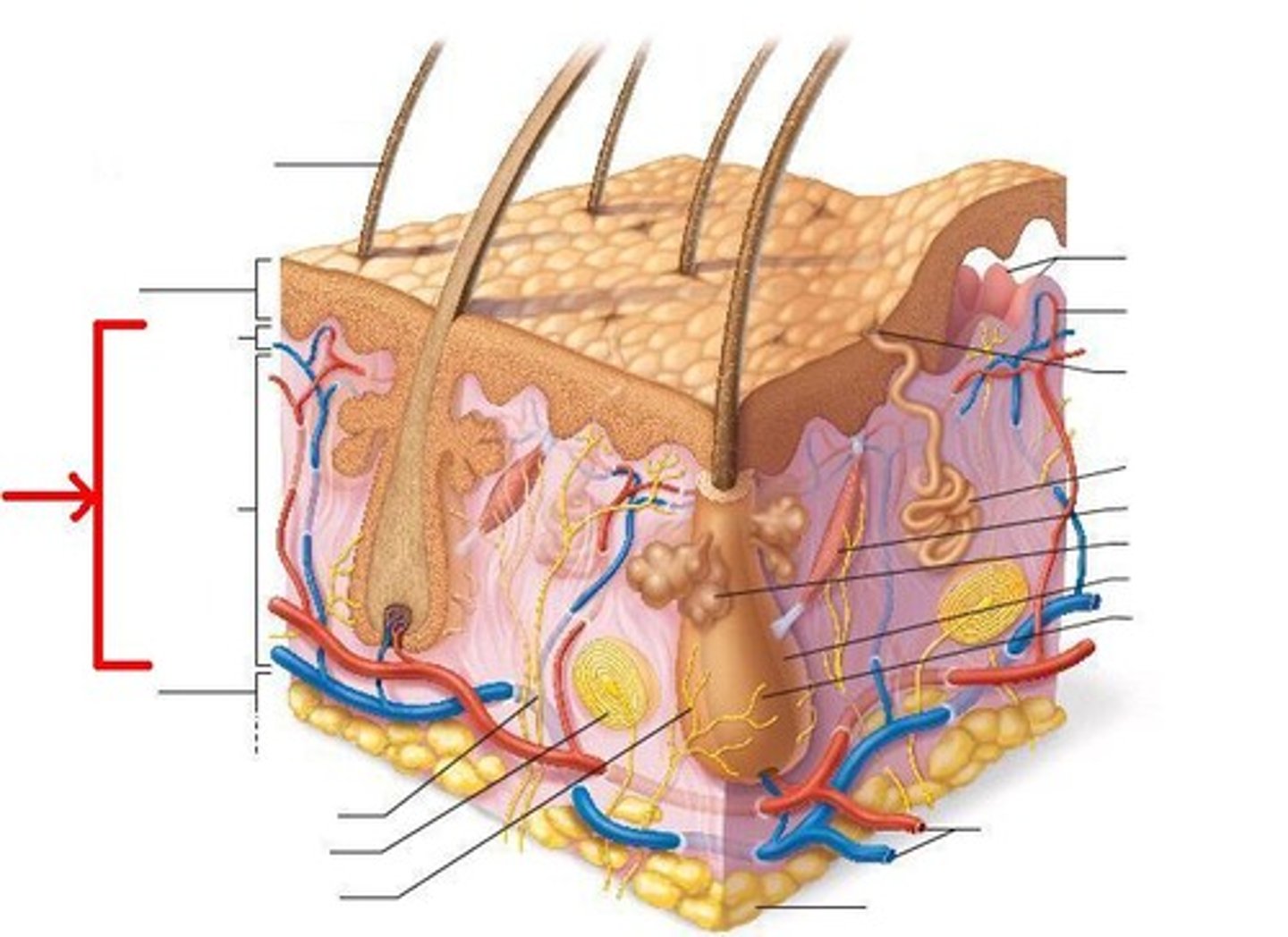
Dermal papilla
Blood vessels that nourish all hair follicles and bring nutrients and oxygen to the lower layers of epidermal cells. Arranged in ridgelike lines. [PAPILLA HAS RIDGES]
![<p>Blood vessels that nourish all hair follicles and bring nutrients and oxygen to the lower layers of epidermal cells. Arranged in ridgelike lines. [PAPILLA HAS RIDGES]</p>](https://assets.knowt.com/user-attachments/36cde864-16d4-4983-9c8c-dd32733e5755.jpg)
Hair follicle
Small cavity in dermis and corium of the skin from which a hair develops.
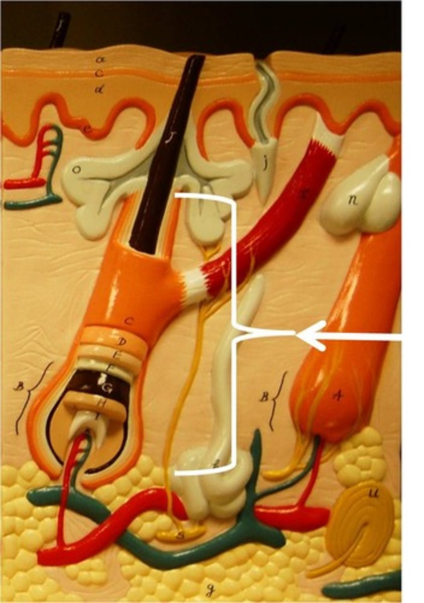
Hair root
Anchors the hair into the skin (extends from base of follicle to where hair shaft loses connection w/follicle walls)
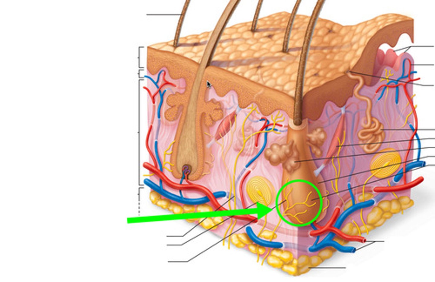
Arrector pili muscle
smooth muscle attached to hair follicle; contractions pull hair erect. [LOOKS LIKE MUSCLE IN DERMIS]
![<p>smooth muscle attached to hair follicle; contractions pull hair erect. [LOOKS LIKE MUSCLE IN DERMIS]</p>](https://assets.knowt.com/user-attachments/b0addadf-5abc-4ce7-b1cb-211aab15560e.jpg)
Sebaceous gland
holocrine glands that discharge oily lipid secretions. contractions of arrector pili muscle causes release of sebum. [LOOKS LIKE A GLAND]
![<p>holocrine glands that discharge oily lipid secretions. contractions of arrector pili muscle causes release of sebum. [LOOKS LIKE A GLAND]</p>](https://assets.knowt.com/user-attachments/8dac5cca-e34a-406c-9869-320e0e308f12.jpg)
Sweat glands
a small gland that secretes sweat, situated in the dermis of the skin. Such glands are found over most of the body, and have a simple coiled tubular structure. Apocrine glands produce odor and starts secreting at puberty. Merocrine glands is the most common. [COILED LIKE WRINGING OUT SWEATY TOWEL]
![<p>a small gland that secretes sweat, situated in the dermis of the skin. Such glands are found over most of the body, and have a simple coiled tubular structure. Apocrine glands produce odor and starts secreting at puberty. Merocrine glands is the most common. [COILED LIKE WRINGING OUT SWEATY TOWEL]</p>](https://assets.knowt.com/user-attachments/5de4709e-a649-458c-a545-f8f510bd0c60.jpg)
Hypodermis
subcutaneous layer of superficial fascia. Separates integument from deep fascia. Deepest layer that is NOT a part of the skin.
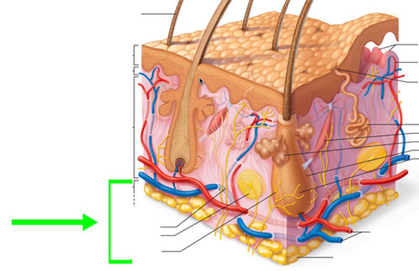
Axial Skeleton
126 bones. Bones of skull, thorax, and vertebral column. Longitudinal axis of body.
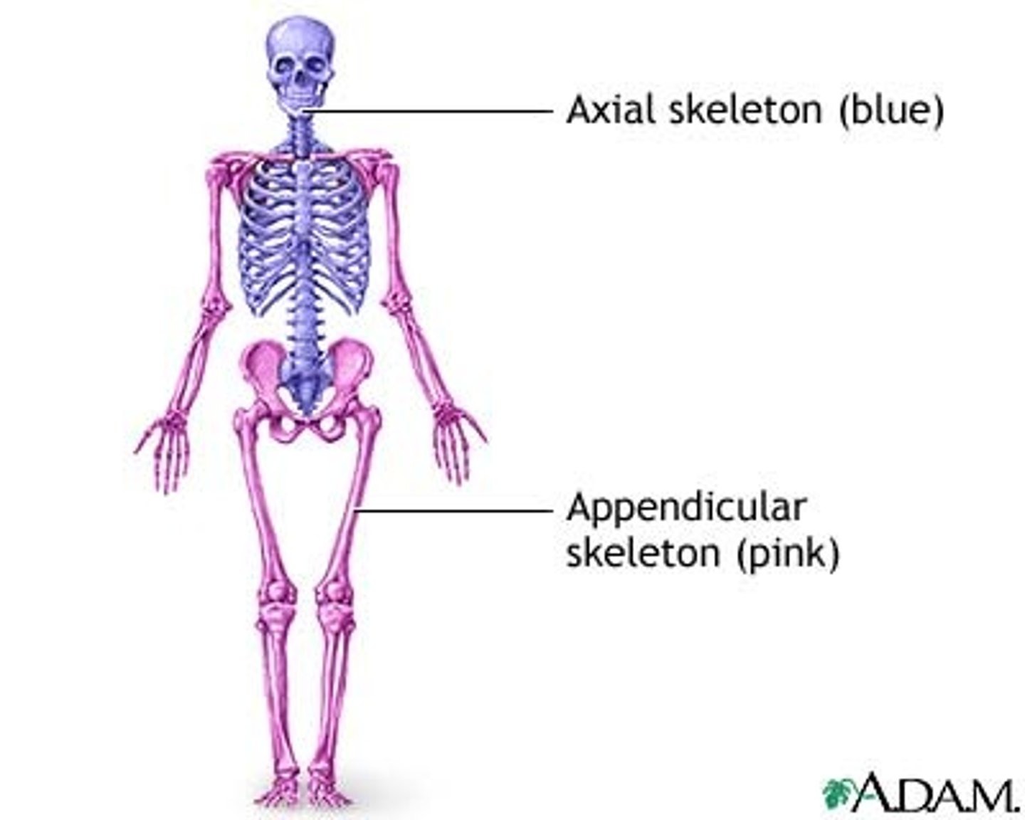
Skull
head
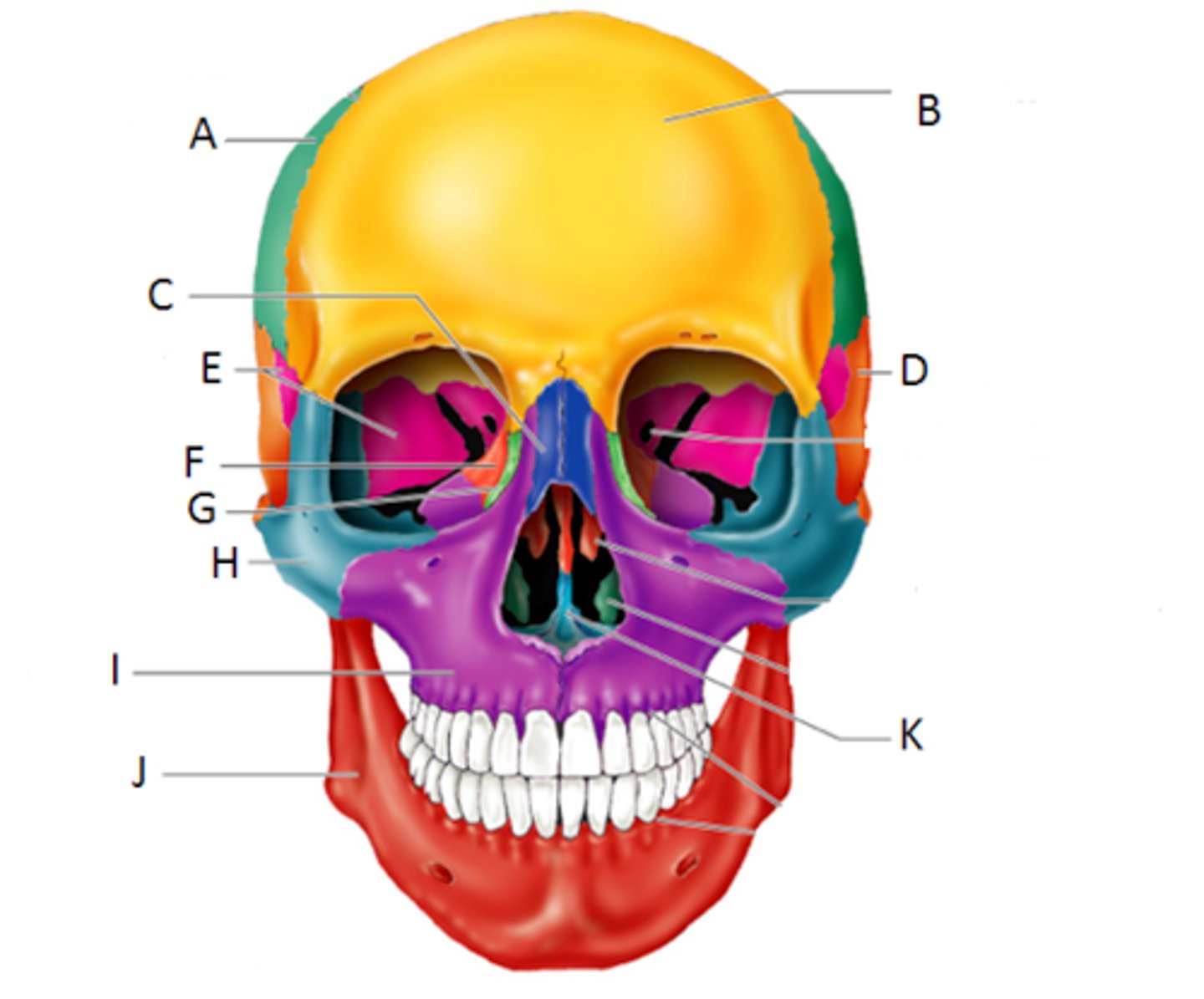
Frontal
Forehead to beginning of nose [FRONT OF HEAD]
![<p>Forehead to beginning of nose [FRONT OF HEAD]</p>](https://assets.knowt.com/user-attachments/cf6102e9-1ed3-4140-bd19-4b8914f0b58f.jpg)
Parietal
back of skull [PARENTS ARE ALWAYS BEHIND CHILDREN]
![<p>back of skull [PARENTS ARE ALWAYS BEHIND CHILDREN]</p>](https://assets.knowt.com/user-attachments/aabc193f-056c-4fc0-bd4f-dbc5c9b155d2.jpg)
Occipital
bottom of skull [OCTOPUS ON THE BOTTOM OF THE OCEAN]
![<p>bottom of skull [OCTOPUS ON THE BOTTOM OF THE OCEAN]</p>](https://assets.knowt.com/user-attachments/b09fc39a-78f8-4dc3-a928-691f8b8cb8c6.jpg)
Foramen magnum
large hole where spinal cord enters the cranial cavity. [MAGNUMS ARE FOR-A-MEN/HOLE]
![<p>large hole where spinal cord enters the cranial cavity. [MAGNUMS ARE FOR-A-MEN/HOLE]</p>](https://assets.knowt.com/user-attachments/3c1bf93b-fba1-4ca3-b0f1-562fdf8687d6.jpg)
Occipital condyle
2 oval shaped bumps on either side of the foramen magnum [ CC: COND(OM)YLE NEXT TO YOUR MAGNUM]
![<p>2 oval shaped bumps on either side of the foramen magnum [ CC: COND(OM)YLE NEXT TO YOUR MAGNUM]</p>](https://assets.knowt.com/user-attachments/bde70083-eb95-44aa-bffa-a88d9c5f9d6d.jpg)
temporal
side of skull connected to parietal and occipital bone [WHEN YOU HAVE A TEMPER YOU RUB THE SIDE OF YOUR HEAD]
![<p>side of skull connected to parietal and occipital bone [WHEN YOU HAVE A TEMPER YOU RUB THE SIDE OF YOUR HEAD]</p>](https://assets.knowt.com/user-attachments/988bb4d8-b584-4806-80b0-c4c572262e90.jpg)
mastoid process
a conical prominence of the temporal bone behind the ear [MASTER LARGEST PROCESS]
![<p>a conical prominence of the temporal bone behind the ear [MASTER LARGEST PROCESS]</p>](https://assets.knowt.com/user-attachments/ed89fd84-bd38-43dd-99ac-63c04b038ee7.jpg)
External acoustic meatus
ear canal [MEAT MAKES ME LISTEN TO ACOUSTIC MUSIC EXTERNALLY!]
![<p>ear canal [MEAT MAKES ME LISTEN TO ACOUSTIC MUSIC EXTERNALLY!]</p>](https://assets.knowt.com/user-attachments/2d06bb39-9298-42ba-b1f8-b592a65d92b8.jpg)
styloid process
pole-like process extending downward from the temporal bone on each side of the skull [STYLOID PEN]
![<p>pole-like process extending downward from the temporal bone on each side of the skull [STYLOID PEN]</p>](https://assets.knowt.com/user-attachments/38921145-a587-4954-99db-c63ba4b7501d.jpg)
carotid canal
passageway in the temporal bone through which the internal carotid artery enters the middle cranial fossa from the neck [I WANNA SHOVE A CARROT IN THE HIGHEST SMALLER CANAL]
![<p>passageway in the temporal bone through which the internal carotid artery enters the middle cranial fossa from the neck [I WANNA SHOVE A CARROT IN THE HIGHEST SMALLER CANAL]</p>](https://assets.knowt.com/user-attachments/74654a91-3035-43c3-b470-aacc308c1af1.jpg)
Sphenoid Bone
Behind eyes
[WHEN YOUR ANNOYED YOU RUB THIS BONE]
![<p>Behind eyes</p><p>[WHEN YOUR ANNOYED YOU RUB THIS BONE]</p>](https://assets.knowt.com/user-attachments/992a2b2b-178b-4357-99a8-3b878917ec9d.jpg)
Sella turcica
small depression of the sphenoid bone that contains the pituitary gland
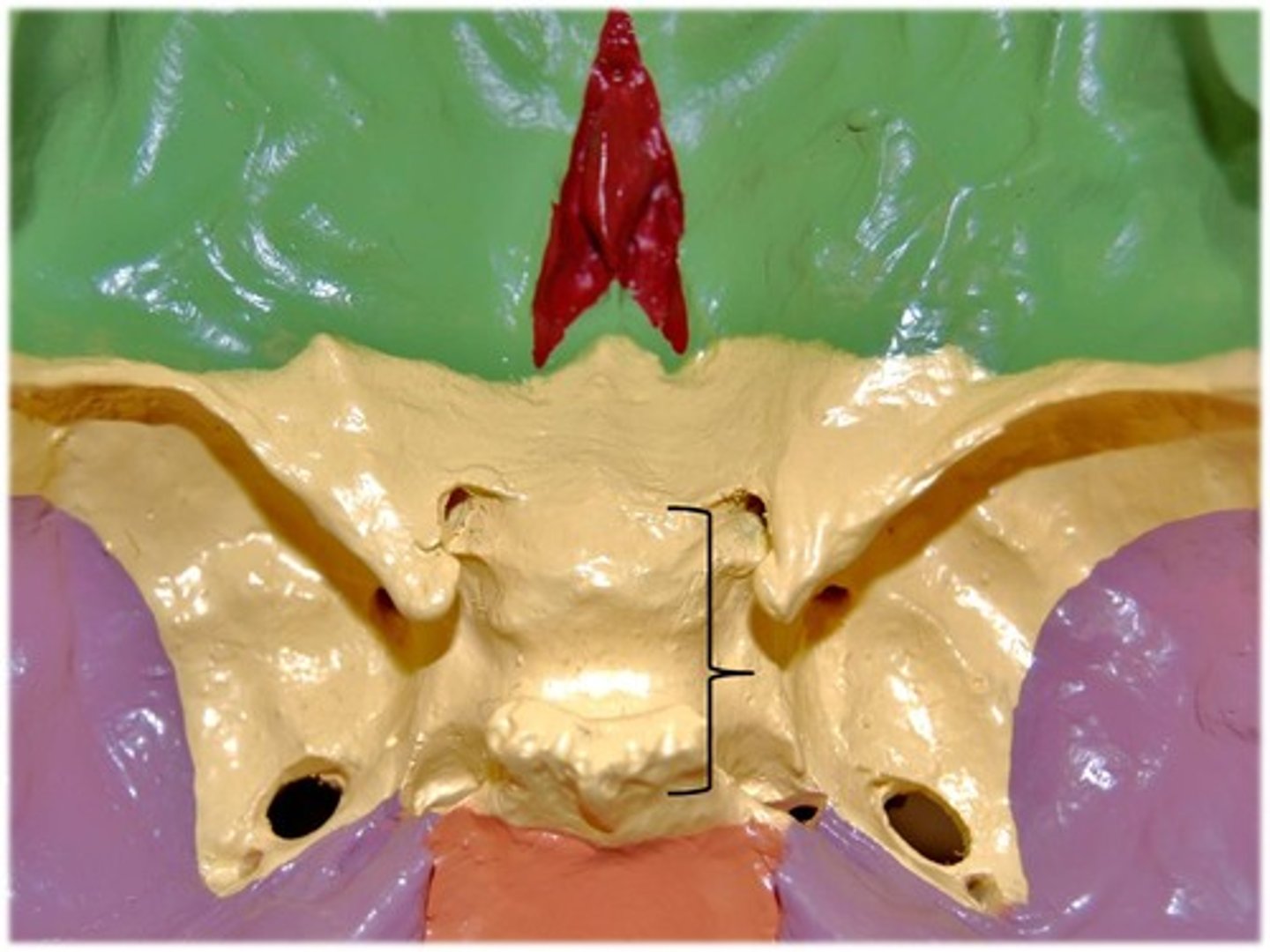
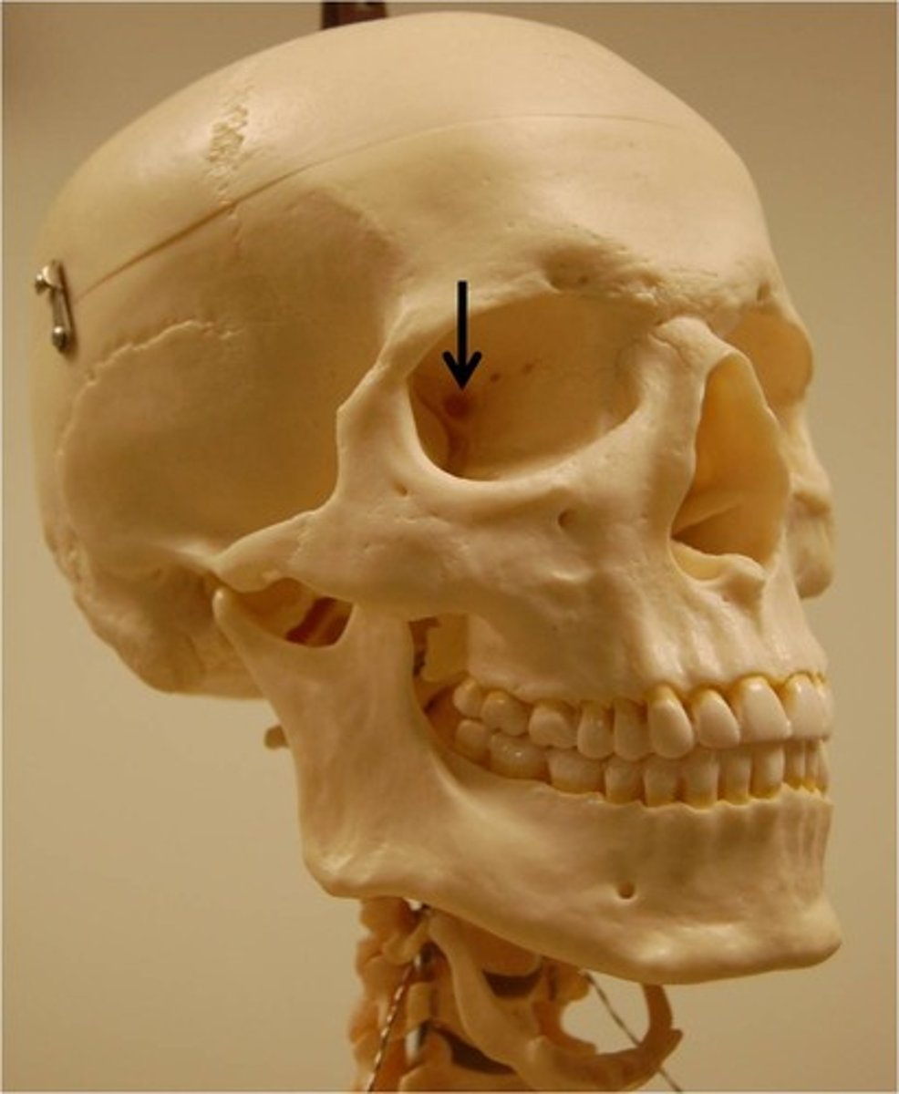
optic canal
Allows the optic nerve to pass to the eye
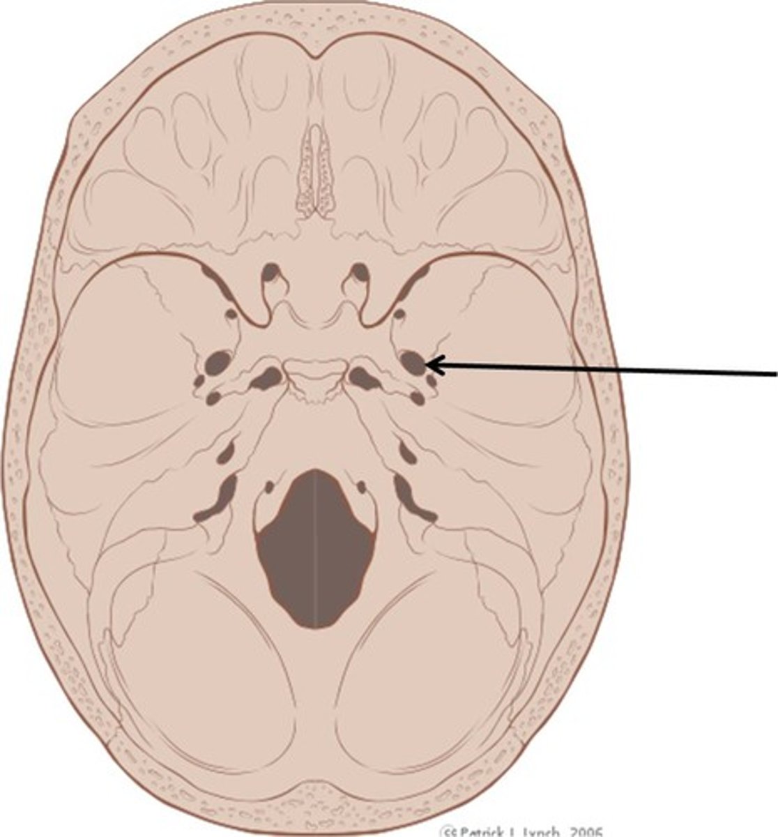
foramen ovale
oval shaped opening in the middle cranial fossa located at the posterior base of the greater wing of the sphenoid bone,
Ethmoid Bone
forms part of the posterior portion of the nose, the orbit, and the floor of the cranium
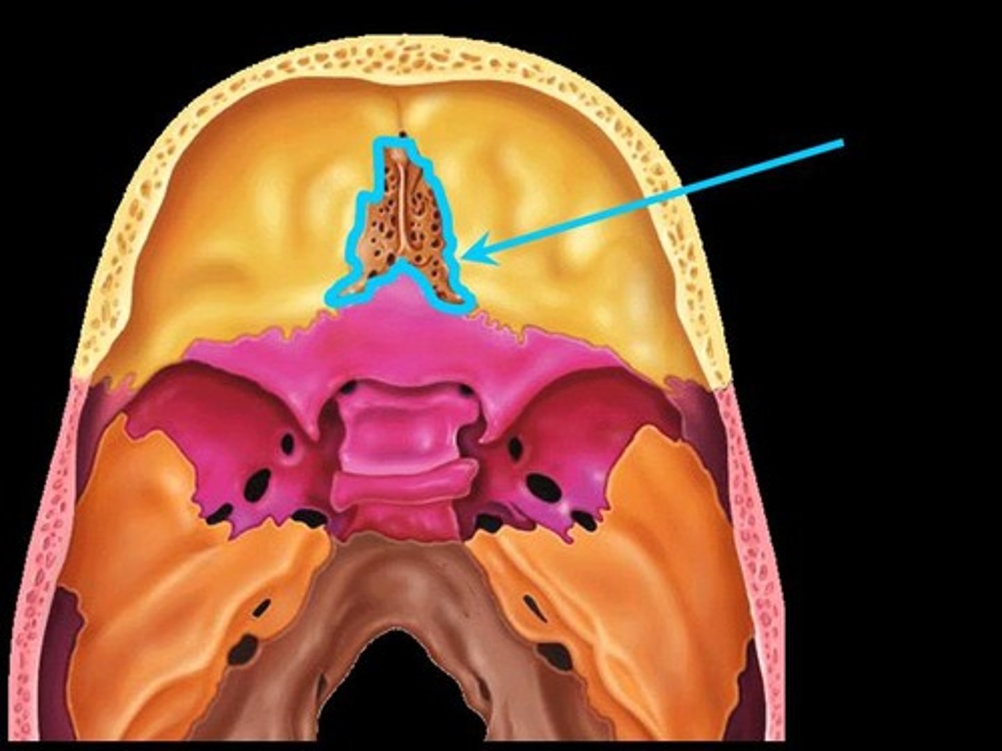
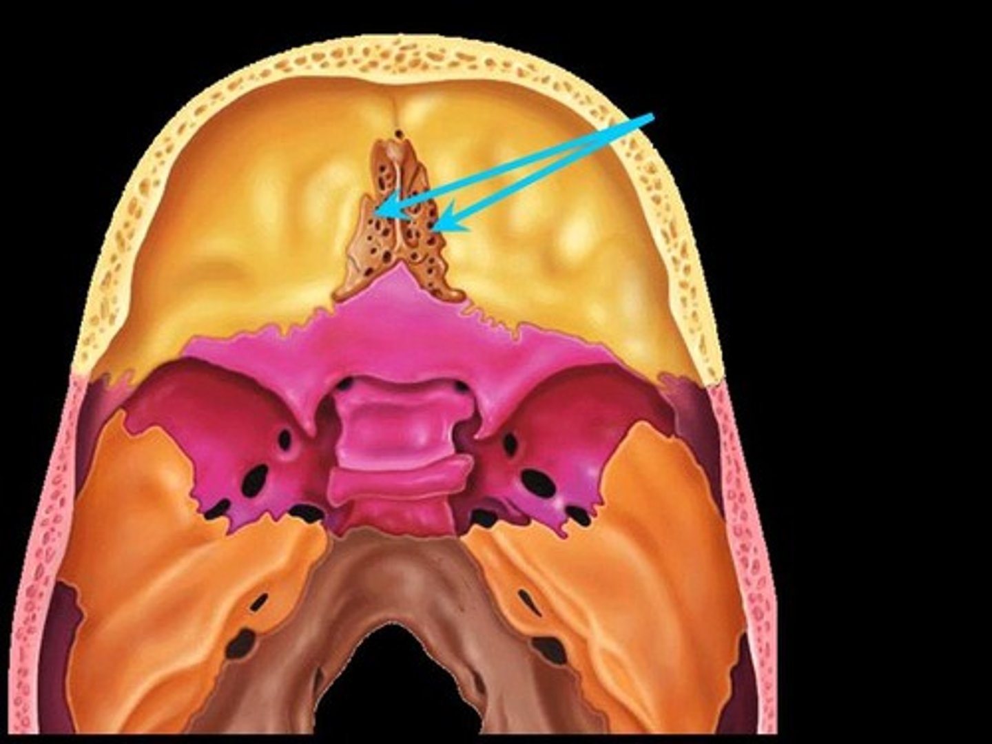
cribriform plate
form roof of nasal cavity
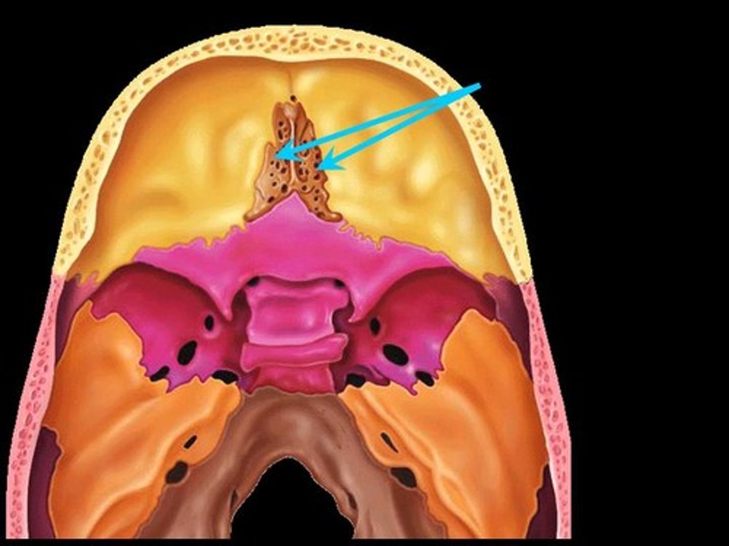
crista galli
is a median ridge of bone that projects from the cribriform plate of the ethmoid bone
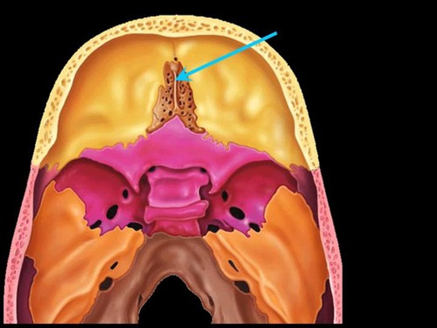
olfactory foramina
Where the nerves with sense of smell pass thru, on ethmoid bone
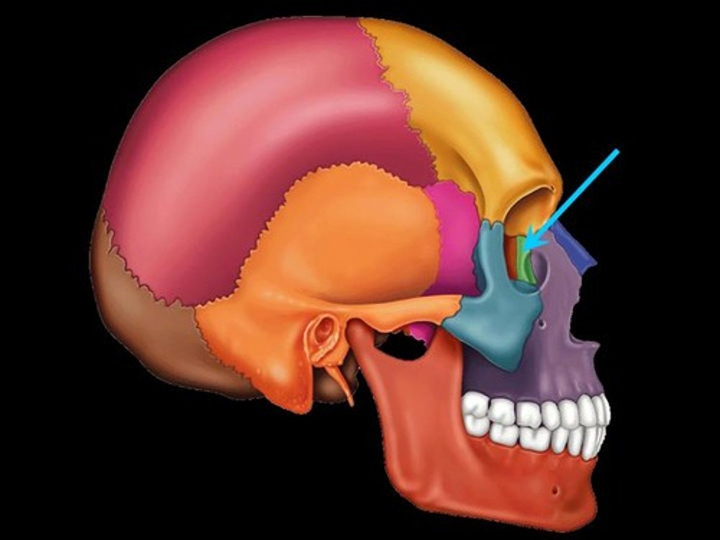
Lacrimal
lacrimal bone forms part of the medial wall of the orbit, or pertaining to tears
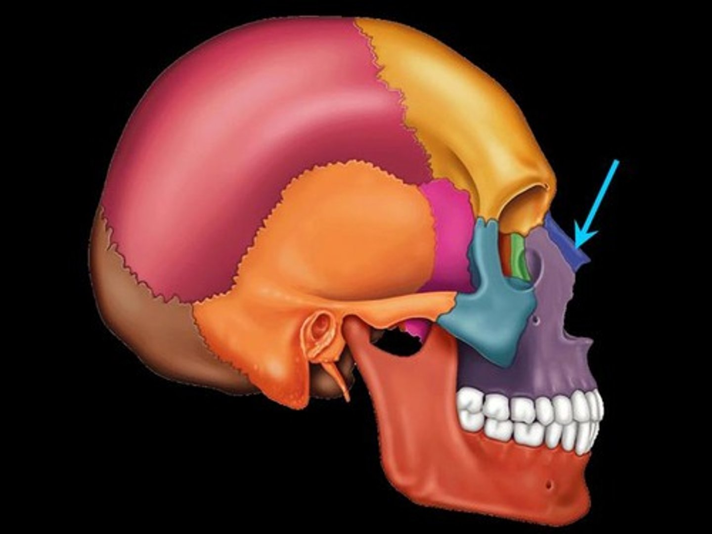
Nasal
Bridge of nose
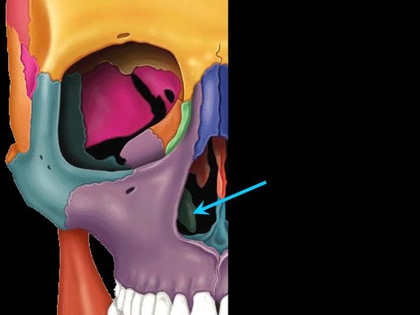
Vomer
Flat, thin bone that forms part of the nasal septum.
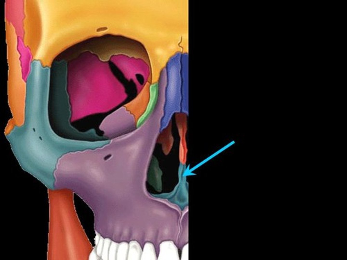
Inferior nasal conchae
extend horizontally along the lateral wall of the nasal cavity
Zygomatic
Cheek bone
[YOUR ZYGOMATICS ARE PINK]
![<p>Cheek bone</p><p>[YOUR ZYGOMATICS ARE PINK]</p>](https://assets.knowt.com/user-attachments/40136e29-6827-42a5-8aaf-e9a1fa3cfeee.jpg)
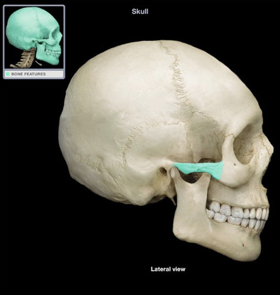
Zygomatic arch
the processes on the temporal and zygomatic bones; determines the widest part of the face
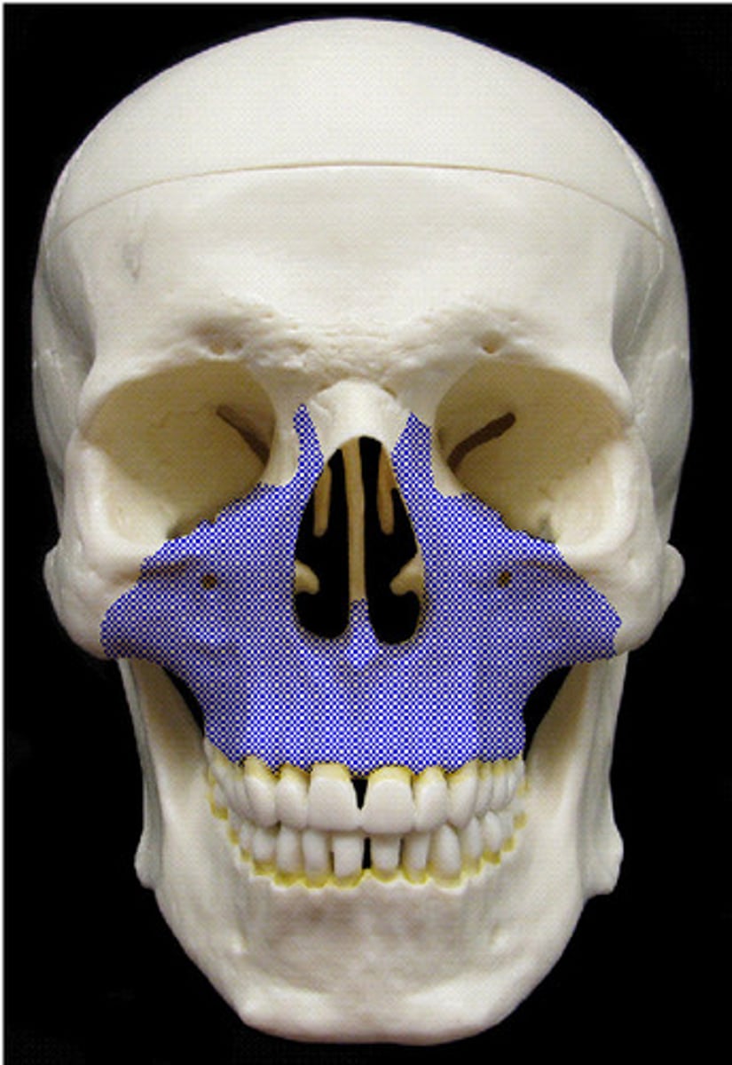
Maxilla
bone located beneath the nose
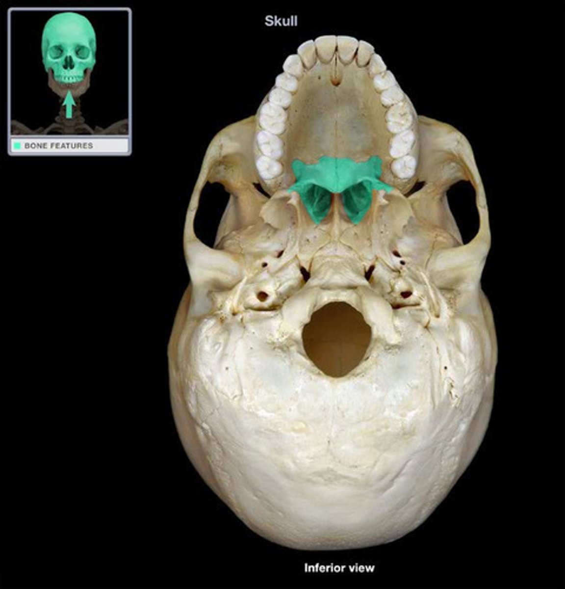
Palatine
posterior, makes up hard palette
Mandible
lower jaw bone [YOU NEED THIS BONE TO SAY MANNNN]
![<p>lower jaw bone [YOU NEED THIS BONE TO SAY MANNNN]</p>](https://assets.knowt.com/user-attachments/bc3c5119-c8c9-43ce-9095-aca7f994a17c.jpg)
Mental foramen
opening in the mandible for passage of chin artery/nerve
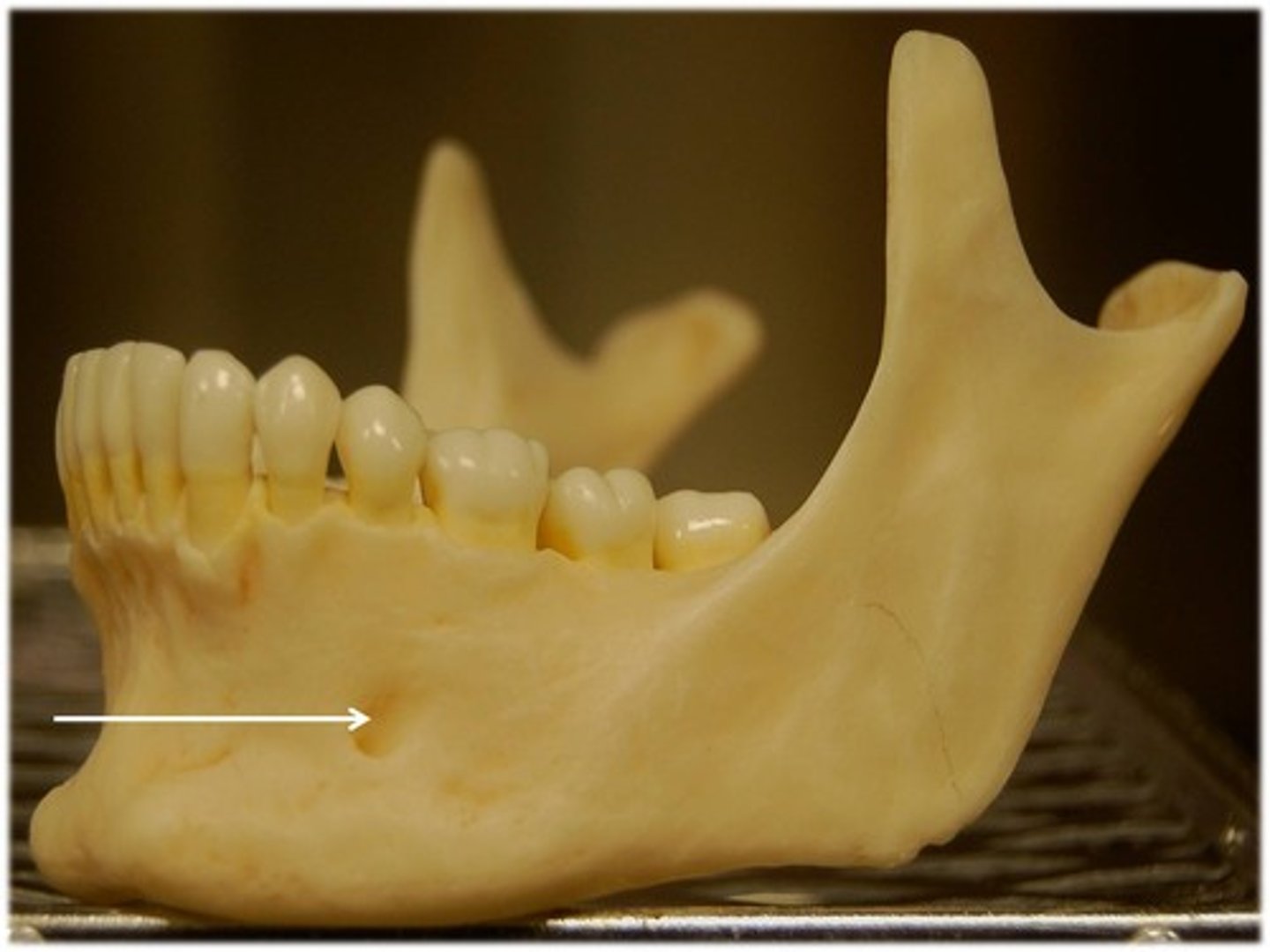
Mandibular foramen
Mandible
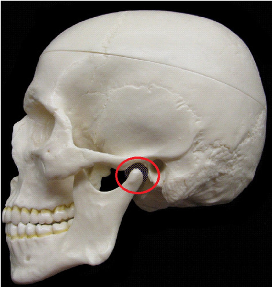
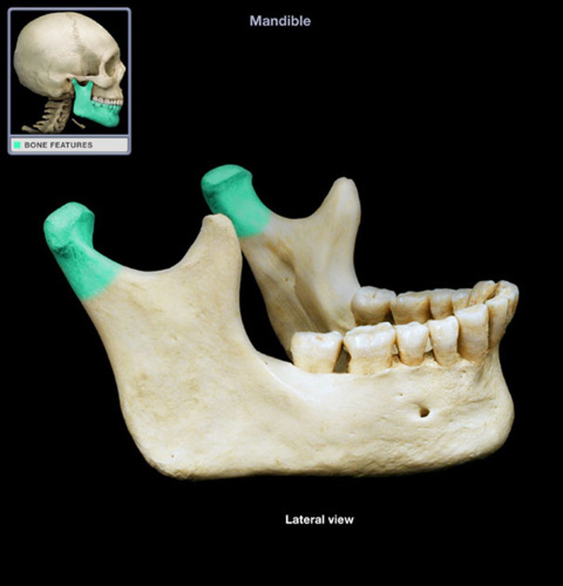
Condylar process
head of condyle, elliptical in shape, articulates with temporal bone, neck of condyle
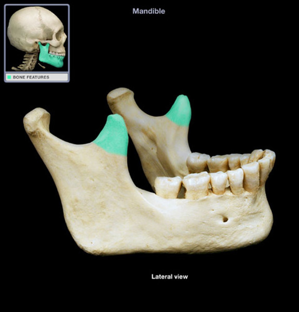
Coronoid process
the anterior projection at the top of the rami
Hyoid
small U-shaped bone in neck, where many tongue muscles attach. Not attached to any other bones. [HY OID YOU LIKE MY FLOATING BONE?]
![<p>small U-shaped bone in neck, where many tongue muscles attach. Not attached to any other bones. [HY OID YOU LIKE MY FLOATING BONE?]</p>](https://assets.knowt.com/user-attachments/1da8f423-1579-4548-a930-55332349ccf8.jpg)
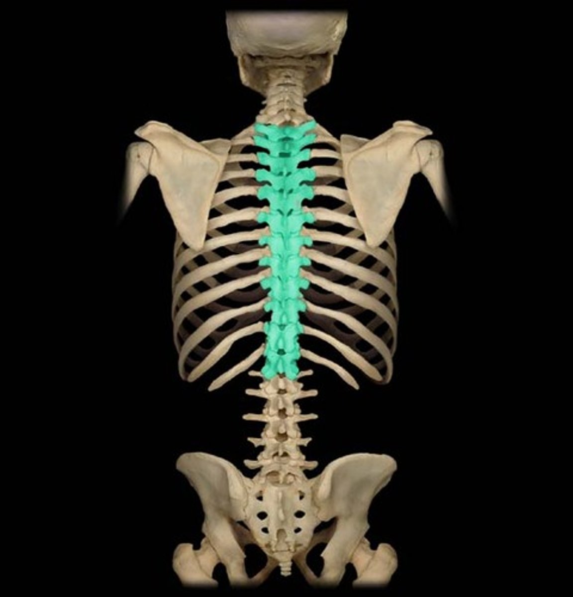
Vertebrae Body
provides support for body by forming column with other bodies known as centrum. Thoracic region.
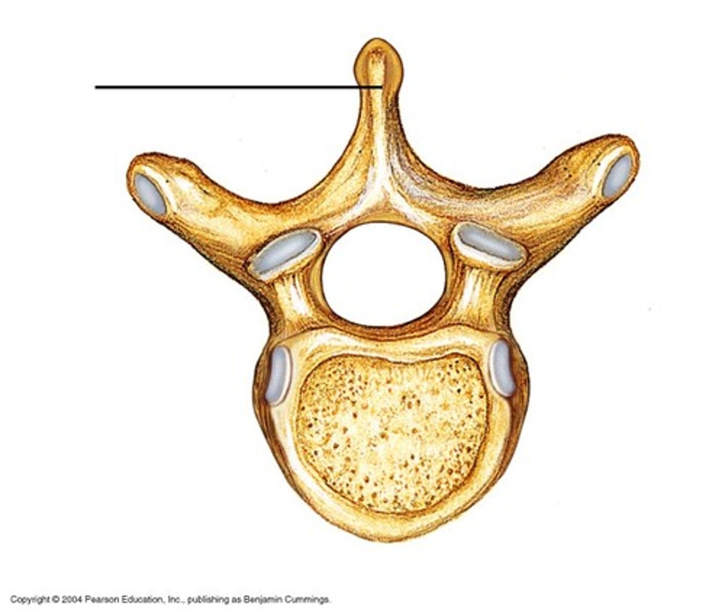
Spinous process
a bony projection off the posterior (back) of each vertebra
Transverse process
small bony projection off the right and left side of each vertebrae.
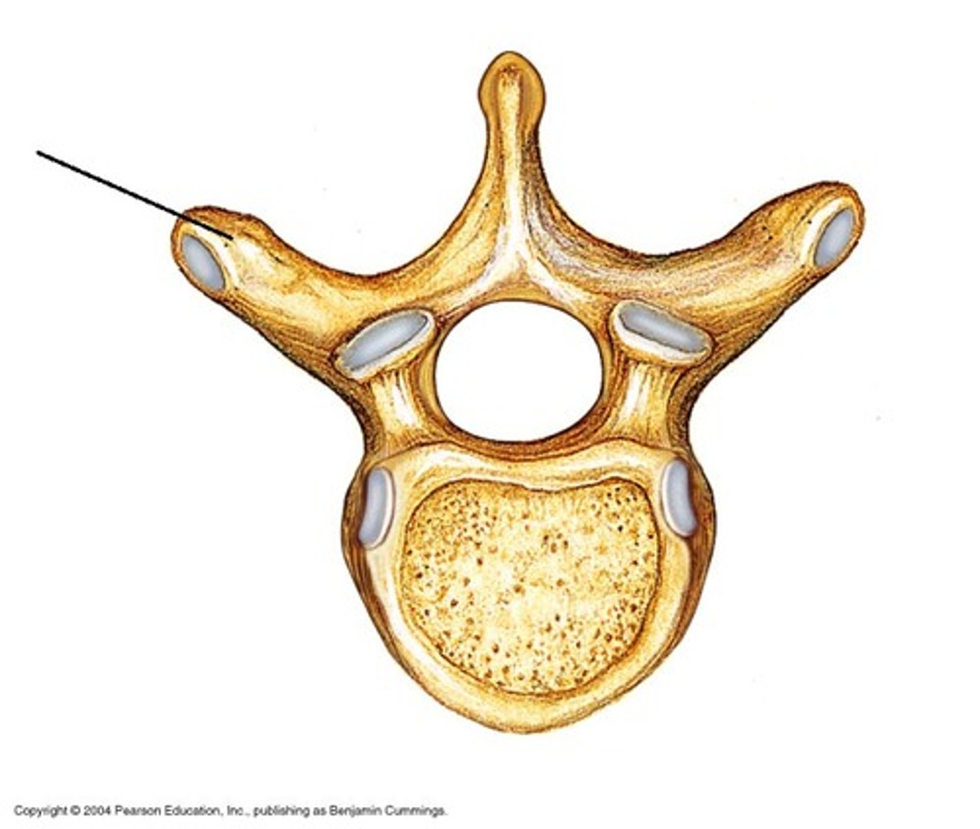
Vertebral foramen
canal through which the spinal cord passes [THIS VERTEBRAL HOLE IS FOR A MAN]
![<p>canal through which the spinal cord passes [THIS VERTEBRAL HOLE IS FOR A MAN]</p>](https://assets.knowt.com/user-attachments/9a14594d-038c-4691-ae0b-f9cca2526080.jpg)
Pedicle
Short stalks that form sides of vertebral foramen, projecting from bodies
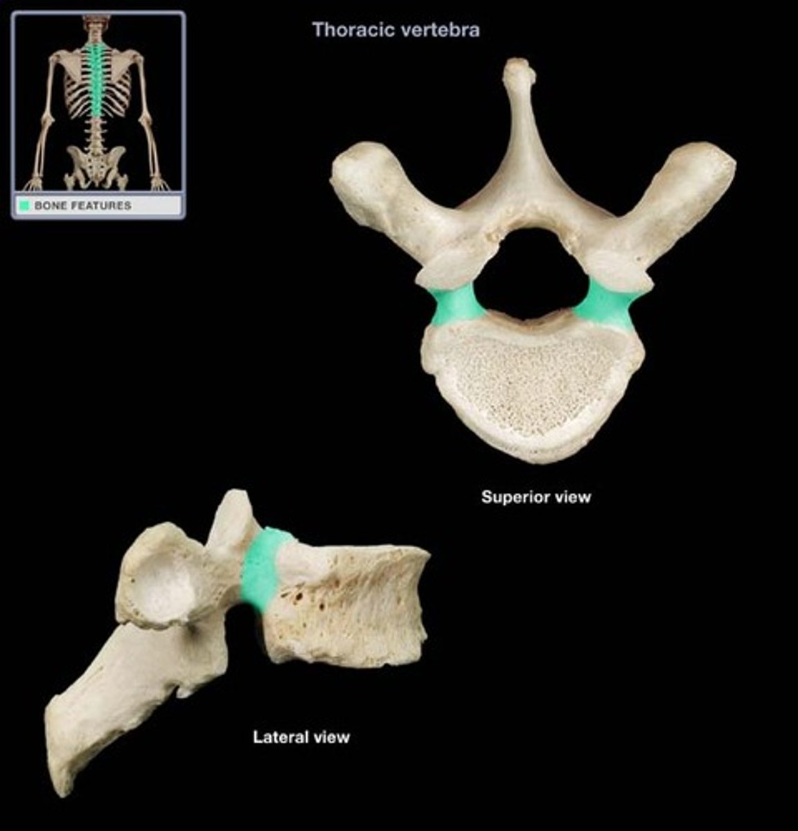
lamina
part of the vertebral arch
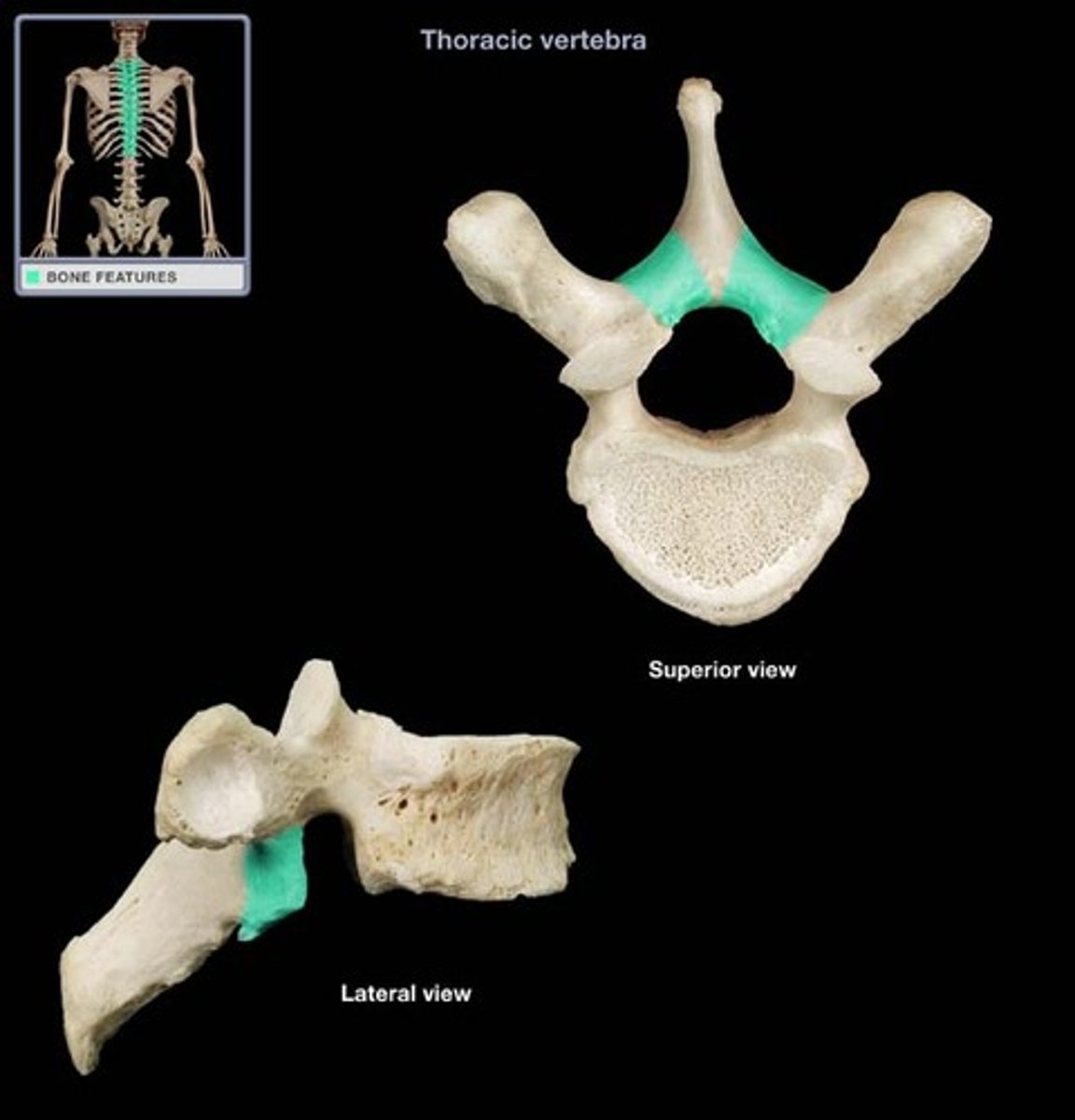
intervertebral foramen
opening between vertebrae for passage of a spinal nerve
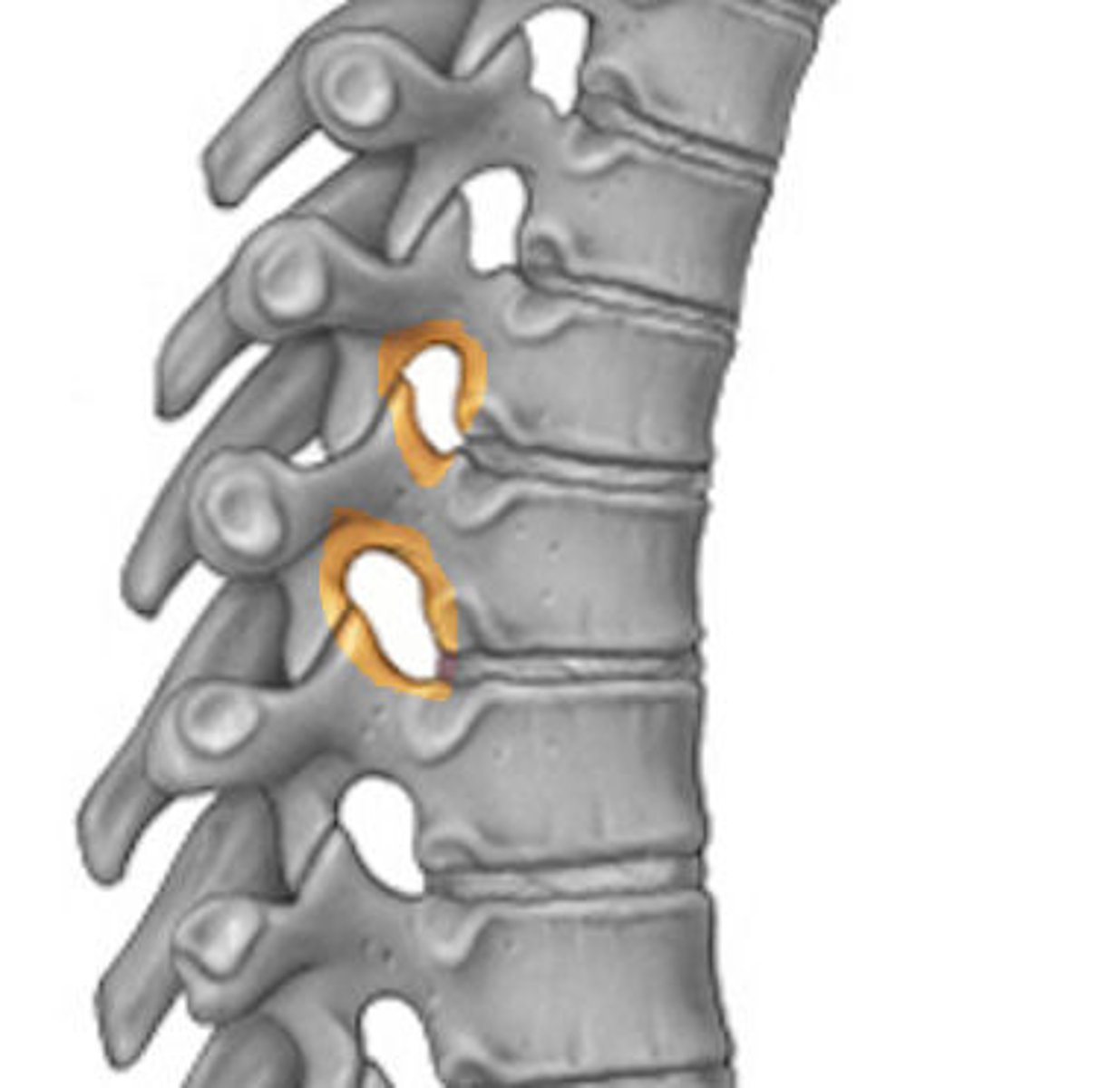
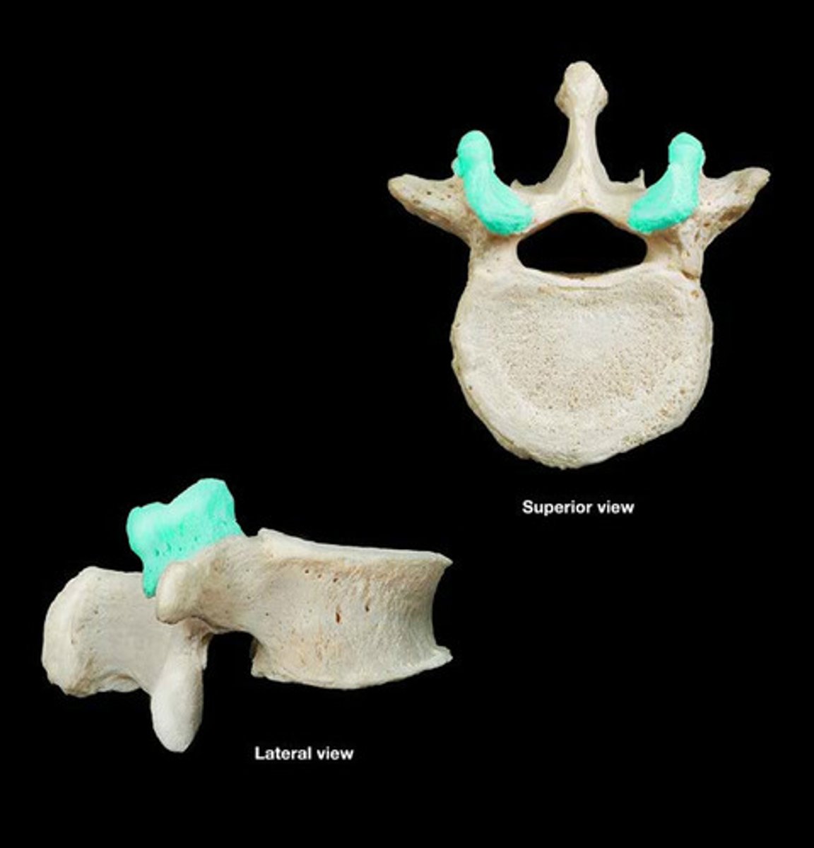
superior articular facet (process)
A flat or nearly flat surface on a bone; where two vertebrae articulate; superior side
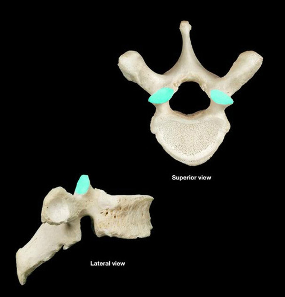
inferior articular facet (process)
A flat or nearly flat surface on a bone; where two vertebrae articulate; inferior side
Cervical Vertebrae (C1-C7)
are the smallest, lightest vertebrae.
[COFFEE FOR BREAKFAST/FIRST]
![<p>are the smallest, lightest vertebrae.</p><p>[COFFEE FOR BREAKFAST/FIRST]</p>](https://assets.knowt.com/user-attachments/cf5c459b-66cc-4faf-963d-f687572240d9.jpg)
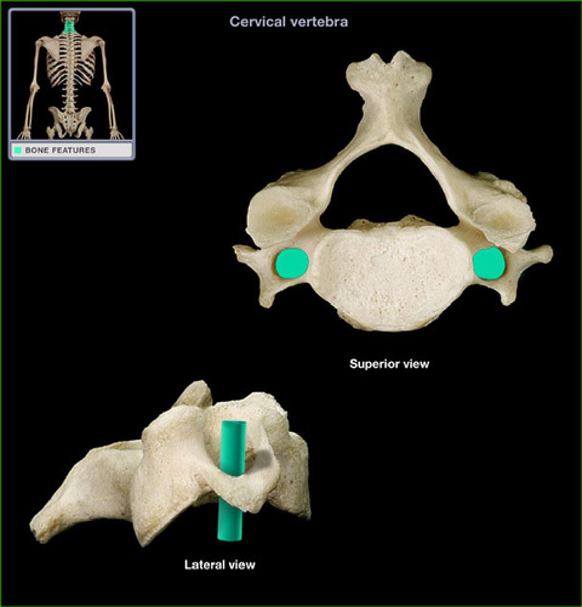
transverse foramen (cervical vertebrae)
opening in the transverse process of a cervical vertebra for the passage of the vertebral artery and vein
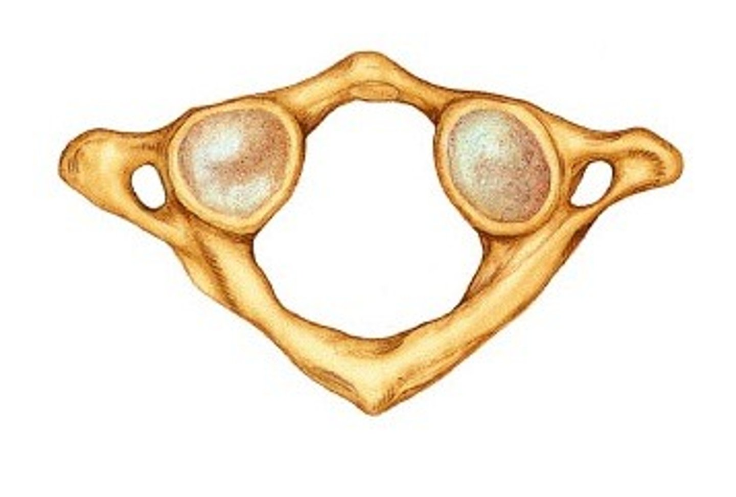
Atlas (C1)
most superior (first) cervical vertebra of the spine
Axis (C2)
supports the atlas
-has heavy spinous processes to attach muscles of head and neck
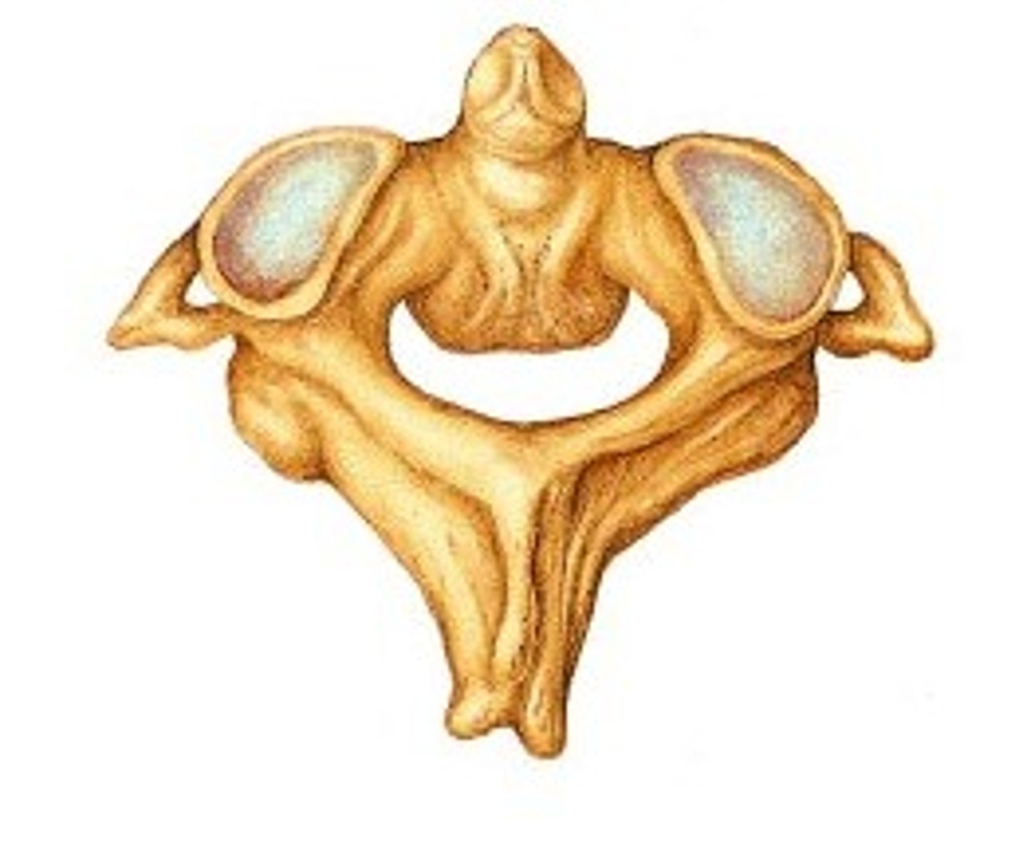
Dens
A toothlike process projecting upward from the body of the axis around which the atlas rotates.
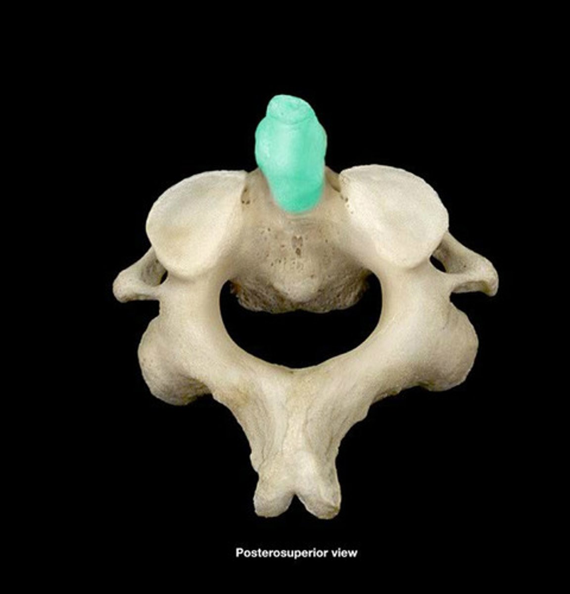
Vertebra prominens (C7)
distinctive long and prominent spinous process, which is palpable from the skin surface
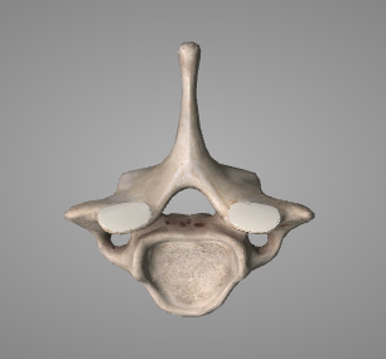
Thoracic vertebrae (T1-T12)
middle segment of the vertebral column, between the cervical vertebrae and the lumbar vertebrae. [TURKEY FOR LUNCH/SECOND]
![<p>middle segment of the vertebral column, between the cervical vertebrae and the lumbar vertebrae. [TURKEY FOR LUNCH/SECOND]</p>](https://assets.knowt.com/user-attachments/c317b48c-18a2-4a83-be43-4b9f4f92e3c8.jpg)
transverse costal facets
grooves on the side of a thoracic vertebra that helps the vertebra attach to the rib cage.
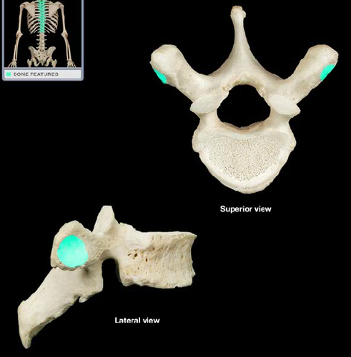
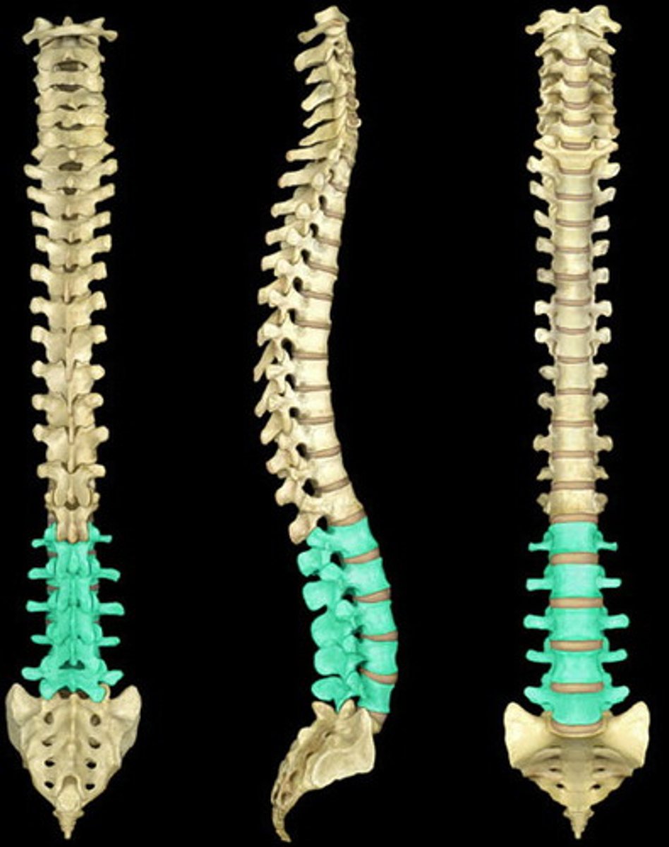
Lumbar vertebrae (L1-L5)
he five vertebrae between the rib cage and the pelvis. They are the largest segments of the vertebral column
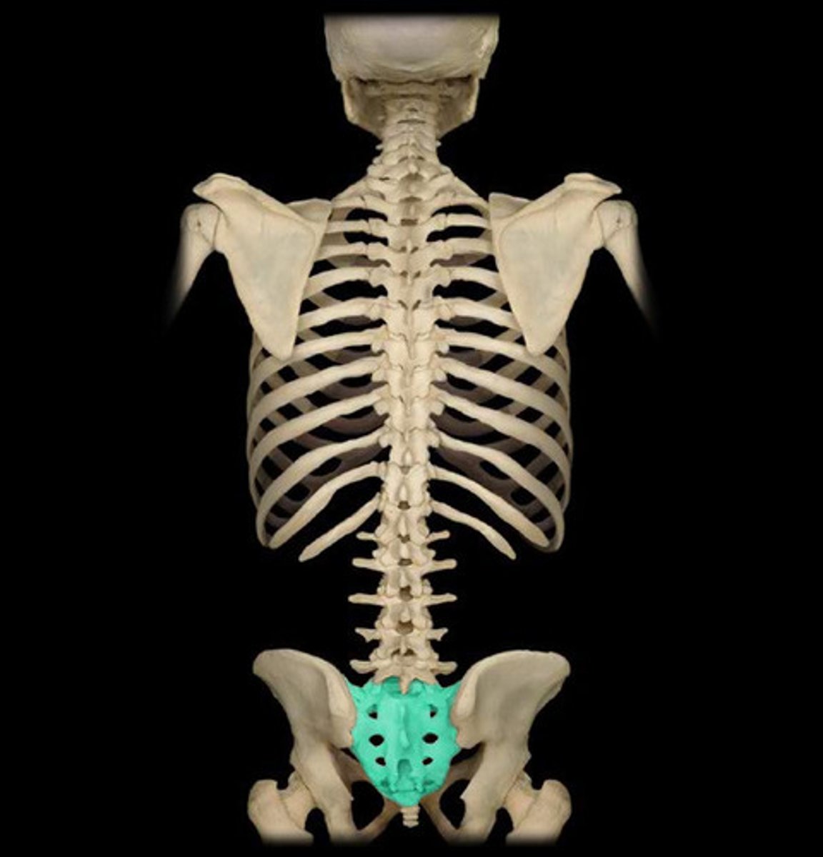
Sacrum
five fused vertebrae that form the posterior of the pelvic girdle
Coccyx
four vertebrae fused together to form the tailbone [THE TAILLLBONNNESS THE COCCYX]
![<p>four vertebrae fused together to form the tailbone [THE TAILLLBONNNESS THE COCCYX]</p>](https://assets.knowt.com/user-attachments/d54ccf52-5374-4be3-a774-7b0252018723.jpg)
True Ribs
first seven pairs of the rib cage
[FIRST IS ALWAYS TRUE]
![<p>first seven pairs of the rib cage</p><p>[FIRST IS ALWAYS TRUE]</p>](https://assets.knowt.com/user-attachments/04ed5af2-358f-43bb-80d5-ecceb89f472a.jpg)
False Ribs
last 5 pairs of ribs; attach indirectly to sternum [LAST IS ALWAYS FALSE]
![<p>last 5 pairs of ribs; attach indirectly to sternum [LAST IS ALWAYS FALSE]</p>](https://assets.knowt.com/user-attachments/f0bc1c7b-8899-4b48-8a26-13a331ca2d77.jpg)
Floating Ribs
last two pairs of ribs not joined to the sternum at all, attach to the thoracic vertebrae [ALWAYS FLOAT WHEN YOU'RE IN THE BACK]
![<p>last two pairs of ribs not joined to the sternum at all, attach to the thoracic vertebrae [ALWAYS FLOAT WHEN YOU'RE IN THE BACK]</p>](https://assets.knowt.com/user-attachments/5b18da9f-737c-4d97-a76e-048885ea800b.jpg)
Sternum
The breast bone [CHEST IS STERN]
![<p>The breast bone [CHEST IS STERN]</p>](https://assets.knowt.com/user-attachments/0c777c29-87e4-4a2b-9ab1-281801518ea6.jpg)
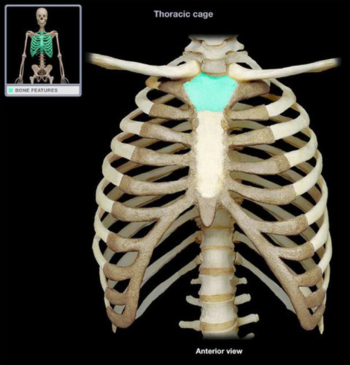
Manubrium
the bony structure that forms the upper portion of the sternum
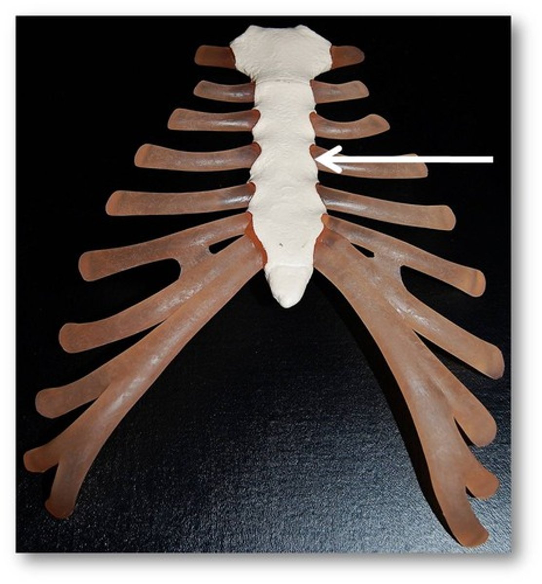
Sternum body
main long part of sternum
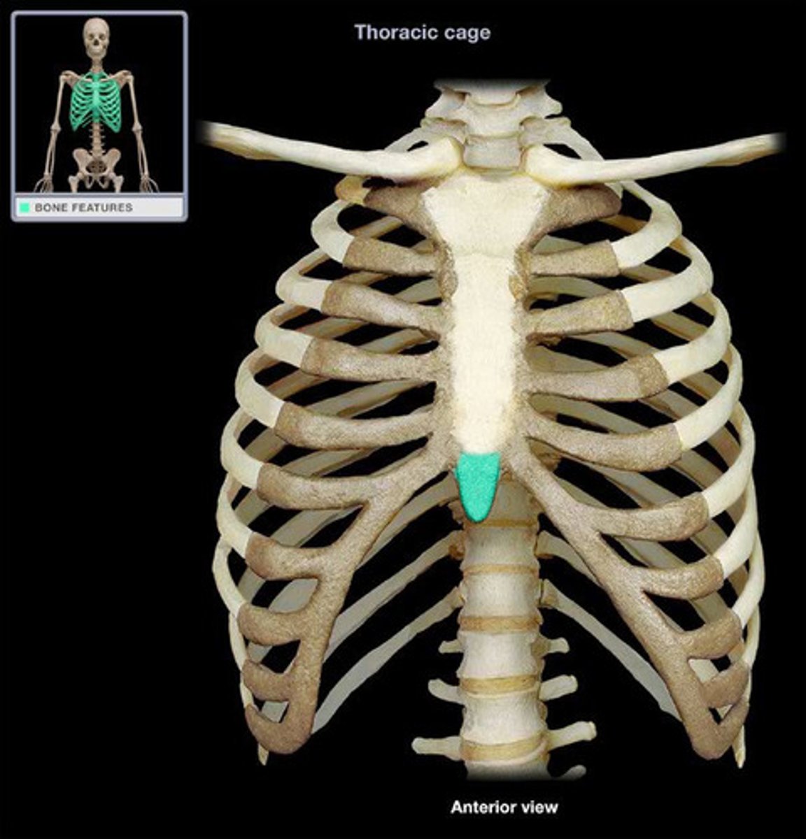
Xiphoid process
sword-shaped lower tip of the sternum
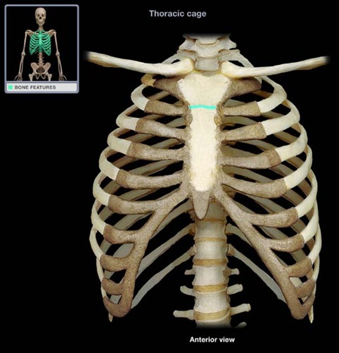
Sternal angle
synarthrotic joint formed by the articulation of the manubrium and the body of the sternum
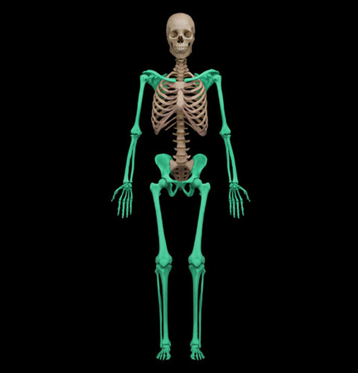
Appendicular Skeleton
80 bones, bones of the limbs and girdles that attach them to the axial skeleton
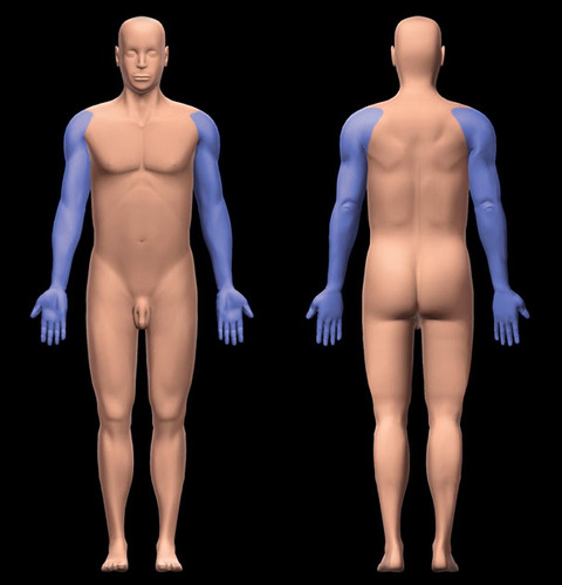
Upper Limb
arm, forearm, hand
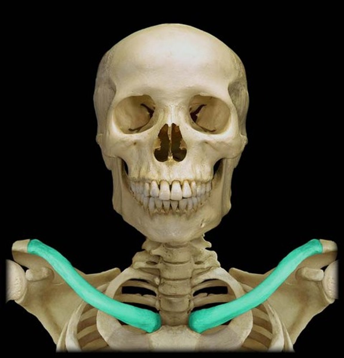
Clavicle
Collar bone
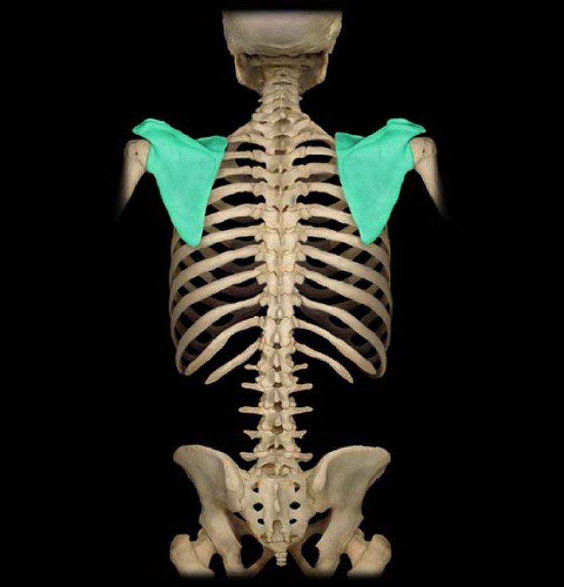
Scapula (right or left)
shoulder blade or wing bone, is the bone that connects the humerus (upper arm bone)
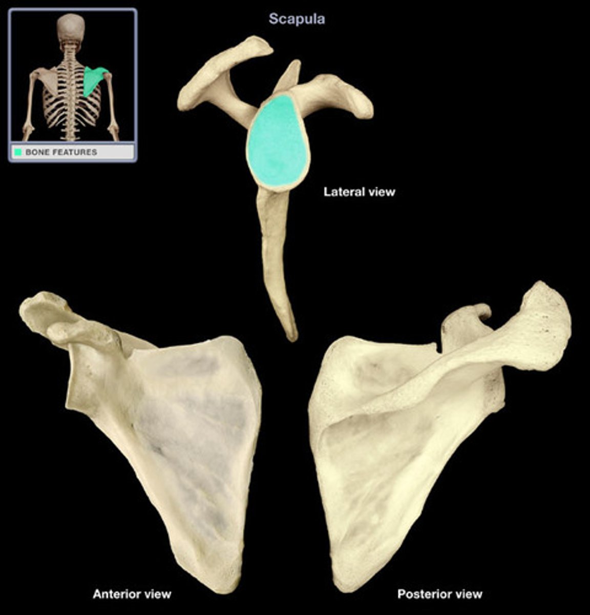
Glenoid cavity
Depression in the scapula that articulates with the humerus
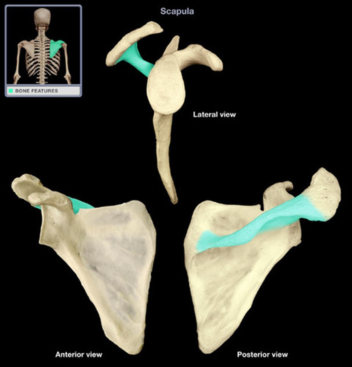
Scapular spine
long ridge running down the scapula
Supraspinous fossa
depression above spine
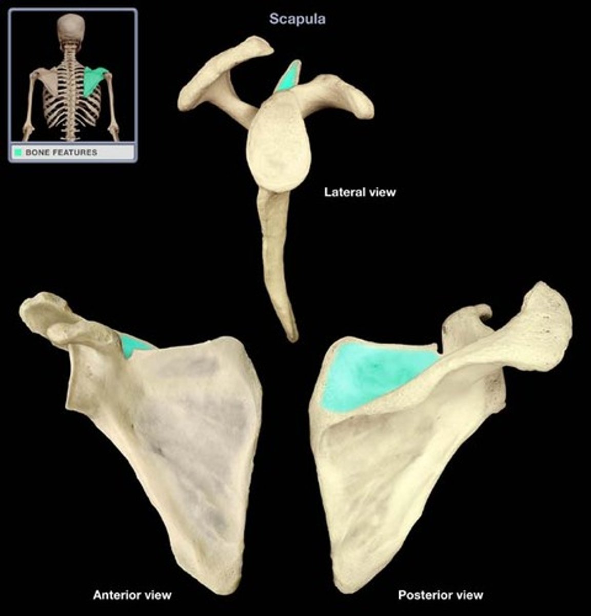
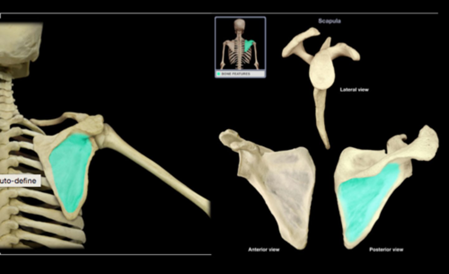
Infaspinous fossa
much larger than the supraspinatous fossa; toward its vertebral margin a shallow concavity is seen at its upper part
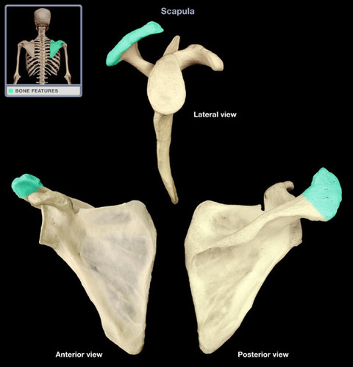
Acromion
lateral end of the scapula, extending over the shoulder joint
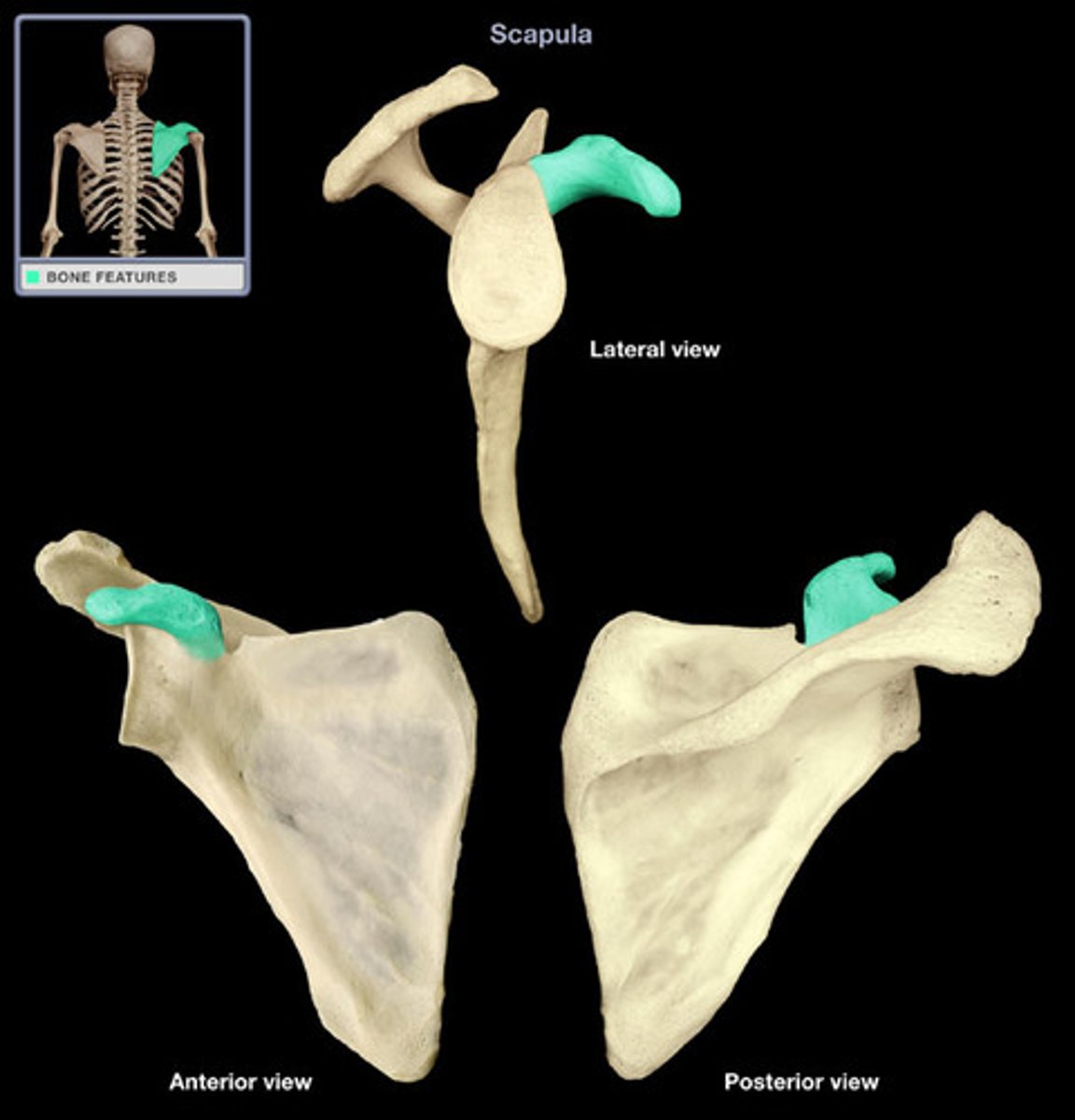
Coracoid process
Process above the glenoid cavity that permits muscle attachment
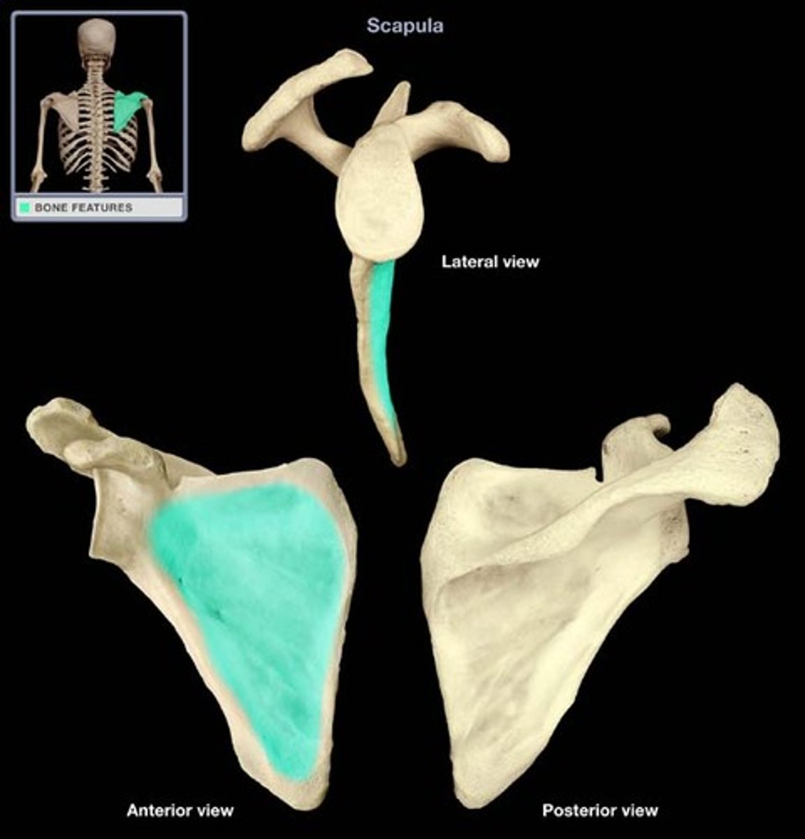
Subscapular fossa
The entire anterior surface of the scapula.
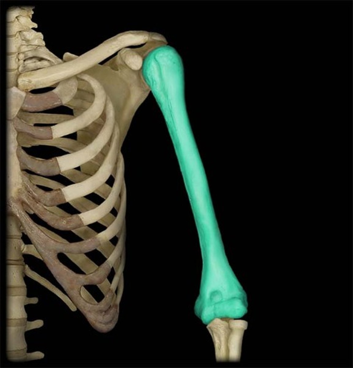
Humerus Bone (right or left)
long bone in the upper arm
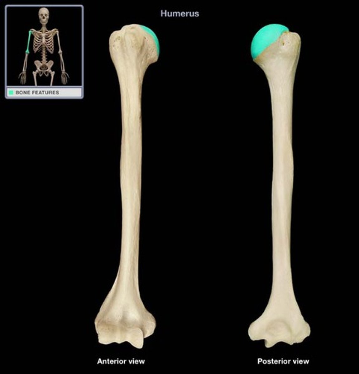
Humerus head
Semi round proximal end articulates with scapula
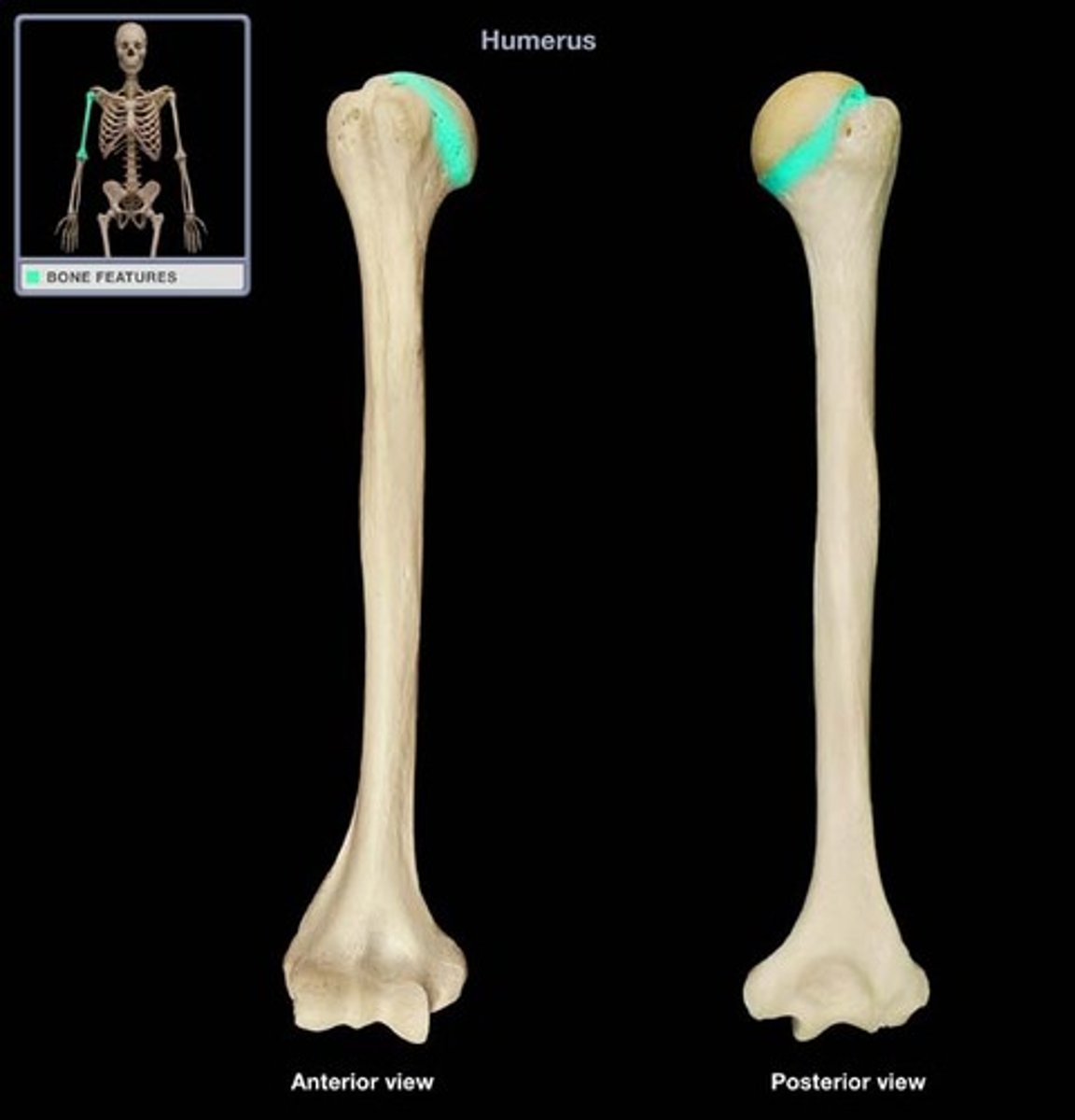
Anatomical neck
The neck of the humerus that narrows behind the head of the humerus.
Surgical neck
The neck of the humerus that is prone to fractures.
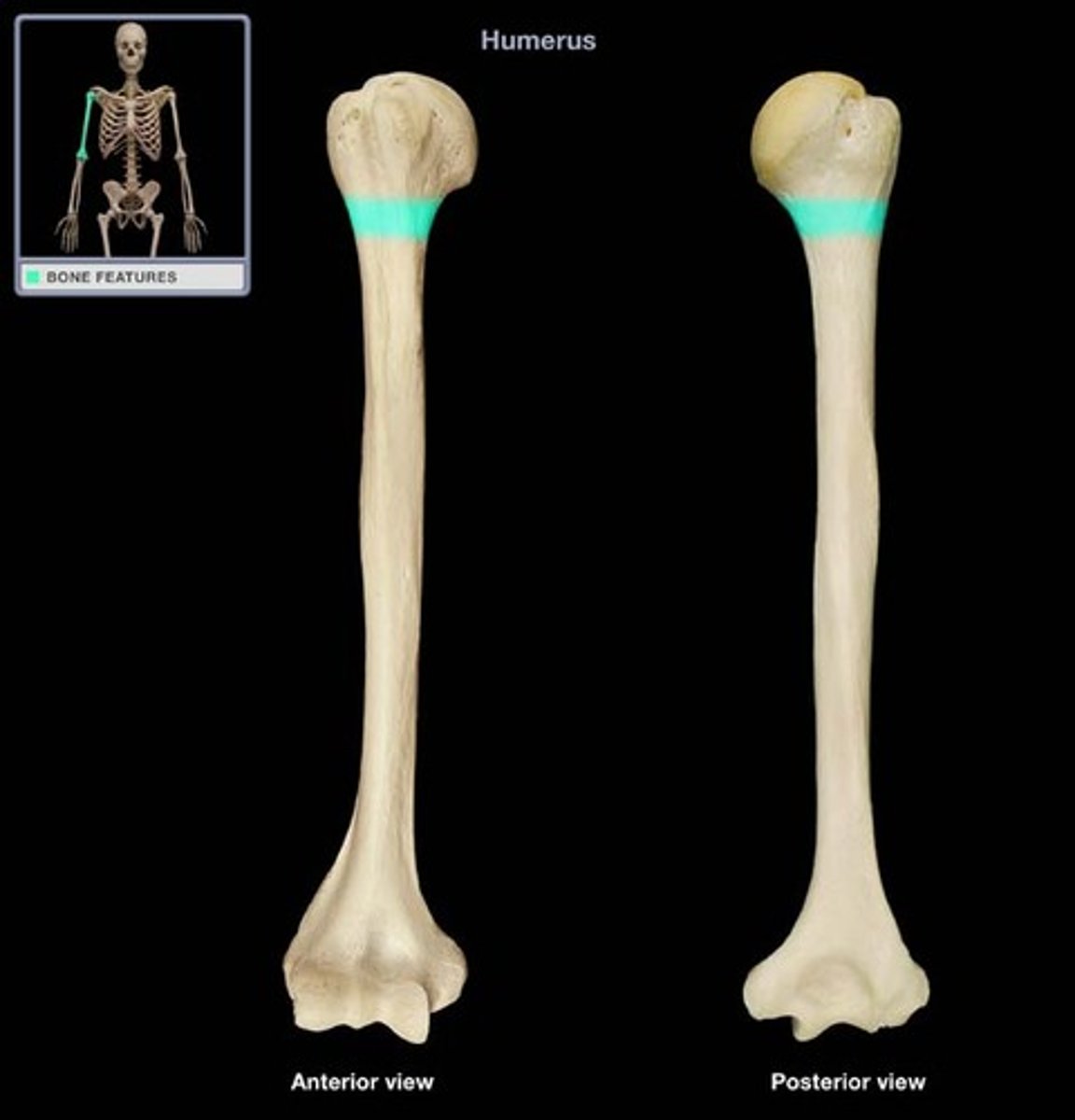
Greater tubercle
situated lateral to the head of the humerus and posterolateral to the lesser tubercle
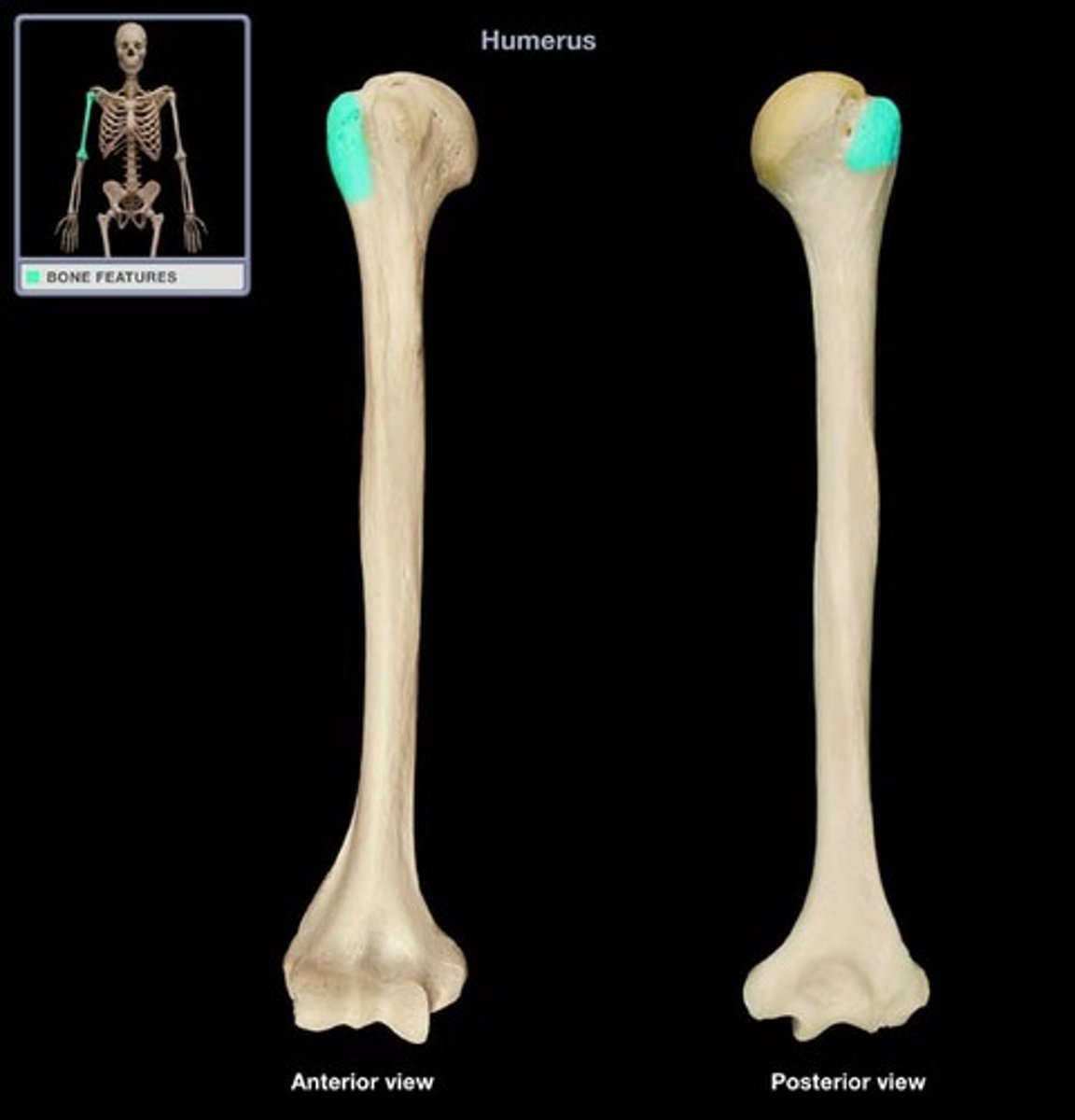
Lesser tubercle
more prominent than the greater tubercle: it is situated in front, and is directed medially and anteriorly.
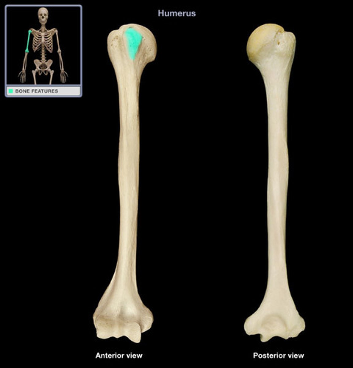
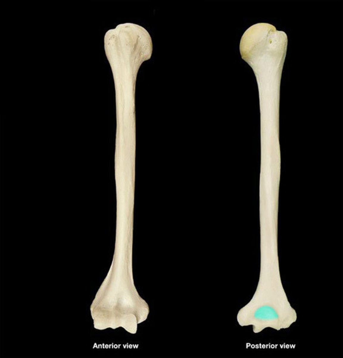
Olecranon fossa
posterior. hole where the lower limb bones articulates with humerus
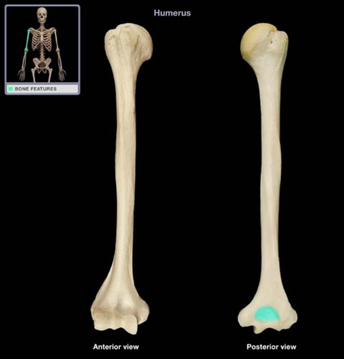
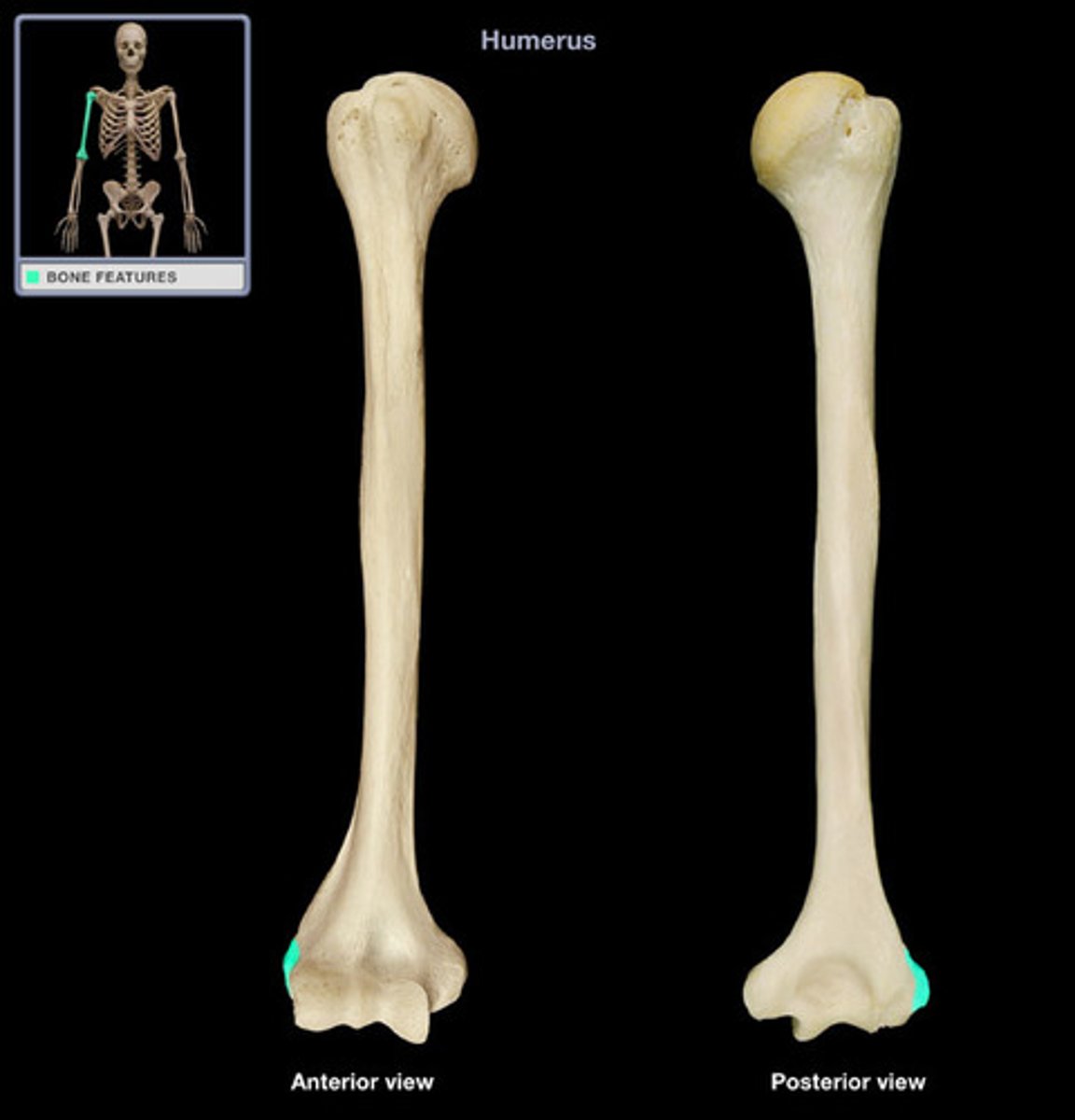
Medial epicondyle
larger and more prominent than the lateral epicondyle
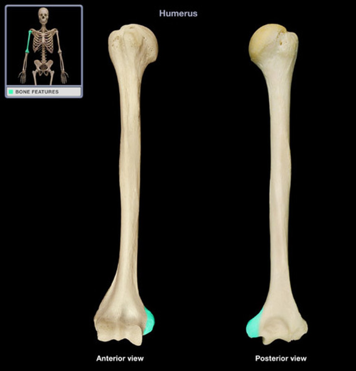
Lateral epicondyle
small, tuberculated eminence, curved a little forward, and giving attachment to the radial collateral ligament of the elbow joint
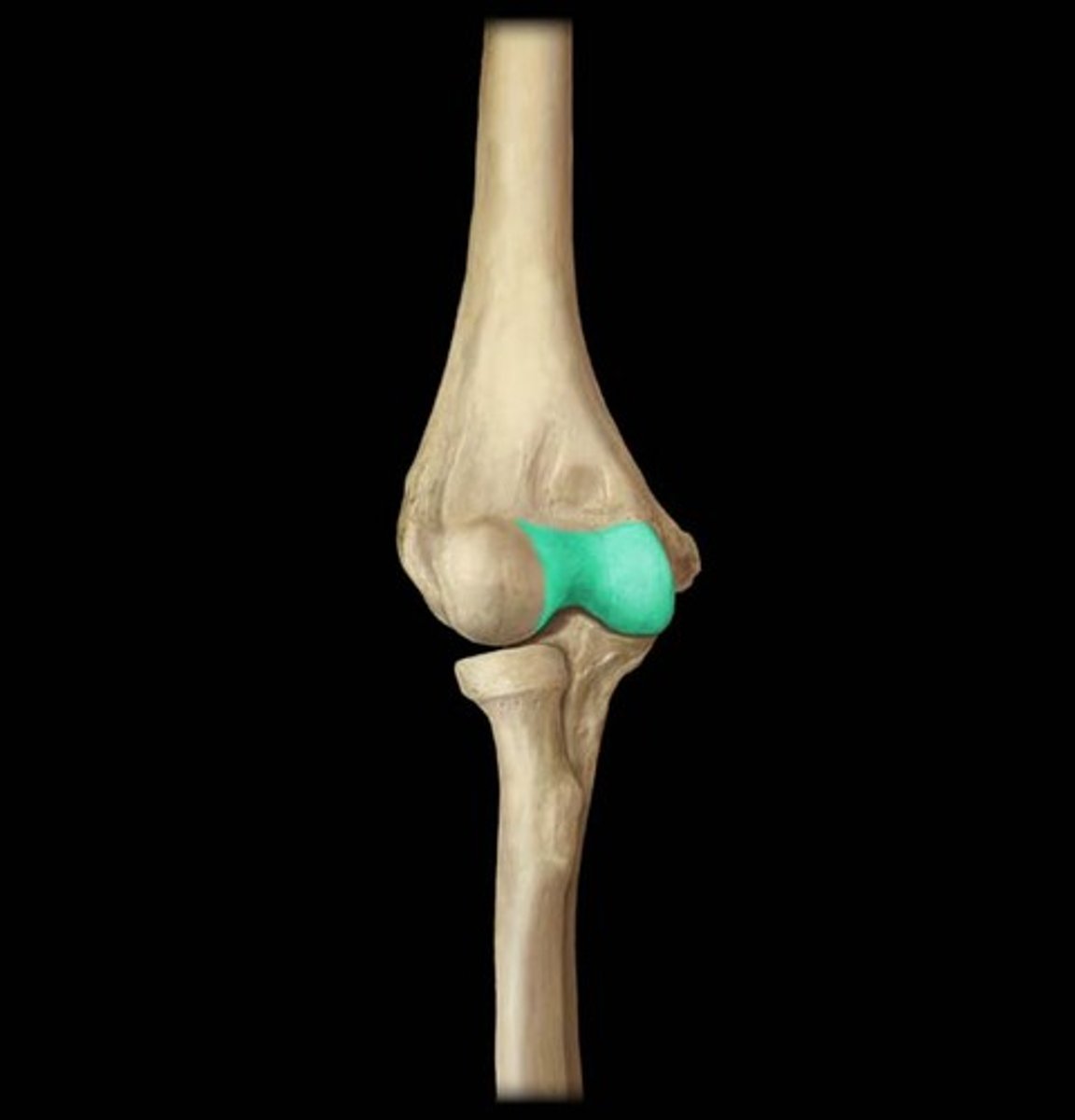
trochlea
a smooth, grooved articular process shaped like a pulley
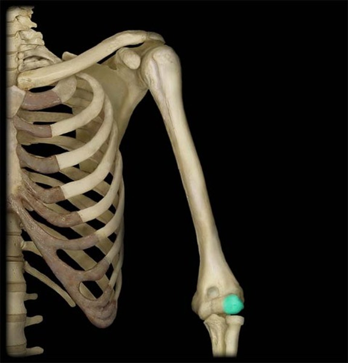
capitulum
rounded knob on the humerus; adjoins the radius
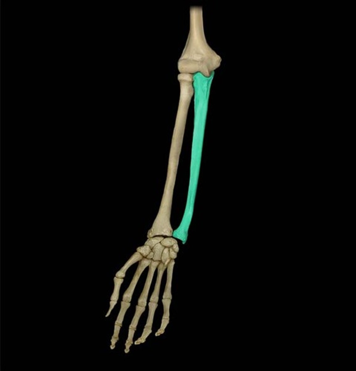
Ulna
medial bone of the forearm
olecranon (process)
forms the pointed portion of the elbow.
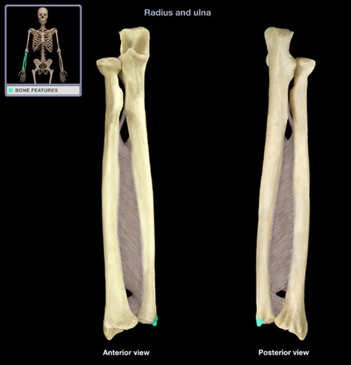
Ulna styloid process
the pointer at the tip of ulna
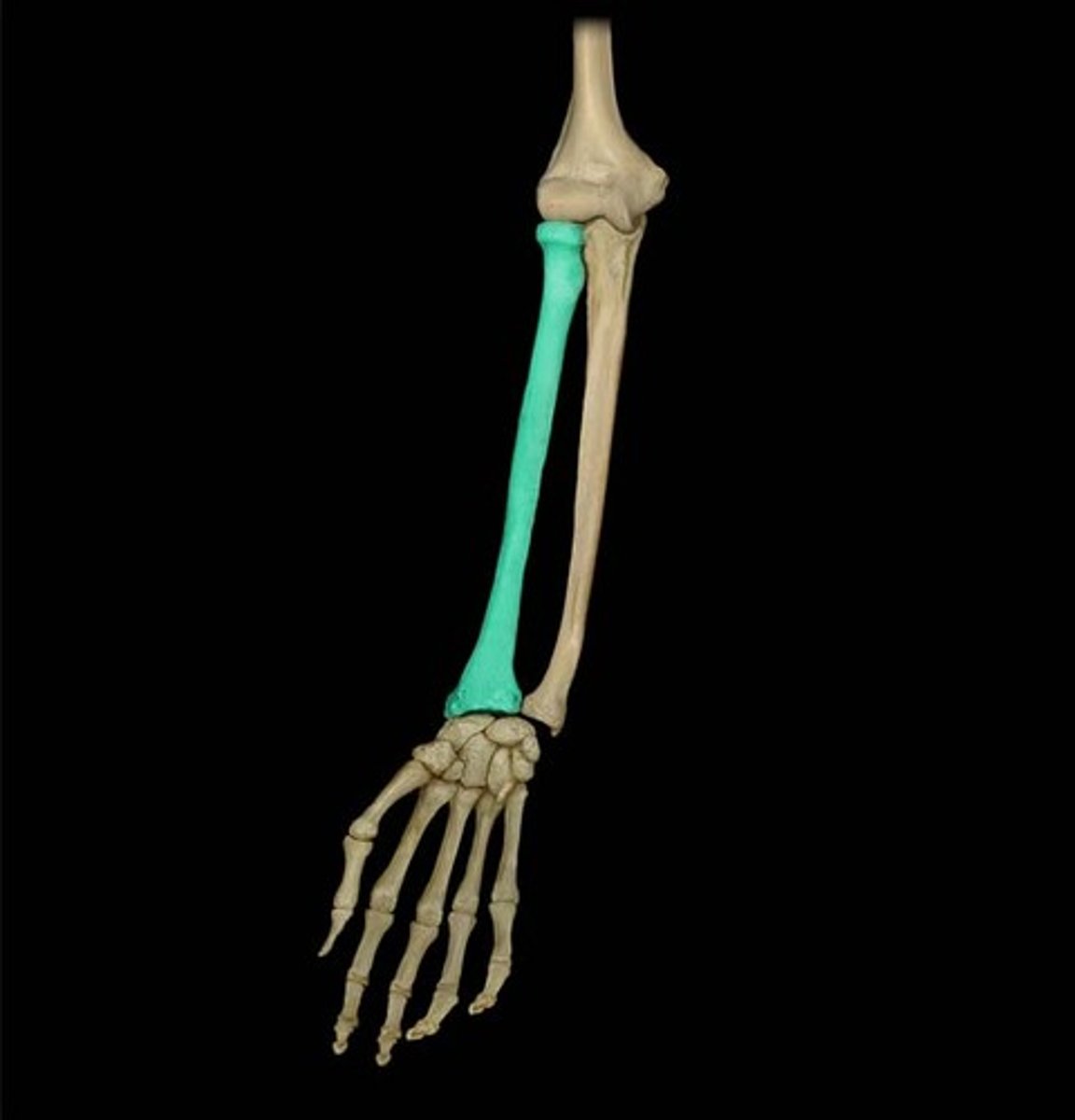
Radius bone
smaller bone in the forearm (lower arm) on the same side as the thumb
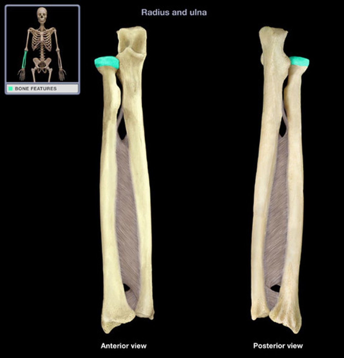
Radius head
cylindrical form, and on its upper surface is a shallow cup or fovea for articulation with the capitulum of the humerus.