biopsychology
1/65
There's no tags or description
Looks like no tags are added yet.
Name | Mastery | Learn | Test | Matching | Spaced |
|---|
No study sessions yet.
66 Terms
what is the nervous system?
specialised network of cells, neutrons, makes use of electrical and chemical signals to relay information from one place to another in the body
consists of the PNS and CNS
structure and functions of the nervous system
the 2 main functions are:
collect, process and respond to environmental stimuli
coordinate the working of different organs and cells in the body
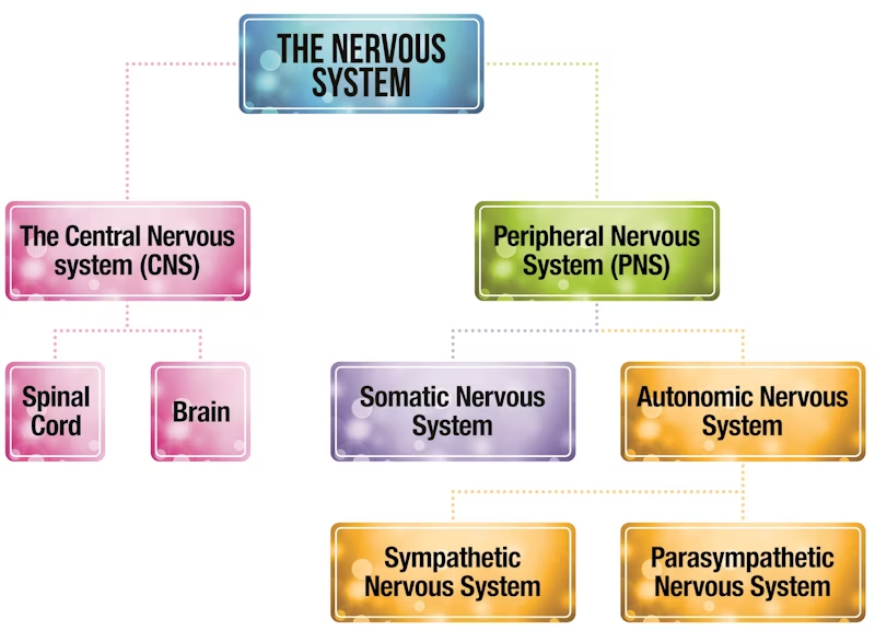
central nervous system (CNS)
consists of the brain and spinal chord
origin of complex commands and decisions
peripheral nervous system (PNS)
relays information to CNS via neurones from outside world and back to muscles and glands
divided into somatic and autonomic systems
somatic: controls vital functions eg breathing, heart rate, stress and sexual arousal, controls voluntary muscles and transmits sensory information to CNS
autonomic: controls involuntary body functions, muscle movement and receives information from sensory receptors
somatic nervous system (SNS)
transmits information from receptor cells in sense organs to CNS, and from CNS to muscles
autonomic nervous system (ANS)
transmits information to and from internal bodily organs, with 2 divisions, sympathetic and parasympathetic
structure of the brain
divided into left and right hemisphere
outer layer, cerebral cortex, only 3mm thick, covers mammals brains
the ridges and valleys of the cerebral cortex increase the brains SA
the human brain is very developed, distinguishing us from animals as we have superior mental functions
the brain developed in 3 stages: reptilian brain is least developed (insinct, survival), then the limbic system (emotions) followed by the neocortex (speech, logic, higher thinking skills)
structure of the spinal chord
extension of the brain, passes messages to and from the brain
connects nerves to the peripheral nervous system and responsible for reflex actions
protected by the spine (vertebrae)
endocrine system
major information system instructing gland to release hormone into the bloodstream to carry to target organs, communicates via chemicals
works alongside the nervous system to control vital functions
acts more slowly than the nervous system but has more widespread effects
glands
an organ that synthesises substances eg hormones, glands in the body produce hormones
thyroid gland: produces thyroxine
pituitary gland: 'master gland' located in the brain, controls release of hormones from all other endocrine glands
hormones
a biochemical substance that circulates in the blood affecting target organs, produced in large quantities but disappear quickly
function of thyroxine (hormone)
affects cells in the heart, increasing heart rate
increases: metabolic rates and affects growth rate, protein synthesis, glucose metabolism, oxygen consumption
regulates: digestion, reproduction, bone growth, muscle tone, development of nerve cells
fight or flight response
how animals respond when stressed, the endocrine system and autonomic nervous system work together during a stressful event
a stressor is perceived eg an approaching car
a part of the brain called the hypothalamus activates the pituitary gland
triggers activity in the sympathetic branch of the ANS
ANS switches from parasympathetic state (resting) to sympathetic (aroused) state eg dilated pupils, rapid heart rate and breathing, trembling etc
arenaline
hormone produced by the adrenal glands, affecting the cardiovascular system
released from the adrenal medulla, part of the adrenal gland near the kidneys, and released into the bloodstream
triggers physiological changes in the body, increases heart rate and deeper breathing
creates the physiological arousal necessary for fight or flight responses
immediate sympathetic responses
as soon as a threat is detected, there is an acute and automatic sympathetic response
this includes: increased breathing and heart rate, constriction of blood vessel, high blood sugar, inhibition of stomach acid/ digestion/ saliva production, converts glycogen to glucose, contracts rectum
parasympathetic action
once the threat is gone, the parasympathetic nervous system kicks back in, returning the body to its resting state
the PNS works in opposition to the ANS (antagonistic) acting like a break to reduce the activities, the reset and digest phase
includes: decreasing heart and breathing rates, constricts pupils, lungs reduce oxygen intake, stomach contracts, stimulates digestion and saliva production, relaxes rectum
structure of a neuron
the basic building blocks of the nervous system, nerve cells that process and transmit messages through electrical and chemical systems, provide the nervous system with its primary means of communication
can be less than 1mm, shortest is trochlear nerve (moves eyes towards nose), longest is sciatic nerve (from lower back down each leg)
there are 100bn nerve cells in the human nervous system, 80% in brain
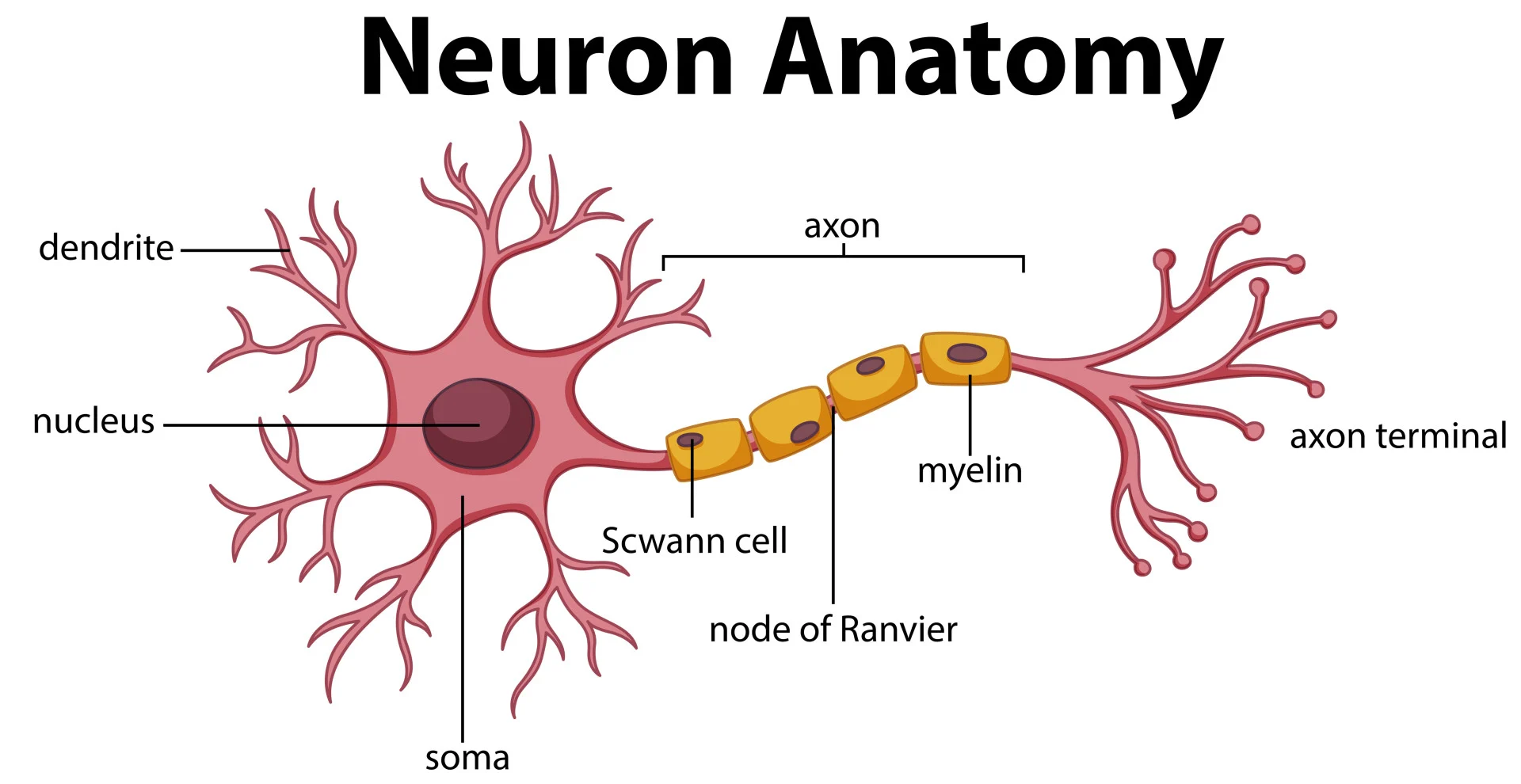
roles of neuron structures
cell body (soma): includes a nucleus, contains genetic material
dendrites: branch like structures off cell body, carry impulses from nearby neurons to cell body
axon: carry impulses away from cell body
myelin sheath: fatty layer covering axon, protects it and speeds up electrical transmission
nodes of ranvier: gaps in myelin sheath that speeds up transmission, impulses can jump
terminal buttons: communicate with next neuron across synapse
sensory neuron
takes information from the environment (senses) towards the CNS, carry messages from PNS to CNS
when you touch something hot, this neuron will be activated
long dendrites and short axons, in PNS in clusters called ganglias
relay neuron
found in the CNS between sensory and motor neurons
the electrical impulse from the sensory neuron detailing the hot surface is passed to a relay neuron which passes it to a motor neuron
found in CNS, short dendrites and axons, 97% of all neurons, mostly found in the brain and visual system
motor neuron
start in CNS, receive electrical impulse from relay neurons and take it to effectors eg muscles and glands
short dendrites and long axons (long axons from part of PNS)
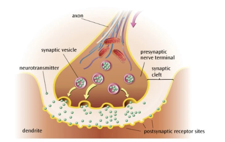
synaptic transmission
the process by which neighbouring neurons communicate with each other by sending chemical messages across the gap that separates them
neurotransmitters
brain chemicals released from synaptic vesicles that relay signals across the synapse from one neuron to another, can be excitatory or inhibitory, diffuse across the synapse to the next neuron
can only travel in one direction as receptors are only on the postsynaptic neuron, each neurotransmitter has a unique molecular structure and fits perfectly into the receptor site (induced fit model)
each neurotransmitter has a specialist function eg Acetylcholine (Act) found where motor neuron meets a muscle, causes muscle to contract
excitation
excitatory neurotransmitters eg glutamate, noradrenaline adrenaline (both a hormone and a neurotransmitter)
increases the positive charge of the postsynaptic neuron, increases the likelihood that the postsynaptic neuron will pass on the electrical impulse
inhibition
inhibitory neurotransmitters eg GABA, Glycine, serotonin (affects mood and social behaviour, cause of depression)
increases the negative charge of the postsynaptic neuron, decreases the likelihood that the postsynaptic neuron will pass on the electrical impulse
chemical transmission
neurons talk to other neurons in groups called neural networks
neurons are separated by a small gap called the synapse
inside the neuron, signals are transmitted electrically, between neurons, signals are transmitted chemically
stages of synaptic transmission
action potential (electrical impulse) travels down the axon
action potential reaches the presynaptic terminal
triggers neurotransmitters to be released from synaptic vesicles into synaptic cleft
neurotransmitter diffuses across the synaptic cleft
neurotransmitters are taken up by the postsynaptic receptors
neurotransmitter is converted back to an action potential
electrical transmission
resting potential: the inside of a neuron is negatively charged compared to the outside of it
when activated by a stimulus, the inside becomes positively charged for a short period, triggers an action potential which travels down the axon towards the neuron
why is dopamine an unusual neurotransmitter?
it is equally likely to have excitatory or inhibitory effects on the postsynaptic neuron
summation
excitatory and inhibitory influences are added up (summed)
if the overall effect on the postsynaptic neuron is inhibitory, its less likely to fire, if its excitatory, its more likely to fire
an action potential in the postsynaptic neuron will only be triggered if the sum of signals reaches the threshold
localisation of function
the theory that different areas of the brain are responsible for specific behaviours, processes or activities
localisation vs holistic theory
scientists historically supported the holistic theory of the brain, all parts of the brain involved in the processing of thought and action
when Broca and Wernicke discovered specific areas of the brain are associated with particular functions, it supported localisation of function, different parts of the brain perform different tasks and control different body parts
if a certain area of the brain gets damaged, the theory suggests the specific function is also affected
hemispheres of the brain
cerebrum is divided into 2 symmetrical halves, left and right
some functions are controlled by a particular hemisphere (lateralisation), activity on the left side of the body is controlled by the right of the body and vice versa
functions of the left hemisphere
sensory stimulus from right side
motor control of right side
speech, language, comprehension
analysis and calculations
time and sequencing
recognition of words, letters, numbers
functions of the right hemisphere
sensory stimulus from left side
motor control of left side
creativity
spatial ability, context, perception
recognition of faces, places, objects
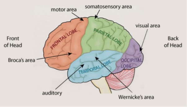
centres of the cortex
cerebral cortex: outer layer of both hemispheres-cerebral cortex: outer layer of both hemispheres
divided into 4 centres, the lobes of the brain: frontal, parietal, occipital, temporal
each lobe has a different function
motor cortex
at the back of the frontal lobe in both hemispheres
controls voluntary movement on the opposite side of the body
damage here could cause a loss of control over fine movements
somatosensory cortex
at the front of both parietal lobes
separated from the motor cortex by a valley, the central sulcus
represents sensory information from the skin, the amount of area devoted to a specific body part denotes its sensitivity
eg receptors for face and hands occupies over half the area
visual cortex
in the occipital lobe
each eye sends information from the RVF to the left visual cortex and from the LVF to the right visual cortex
damage to the left hemisphere could cause blindness in the RVF of both eyes
auditory cortex
in the temporal lobe
analyses speech based information , damage could produce partial or more extensive hearing loss
damage to Wernicke's area could affect language comprehension
language centres- Broca's area
language is usually restricted to the left hemisphere
1880s Broca identified a small area in the left frontal lobe responsible for speech production
damage here causes Broca's aphasia, slow speech, lacking in fluency, patient 'tan' could only say the word 'tan'
struggle with prepositions and conjunctions (a, the, and)
language centres- Wernicke's area
Wernicke's area in the left temporal lobe, results in Wernicke's aphasia when damaged, often produce nonsense words (neologisms)
1880s Wernicke discovered patients with no problem producing language but difficulties understanding it, speech produced is fluent but meaningless
case study evidence of localisation of function
Phineas Gage had damage to his left frontal lobe, it changed him from a calm person to someone rude, supports localisation theory as personality is localised in the frontal lobe
however, case studies are difficult to generalise and researcher may interpret them subjectively
strength of localisation of function- support from neurosurgery
neurosurgery is used to treat mental disorders eg a cingulotomy involves isolating the cingulate gyrus, disfunction of this area may cause OCD
Dougherty et al. (2002) studied 44 people with OCD who had a cingulotomy, at follow up, 30% met the criteria for successful response and 14% for partial
the success of such procedures strongly suggests behaviours associated with serious mental disorders may be localised
strength of localisation of function- brain scan evidence to support
Petersen et al. (1988) used brain scans to show activity in Wernicke's area during a listening task and in Broca's during a reading task
also, a study of LTM by Tulving et al. (1994) revealed semantic and episodic memories are located in different parts of the prefrontal cortex
theres now a number of sophisticated and objective methods for measuring activity in the brain, providing scientific evidence of localisation of function
counterpoint of brain scan evidence to support localisation of function
Lashley removed areas of the cortex, up to 50%, in rats learning the route through a maze, learning required all the cortex rather than being confined to a specific area
suggests higher cognitive processes eg learning aren’t localised but distributed in a more holistic way in the brain
limitation of localisation of function- the language localisation model has been questioned
Dick + Tremblay (2016) found very few researchers still believe language is still only in Broca's and Wernicke's area
advanced techniques eg fMRI have identified regions in the RH and the thalamus
suggests rather than being confined to a few key areas, language may be organised more holistically in the brain, contradicts localisation theory
evaluation of localisation of function- case study evidence
unique cases of neurological damage support localisation theory eg Phineas Gage
however, its difficult to make meaningful generalisations from a single individual and conclusions may depends on the researchers subjective interpretation
suggests some evidence supporting localisation may lack validity, oversimplifying brain processes and undermining the theory
hemispheric lateralisation
the idea that the 2 brain hemispheres are functionally different and certain mental processes and behaviours are mainly controlled by one hemisphere
what's the difference between localisation and lateralisation?
localisation: some functions being controlled by different areas in the brain
lateralisation: the 2 hemispheres are functionally different
in some functions, the localised areas are in both hemispheres eg vision, visual cortex in both left and right occipital lobe
how is language lateralised?
the 2 main language areas, Broca's and Wernicke's, are only in the left hemisphere, language is lateralised as its performed by only one hemisphere
right hemisphere can only produce basic words and phrases but gives emotional context to what it said
left hemisphere is the analyser, right hemisphere is the synthesiser
how are the cortex's lateralised?
vision, motor and somatosensory areas are in both hemispheres
motor cortex has contralateral wiring, the RH controls movement on the left side and vice versa
vision is contralateral and ipsilateral, each eye gets light from the LVF and the RVF, LVF of each eye is connected to the RH, RVF of each eye is connected to the LH
this aids depth perception and provides different perspectives
auditory cortex has a similar cross over, allows us to locate sounds
what is split brain research?
a series of studies beginning in the 1960s (ongoing) involving people with epilepsy who had experienced surgical separation of the brain hemispheres by serving the corpus callosum to reduce the severity of their epilepsy as during a seizure excessive electrical activity travels from one hemisphere to another
allows researchers to test lateral functions of the brain in isolation
Roger Sperry (1968) split brain research- procedure
used 11 people who had a split brain operation, used the set up to project images to the LVF and RVF, either the same or different
normal brain: corpus callosum would share the information between both hemispheres, provides a complete picture
split brain: information can't be conveyed from one hemisphere to another
Roger Sperry (1968) split brain research- findings
image shown to RVF, participant can describe what they see, image shown to LVF, can't describe it + say there's nothing there but could select a matching object with left hand/ most closely related
in normal brain messages from RH would be relayed to language centres in LH, not possible in split brain patients, because the LH is dominant for verbal processing the patients answer matches the word, the RH can't share information with the left so can't say what he saw but can draw it
emotional reaction to images presented to LVF but couldn't report what they'd seen
Roger Sperry (1968) split brain research- conclusions
certain functions are lateralised in the brain
LH is verbal, RH is silent but emotional
strength of hemispheric lateralisation- evidence of lateralised brain functions in 'normal' brains
Fink et al. (1996) used PET scans to see which areas were active in a visual processing task
show when 'normal' participants attend to global elements of an image, the RH is more active, when required to focus on more detail the specific areas of the LH tend to dominate
suggests hemispheric lateralisation is a feature of connected brains and split brains as even in connected brains the 2 hemispheres process information differently
limitation of hemispheric lateralisation- idea of analyser vs synthesiser may be wrong
may be different processes in the RH and LH but research suggests people don't have a dominant side, creating a different personality
Neilson et al. (2013) analysed over 1000 brain scans, finding people did use certain hemispheres for certain tasks but no dominance
suggests the notion of right or left brained people is wrong eg artist brain
limitation of hemispheric lateralisation- lateralisation vs plasticity
lateralisation is adaptive, enabling 2 simultaneous tasks more efficiently eg only lateralised chickens better at finding food while watching for predators (Rogers et al. 2004)
on the other hand, neural plasticity is also adaptive, after damage to brain, language function can 'switch sides' (Holland et al. 1996), functions are taken over by non specialised areas in the opposite hemisphere
suggests lateralisation is first preference but ultimately plasticity is more important
strength of hemispheric lateralisation- support from more recent brain studies
Luck et al. (1989) showed split brain participants are better than normal controls on certain tasks eg 2x as fast at identifying the odd one out in an array of similar objects
in the normal brain, the LH's superior processing abilities are 'watered down' by the inferior RH (Kingstone et al. 1995)
supports Sperry's findings that the left and right brains are distinct in functions and abilities
limitation of hemispheric lateralisation- causal relationships are hard to establish
in Sperry's research, the behaviour of split brain participants was compared to a neurotypical control group
however, none of the control group had epilepsy, any differences between the groups may be due to epilepsy not the split brain (confounding variable)
means that some of the unique features of the split brain participants cognitive abilities may be due to their epilepsy
limitation of hemispheric lateralisation- ethics
Sperry's participants weren't deliberately harmed and procedures were explained in advance to gain informed consent
however, participants may not have understood they would be tested for many years and participation was stressful
suggests there was no deliberate harm but the negative consequences make the study unethical
plasticity
the brains tendency to change and adapt as a result of experience and new learning, generally involves the growth of new connections
functional recovery
a form of plasticity, following damage through trauma, the brains ability to redistribute or transfer functions usually performed by a damaged area to other undamaged areas
how does brain plasticity change the brain?
the brain can change throughout your life
infancy: rapid growth, increasing number of synaptic connections
2/3 years: peaks at 15000 synaptic connections per neurone, adult brain has about half this number
synaptic pruning
as we age we lose synaptic connections, those that are rarely used are removed, those that are frequently used are strengthened
allows lifelong plasticity, new neural connections are formed as demands on the brain change