2: Embryological development
 Call Kai
 Learn
Learn Practice Test
 Spaced Repetition
 Match
Match Flashcards
 Knowt Play
1/79

There's no tags or description
Looks like no tags are added yet.
Name | Mastery | Learn | Test | Matching | Spaced | Call with Kai | Chat |
|---|
No analytics yet
Send a link to your students to track their progress
80 Terms
when is left-right patterning established of the heart
gastrulation
including remodelling and septation, how does the majority off cardiac development occur
while heart is pumping blood
when does the heart start beating
21st day of development
when does the heart start pumping blood
day 24-25
lateral mesoderm derivatives
myocardium, epicardium, blood cells, blood vessles, endothelium, endocardium.
primordial heart, blood and lymphatic cells, spleen, connective tissue and muscle of viscera. serous membrane of pleura, pericardium and peritoneum
morphogenesis
gastrulation. the development of the form and structure of various organs
overview of heart development in (5 stages)
1. specification of cardiac progenitor cells which then migrate towards midline to form cardiac crescent.
2. developing heart forms a linear tube
3. tube undergoes dextral looping to acquire left-right symmetry
4. heart tube is subdivided into four chambers
5. maturation of the endocardial cushions into valves and development of the great vessles to provide for unidirectional blood flow through the chambers
formation of the first heart field
1. cardiac progenitor cells develop into cardiac myoblasts while blood islands undergo vasculogenesis.
2. endoderm, ectoderm and midline mesoderm produce signals to cause cardiogenic precursors to form a cardiac primordium within lateral mesoderm at the cranial end of embryo forming the first heart field
describe the initial structure of cardiogenic field and then its fate
initially horseshoe shaped and surrounded by cardiac myobasts within the cardiogenic field's apex . eventually develops into primitive ventricles along with their respective outflow tracts
formation of endocardial tubes
signals from underlying endoderm cause a subpopulation of cells within first heart field form a pair of lateral endocardial tubes
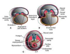
fusion of lateral endocardial tubes
folding of embryo (during week 4) causes fusion of the lateral endocardial tubes into a single primary heart tube
describe the primary heart tube formation
formation of two lateral endocardial tubes as a result of signals induced by endoderm onto the first heart field. these tubes then fuse during folding of the embryo
describe the primary heart tube
single endocardial tube with adjacent mesoderm differentiating into cardiomyocytes (which then form the myocardium which deposits cardiac jelly separating the myocardium from endocardial tube)
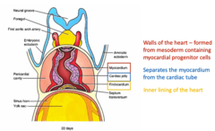
cardiac jelly
deposited by myocardium. acellular extracellular matrix separates the myocardium from the endocardial tube.
dorsal mesocardium:
formation, location, purpose, fate
formed by splanchic lateral mesoderm (located beneath the foregut). suspends the primary heart tube in the pericardial cavity. eventually ruptures over nearly entire length of the heart tube (causing the heart to be suspended in pericardial cavity by developing atrial and venous poles) and the ruptured dorsal mesocardium becomes the transverse pericardial sinus within the pericardial sac of the definitive heart
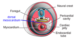
transverse pericardial sinus
ruptured dorsal mesocardium along the length of the heart tube. is within the pericardial sac of the definitive heart
differential growth of the embryo and its effect on the primary heart tube
causes heart to be displaced towards tail of embryo so heart is within the thorax
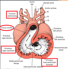
list regions of the primary heart tube
1. aortic roots (arterial poles)
2. truncus arteriosus
3. bulbus cordis
4. primitive ventricle
5. primitive atrium
6. sinus venosus (venous poles)
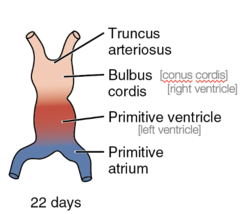
blood flow of primary heart tube
all venous blood flows into the sinus venosus (tail end) and contractions propel blood to truncus arteriosus (head end)
truncus arteriosus
divides and gives rise to ascending aorta and pulmonary trunk
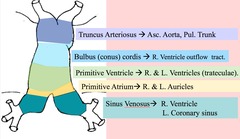
bulbus cordis
develops into right ventricle
primitive ventricle
develops into the left ventricle
primitive atrium
eventually develops into right and left atria
sinus venosus
develops into posterior portion of right atrium, sinoatrial node and coronary sinus
pneumonic for regions of the heart
All (aortic roots)
People (primative ventricle)
Buy (bulbus cordis)
Telephones (truncus arteriosus) and
Sell them (sinus venosus)
Pricy (primative atrium)
All People Buy Telephones and Sell them Pricy
Aortic roots, Primitive ventricles, Bulbus cordis, Truncus arteriosus, Sinus Venosus, Primitive atrium
when does cardial looping happen
day 23
describe cardiac looping to form c-shaped curvature
primary heart tube elongates and simultaneously bends. the ventral surface is displaced towards the right by torsion forces working along craniocaudal axis (this ventral surface forms the right outer curvature of the c-shaped heart)
describe the s-shaped heart formation by looping
c-shaped heart continues to elongate at both arterial end venous poles. primitive right ventricle displaced caudally, ventrally and to the right. primitive left ventricle displaced to the left. primitive atrium acquires a dorsal and cranial position.
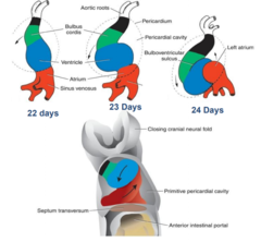
when does elongation of the heart tube complete
day 28
result of looping of heart
-atrioventricular canal aligns with both ventricles.
after looping. atrium communicates with ventrical by atrioventricular canal
-primadorium of right ventricle is positioned closest to outflow tract
-outflow tract lies between presumptive future atria.
-primadorium of left ventricle positioned closest to inflow tract
-atrium dorsal to bulbus cordis so that inflow is dorsal to outflow
ventricular inversion
condition where heart that has an apex inferior to the right (instead of left) side. the
looping of the heart tube is reversed from its normal sinistral pattern.
heterotaxy
any defect ascribing to abnormal left-right formation. partial situs ambiguous (reversal of some organs) or situs inversus totalis (reversal of all viscera). normally few, if any symptoms
visceroatrial heterotaxy syndroms
abdominal viscera and atrial pole are orientated on opposing sides. associated with structural defects. common atrium, malalignment of atrioventricular canal, outflow tract, abnormal venous and arterial vascular connections. symptoms
formation primitive blood vessles associated with the endocardial tube
paired dorsal aortae develops at the same time as the endocardial tube.
paired dorsal aortae
form the primary outflow vessles of the heart, develop at the same time as the endocardial tube
primitive heart tube translocation and the development of blood vessles
translocation from cranial into the thorax area of embryo, the cranial ends of the dorsal aortae are pulled ventrally until they form a dorsal ventral loop. these are the first pair of aortic arch arteries. pharyngeal arches give rise to four aortic arch arteries.
describe venous blood and embryo
enters the heart through a pair
common cardinal veins
formed by the continuance of paired posterior cardinal veins drains the trunk and the paired anterior cardinal veins draining the head region
6 veins inflow to heart (endocardial tube)
enters through pair of common cardinal veins, pair of vitelline veins that drain the yolk sac, pair of umbillical veins that deliver oxygenated blood to heart from placenta.
vitelline veins
drains the yolk sac
umbilical veins
carry oxygenated blood from placenta to heart
blood flow through the heart day 24-25
blood circulates around the embryo. initially, venous return enters the left and right sinus horns via common cardinal, umbilical and vitelline veins. eventually remodelled so that it enters the right sinus horn via superior and inferior vena cava
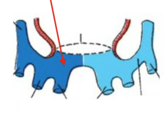
during weeks 4-8 remodelling of the inflow end of the heart occurs so that all systemic blood flows into the future right atrium.
describe the fate of the sinus venosus to form the brachiocephalic vein and the formation of the terminal segment of the inferior vena cava.
left sinus horn is reduced and pulled to the right side and loses its connection with the left anterior cardinal vein becoming the coronary sinus (draining blood only from the heart wall)
the left anterior cardinal vein connects to the right anterior cardinal vein through an anastomosis of thymic and thyroid veins eventually the left brachiocephalic vein. the right vitelline vein becomes the terminal segment of the inferior vena cava
from where does the right atrium develop and from where does it recieve venous drainage
develops from most of the primitive atrium and sinus venosus. the venous drainage is received via the vena cava and the heart via coronary sinus
from where does the left atrium develop and from where does it recieve its venous drainage
develops from a small portion of the primitive atrium. absorbs proximal parts of coronary veins. receives oxygenated blood from the lungs
describe the development of the right atrium during the 5th week
the formation of the trabeculations and the formation of the smooth walled sinus venarum
primitive atrial tissue on left and right sides ventrally displaced to form the trabeculated portion of the atria of the mature heart.
right sinus horn is incorporated into dorsal wall of right side atrium to form the smooth walled sinus venarum which expands within dorsal wall of future atrium
describe the development of the left atrium during the 5th week
movemement and development of pulmonary veins
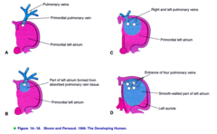
a single pulmonary vein develops in the left side of the primitive atrium and branches twice to produce two right and two left pulmonary veins. Eventually pulmonary vein system undergoes intussusception into left dorsal wall of primitive atrium. these four branches are then incorporated into dorsal wall of left side of primitive atrium, completing formation of the smooth wall.
foetal circulation
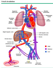
embryo recieves oxygenated blood from the placenta and umbilical vein. lungs are non-functional so circulation by-passes them. deoxygenated blood returns to placenta via umbilical arteries.
name the foetal circulatory shunts
ductus arteriosus, ductus venosus, foramen ovale
ductus arteriosus
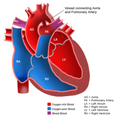
foetal artery that connects aorta to pulmonary artery. blood detours away from lungs before birth
ductus venosus
foetal blood vessle connecting the umbilical vein to the inferior vena cava. carries mostly oxygenated blood
foramen ovale
shunts oxygenated blood from right atrium to left atrium
cardinal system
drains the head, neck, and body wall
day 17 development of blood and vasculature
hemangioblast cells first seen in extraembryonic splanchic mesoderm of yolk sac. earliest multipotent precursor cells for blood and blood vessles. aggregate with endothelial precursors to form blood island
blood islands
clusters of mesodermal cells. aggregations of haematopoietic progenitors and endothelial precursors.
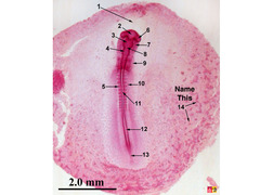
development of the aortic arches in the 4th week. day 22 and 24
body folding, ventral and dorsal development
first pair form which later regress. body folding results in translocation of the heart tube into the thorax and draws cranial ends of the attached aortae into a dorsoventral loop. ventrally, the aortic arch arteries arise from aortic sac expanding at the distal end of the cardiac outflow tract; dorsally connect to left and right dorsal aortae
development of 2nd arch artery
develops by vasculogenesisand angiogenesis. arises in the 2nd pharyngeal arch by day 26. grows to connect aortic sac to dorsal aortae
formation of 3rd and 4th aortic arch arteries on day 28
develop by vasculogenesis and angiogenesis within respective pharyngeal arch. develops while 1st arch is regressing
maxillary artery from 1st aortic arch
1st aortic arch regresses leaves maxillary artery
6th arch arteries formation on day 29
develops as 2nd arch arteries regress
regression of 2nd arch artery
leaves behind stapedial artery
bilateral arch arteries
arteries that arise from first three pairs of aortic arch arteries
asymmetrical vessles arise from
arches four and six. muscles
importance of the asymmetrical vessles
aymmetrical development of 4 and 6th aortic arch arteries are responsible for the asymmetry of the right and left laryngeal nerves (branch from vagus nerves) and innervate laryngeal muscles
6th aortic arch develops asymmetrically. this is important for the asymmetrical development of right and left recurrent laryngeal nerves.
larynx moves towards the cranial end of the embryo, the laryngeal nerves are trapped under the most caudal remaining arch on each side. right recurrent laryngeal nerve therefore loops under right subclavian artery. left laryngeal nerve loops under ductus arteriosus
development of aortic system on day 35 so that the
finish this
disap
1st aortic arch derivative
maxillary artery
2nd aortic arch derivative
stapedial artery and hyoid artery
3rd aortic arch derivative
right and left common carotid arteries and to proximal portion of the right and left internal carotid arteries
right 4th aortic arch artery derivative
gives rise to right subclavian artery
left 4th aortic arch artery derivative
aortic arch and most proximal portion of the descending aorta
right 6th aortic arch artery derivative
pulmonary artery
left 6th aortic arch artery derivative
ductus arteriosus. allows blood to shunt from pulmonary trunk to descending aorta
ductus arteriosus derivative
closes at birth and transforms into ligamentum arteriosum. attaches pulmonary trunk to arch of aorta
right recurrent laryngeal nerve loops around what
right subclavian artery
left recurrent laryngeal nerve loops around what
ductus arteriosus
ventral branches from dorsal aorta derived from what
vitelline arteries (emerge from dorsal aortae during 4th week to supply yolk sac)
ventral branches of dorsal aorta
supply gut tube and its derivatives.
ventral branches of dorsal aorta originate from vitelline arteries. after the paired dorsal aortae fuse at the end of paired dorsal aortae fuse, many vitalline arteries disap
finisj