UNIT 11 TMJ RELATED FOSSA
1/28
There's no tags or description
Looks like no tags are added yet.
Name | Mastery | Learn | Test | Matching | Spaced | Call with Kai |
|---|
No analytics yet
Send a link to your students to track their progress
29 Terms
Temporal fossa
-Shallow depression on the side of the skull
-Communicates with the infratemporal fossa inferior to the zygomatic arch
(ZYGOMATIC CANAL)
BOUNDARIES OF TEMPORAL FOSSA
superior; superior temporal line
anterior: temporal line of the frontal bone
zygommatic process of the frontal bone
frontal process of the zygomatic bone
posterior- sup border of the zygomatic bone
inferior:
sygomatic arc (laterally) of the temporal bone
zygomatic canal
infratemporal crest of the greater wing of the sphenoid (medially)
supramastoid crest
Bones that build up the framework of the fossa
Lateral surface of frontal
Inferior surface of the parietal
Squama of the temporal
Temporal surface of the greater wing of the sphenoid
Temporal surface of the zygomatic
Most important structure housed
Deep and superficial temporal arteries and nerves
Auriculotemporal nerve
Temporal fascia
Temporalis muscle
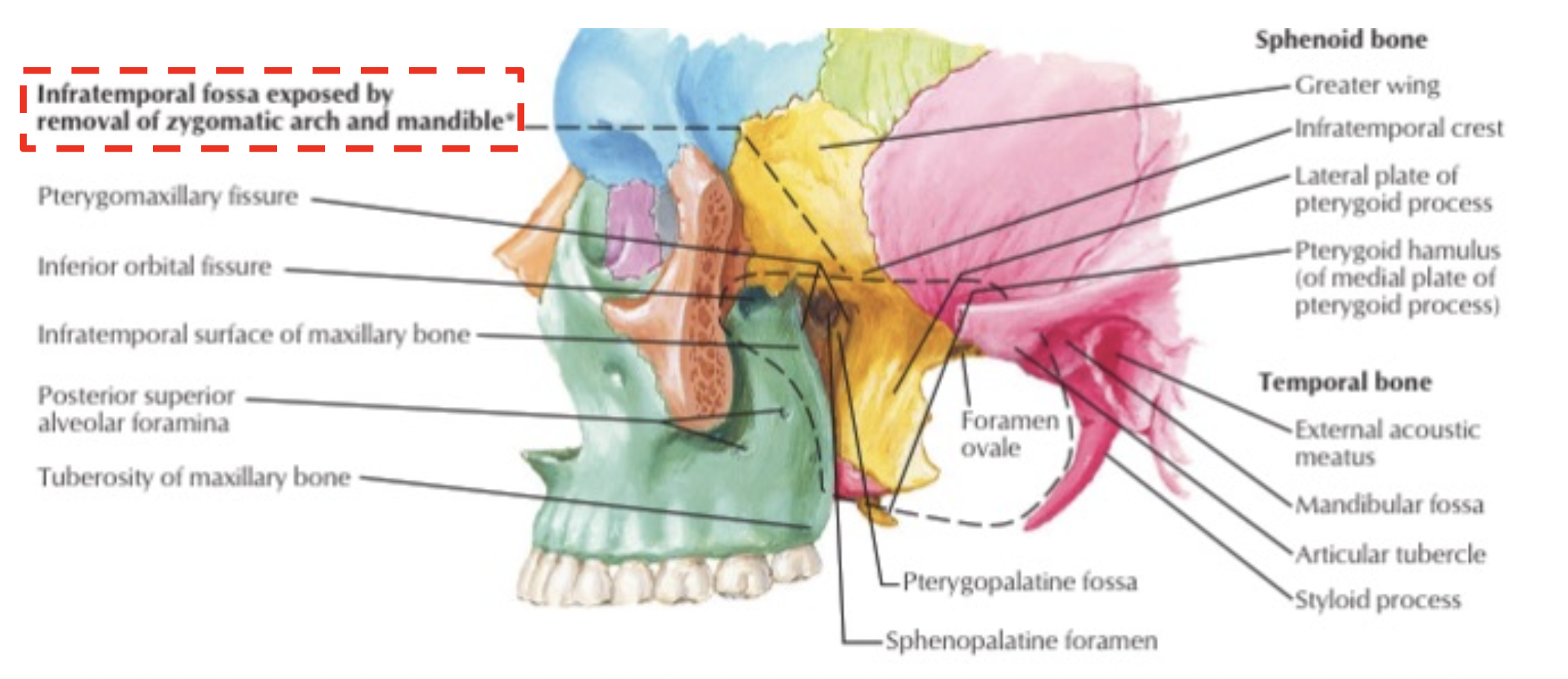
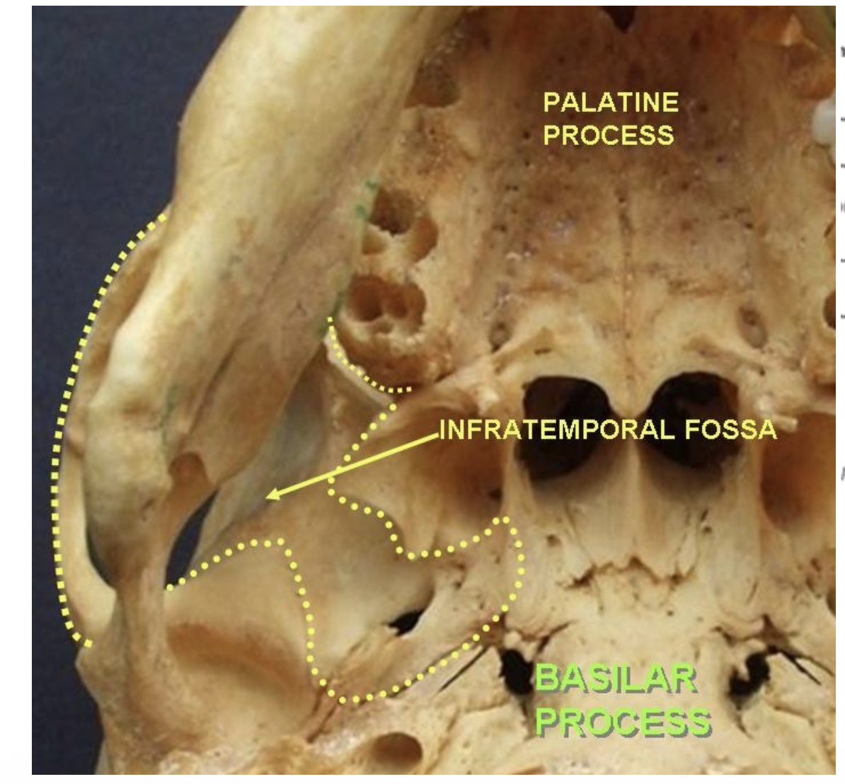
INFRATEMPORAL or PTERYGOMAXILLARY FOSSA
Wide farrow localized:
-Posterior to the maxillary bone
-Inferior to the greater wing of the sphenoid
-Lateral to the pterygoid process of the sphenoid
INFRATEMPORAL or PTERYGOMAXILLARY FOSSA
Lateral or external border: INFRATEMPORAL or PTERYGOMAXILLARY FOSSA
ramus, coronoid process and mandibular notch of the mandible and medial surface of masseter muscle
INFRATEMPORAL or PTERYGOMAXILLARY FOSSA- medial or internal border
pterygomaxillary fissure, lateral pterygoid plate of the sphenoid, pyramidal process of the palatine bone and pharynx
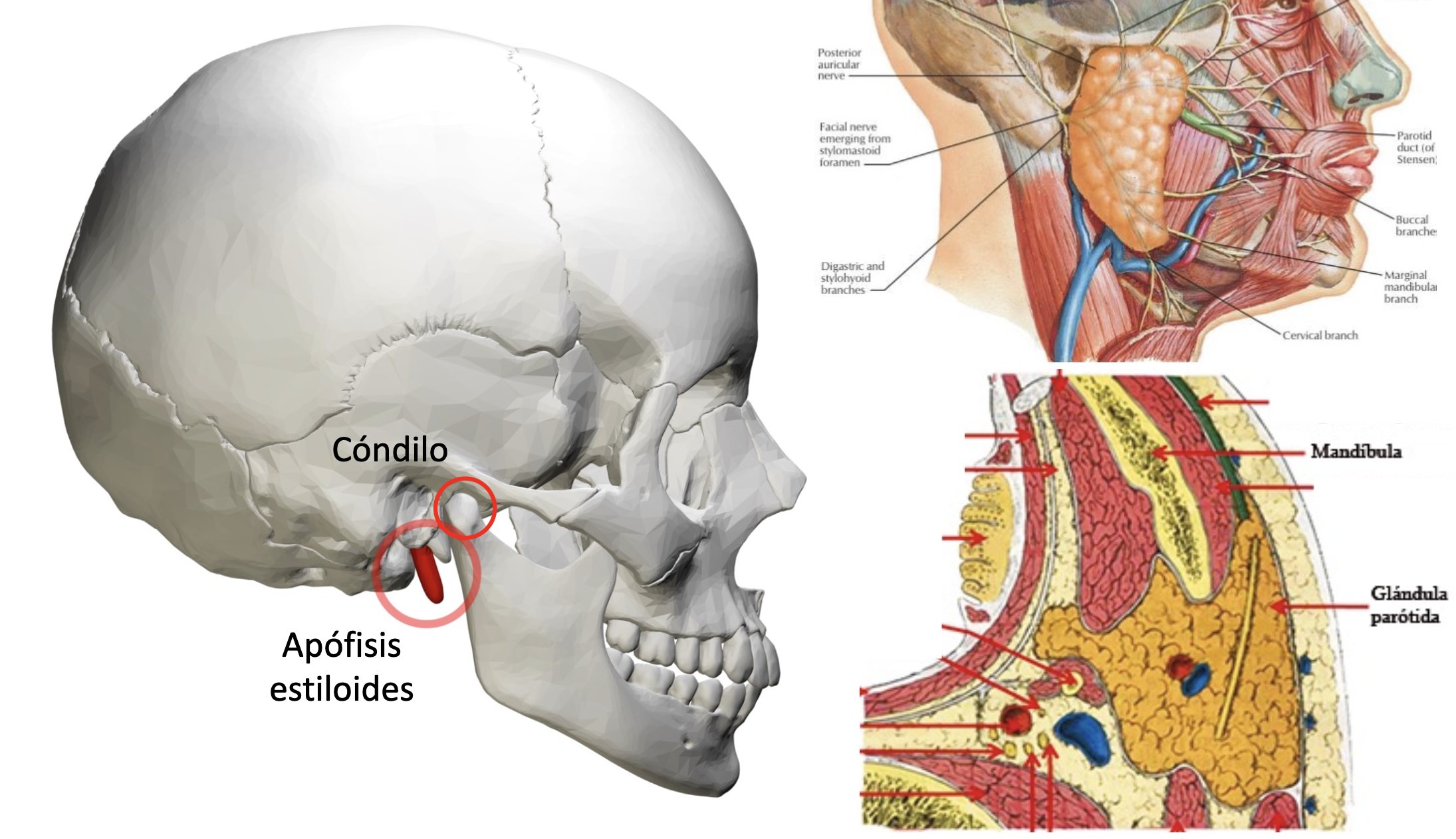
INFRATEMPORAL or PTERYGOMAXILLARY FOSSA- posterior border
styloid process, condylar process of the mandible, and parotid gland
INFRATEMPORAL or PTERYGOMAXILLARY FOSSA- ANTERIOR BORDER
tuberosity of the maxillary bone (communication to the surface below the zygomatic process of the maxillary bone)
INFRATEMPORAL or PTERYGOMAXILLARY FOSSA- SUPERIOR BORDER
zygomatic (infratemporal surface), infratemporal crest of
the lateral surface of the greater wing of the sphenoid (foramen oval and
foramen spinosum), lateral surface of the squama of the temporal
INFRATEMPORAL or PTERYGOMAXILLARY FOSSA- POSTERIOR BORDER
No anatomic floor (an inferior plane to mandible’s inferior border), where the medial pterygoid
muscle attaches to the mandible
INFRATEMPORAL or PTERYGOMAXILLARY FOSSA BOARDERS- Lateral or external border
ramus, coronoid
process and mandibular notch of the mandible
and medial surface of masseter muscle
INFRATEMPORAL or PTERYGOMAXILLARY FOSSA BOARDERS- MEDIAL OR INETERNAL BORDERS
lateral pterygoid plate of the sphenoid, pyramidal process of thepalatine bone, pterygomaxillary fissure, and pharynx
INFRATEMPORAL or PTERYGOMAXILLARY FOSSA BOARDERS- POSTERIOR
styloid process, condylar process of the mandible and parotid gland
INFRATEMPORAL or PTERYGOMAXILLARY FOSSA BOARDERS- ANTERIOR
tuberosity of the maxillary bone (communication to the face below
the zygomatic process of the maxillary bone)
INFRATEMPORAL or PTERYGOMAXILLARY FOSSA BOARDERS- SUPERIOR
zygomatic (infratemporal) portion of the lateral surface of the greater wing of the sphenoid (foramen oval and foramen spinosum)
INFRATEMPORAL or PTERYGOMAXILLARY FOSSA BOARDERS- INFERIOR
no anatomic floor (an inferior plane to mandible’s inferior border), where the medial pterygoid muscle attaches to the mandible
OPENING OF INFRATEMPORAL or PTERYGOMAXILLARY FOSSA
- Temporal fossa (lateral-superior): ZYGOMATIC CANAL
- Masseter region (lateral): MANDIBULAR NOTCH
-Pterygopalatine fossa (medial): PTERYGOMAXILLARY FISSURE
- Middle cranial fossa (medial-superior): FORAMEN OVALE and FORAMEN
SPINOSUM
IMPORTANT STRUCTURES HOUSED:INFRATEMPORAL or PTERYGOMAXILLARY FOSSA
-Inferior part of the temporalis muscle and lateral and medial pterygoid muscles
-Maxillary vessels
-Mandibular nerve (V3)
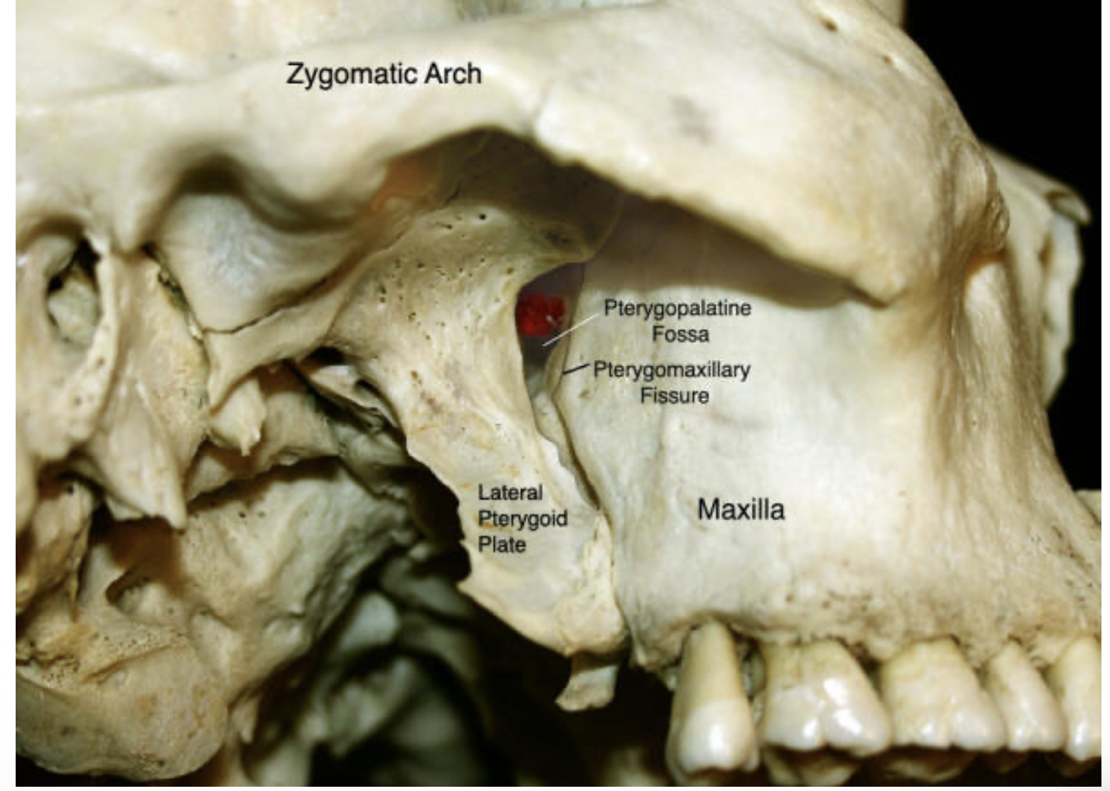
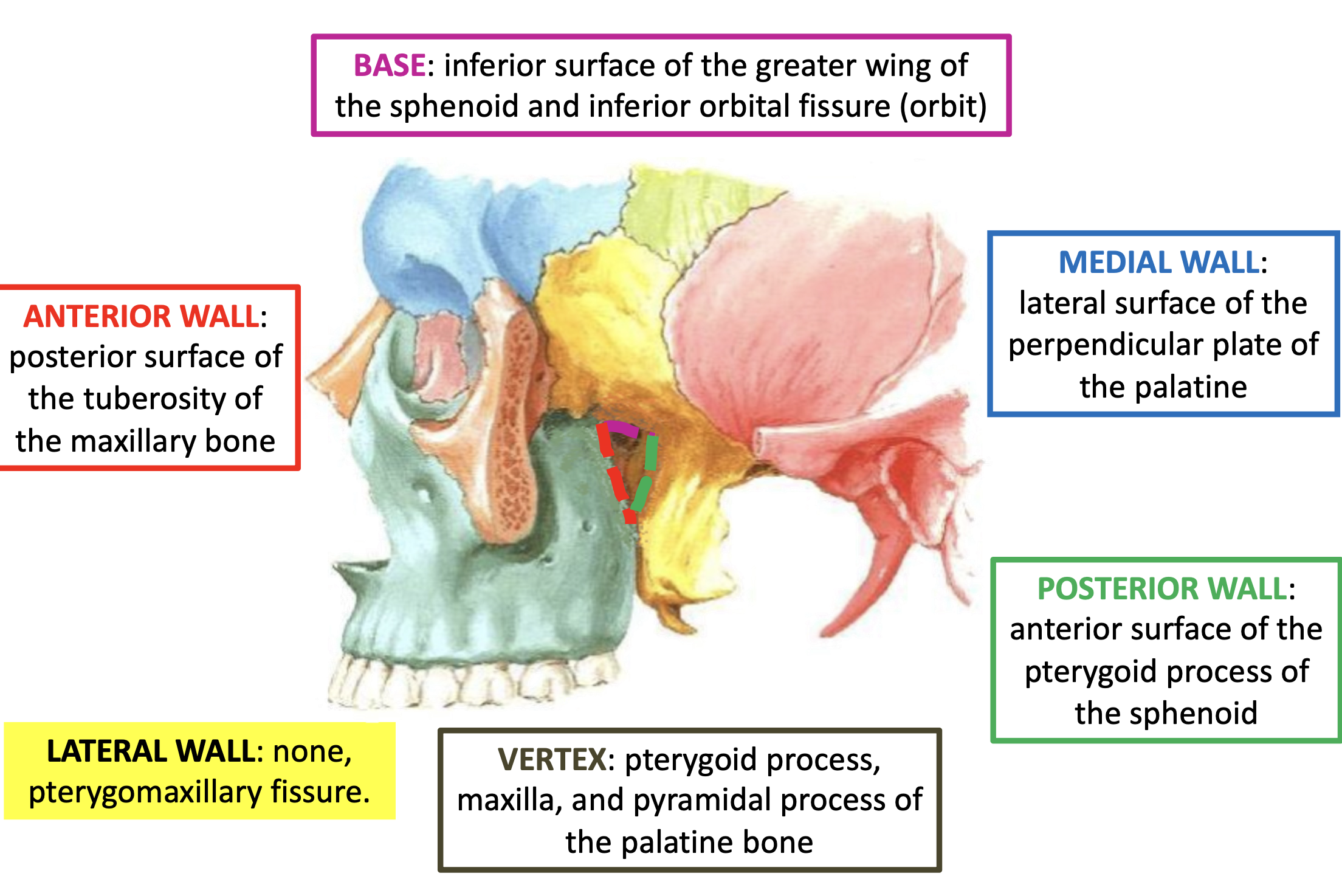
PTERYGOPALATINE FOSSA
Pyramid-shaped fossa on the lateral aspect of the skull between the maxilla’s
posterior surface and the pterygoid process of the sphenoid
Medial to the infratemporal fossa
attention has no lateral walls
PTERYGOPALATINE FOSSA ANTERIOR WALL
tuberosity of the maxillary bone
PTERYGOPALATINE FOSSA- POSTERIOR WALL
anterior-medial surface of the pterygoid process of the
sphenoid (in its superior part, it is located the PTERYGOID or VIDIAN CANAL)
PTERYGOPALATINE FOSSA- MEDIAL WALL
perpendicular plate of the palatine (in its superior part
SPHENOPALATINE NOTCH for the communication with the nasal cavities)
PTERYGOPALATINE FOSSA- BASE
upward; is related anterior with the orbit through the INFERIOR ORBITAL
FISSURE and posterior with the inferior surface of the sphenoid (FORAMEN
ROTUNDUM)
PTERYGOPALATINE FOSSA- Vertex
downward; it’s the joint between the pterygoid process, the
tuberosity of the maxillary bone, and pyramidal process of the palatine
PTERYGOPALATINE FOSSA
PTERYGOPALATINE FOSSA - OPENINGS
Pterygomaxillary fossa (lateral): PTERYGOMAXILLARY FISSURE
Orbit (antero-superior): INFERIOR ORBITAL FISSURE (infraorbitary v&n)
Nasal cavity (medial): SPHENOPALATINE FORAMEN (sphenopalatine v&n)
Middle cranial fossa (antero-posterior): FORAMEN ROTUNDUM (V2 trigeminal)
Pterygoid fossa (posterior): PTERYGOID CANAL (vidian nerve)
Oral cavity (inferior): PALATINE CANAL (greater palatine nerves and vessels)
PTERYGOPALATINE FOSSA-IMPORTANT STRUCTURES HOUSED
Maxillary nerve V2 (foramen rotundum)
Pterygopalatine ganglion
Vidian nerve (pterygoid canal)
Sphenopalatine canal, sphenopalatine nerves and vessels
Palatine and alveolar nerves