non-traumatic joint pain: lower extremity
 Call Kai
 Learn
Learn Practice Test
 Spaced Repetition
 Match
Match Flashcards
 Knowt Play
1/47

There's no tags or description
Looks like no tags are added yet.
Name | Mastery | Learn | Test | Matching | Spaced | Call with Kai | Chat |
|---|
No analytics yet
Send a link to your students to track their progress
48 Terms
lateral ankle sprain of ATFL
most common ankle sprain
valgus/varus stress
test for MCL/LCL
McMurray and Thessaly
tests for meniscal tear
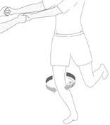
anterior drawer ankle test
test for tear or laxity of anterior talo-fibular ligament
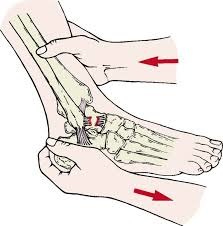
Squeeze test (ankle)
test for syndesmosis ankle sprain (high ankle sprain)
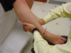
L2-3
nerve root of hip flexion
L4
nerve root of knee extension
L5
nerve root of hip abduction
S1
nerve root of hip extension
L3-4
dermatome of anterior knee
L5-S1
dermatome of posterior/lateral knee
L4
dermatome of medial ankle
L5
dermatome of dorsum/great toe
S1
dermatome of lateral foot/sole
T10-L2
sympathetics of the lower extremity
Q angle
degree of angle when measureing from midpatella to ASIS and tibial tubercle. normal q angle is 13 degrees for man and 18 degrees for a woman.
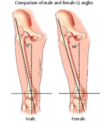
J sign
lateral patellar deviation during terminal knee extension
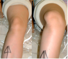
gluteus medius weakness (L5, superior gluteal n.), trochanteric pain.
Trendelenburg gait suggests
Steppage gait
A gait pattern in which the feet and toes are lifted through hip and knee flexion to excessive heights; usually secondary to dorsiflexor weakness. The foot will slap at initial contact with the ground secondary to the decreased control.
L4-L5 radiculopathy or common peroneal neuropathy\\
ddx of steppage gait
circumduction gait
A gait pattern characterized by a circular motion to advance the leg during swing phase; this may be used to compensate for insufficient hip or knee flexion or dorsiflexion.
meniscal tear, post-op stiffness, spasticity.
ddx of circumduction gait
cerebellar or sensory neuropathy
ddx of ataxic/wide-based gait
ober test
pain or resistance to adduction of leg parallel to the table in neutral position
test for iliotibial band sydrome
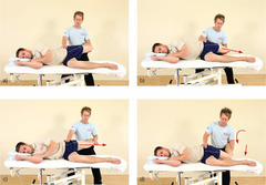
thomas test
tests for Hip flexion contracture (iliopsoas; rectus femoris)
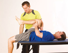
lachman test
application of anterior and posterior force to the proximal posterior tibia to determine the stability of the ACL and PCL
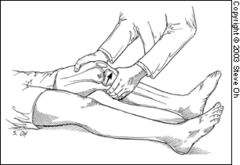
-correct proximal somatic dysfunctions first
priorities for lower extremity OMT
ottawa rules
X-rays required if there is any pain in malleolar zone and any bone tenderness at lateral or medial edge of malleolus
OR X-rays required if there is any pain in mid-foot zone and any bone tenderness at base of fifth metatarsal or the navicular
apley test
helps differentiate between meniscal tears and ligamentous lesions.
-positive for meniscal tears
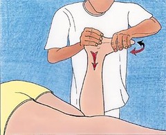
anterior dysfunction (restricted posterior motion)
fibular head dysfunction associated with ankle dorsiflexion injury
posterior dysfunction (restricted anterior motion)
fibular head dysfunction associated with ankle inversion sprain
Patient supine, knee flexed to 90.
Physician induces dorsiflexion + eversion (to encourage anterior glide).
Patient plantarflexes/inverts against resistance
muscle energy for posterior fibular head
Patient supine, knee flexed to 90.
Physician induces plantarflexion + inversion (to encourage posterior glide).
Patient dorsiflexes/everts against resistance.
muscle energy for anterior fibular head
Patient prone, knee flexed, fine-tune with hip rotation.
counterstrain position for hamstring (posterior thigh points )
Patient prone, knee flexed, ankle plantarflexed
counterstrain position for gastrocnemius tenderpoint
patellofemoral syndrome
anterior knee pain, worse with prolonged sitting ("theater sign"), stairs, squatting.
Pain with patellar grind test.
Maltracking or lateral patellar glide.
Weak hip abductors/quadriceps imbalance.
exam findings associated with patellofemoral syndrome
malalignment, muscle imbalance (weak VMO, tight IT band), overuse.
etiology of patellofemoral syndrome
Conservative first-line: activity modification, NSAIDs, ice.
Physical therapy: strengthen quadriceps (esp. VMO), gluteus medius, core. Stretch IT band, hamstrings, hip flexors.
Supportive: patellar taping, orthotics for overpronation.
management of patellofemoral syndrome
correct hip/pelvic/ankle dysfunctions that alter patellar tracking.
-indirect MFR of patella
OMT recommended for patellofemoral syndrome
plantar fasciitis
-Heel pain, worst with first steps in the morning or after rest.
-Improves with activity, worsens with prolonged standing.
pain localized at medial calcaneal tubercle (heal)
where is the pain localized in plantar fasciitis
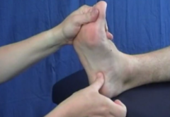
windlass test
Purpose: test for Plantar fascitis
Procedure:Pt is seated. -NWB: pt sits and examiner stabilizes the ankle while 1st MTP joint is extended, while allowing the IP jt to flex
+Result:pain is reproduced at end range of MTP extension
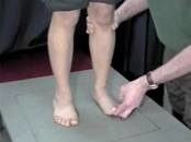
anteromedial calcaneus
plantar fascia origin
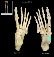
avoid prolonged standing, high-impact running until symptoms improve.
activity modification in management of plantar fascitis
Plantar fascia stretch (pull toes dorsiflexed).
Calf stretch (gastrocnemius & soleus).
Towel stretch in morning before first steps.
stretching routines recommended in management of plantar fasciitis
Medial Calcaneal Tubercle
tenderpoint of plantar fascia
Patient prone, knee flexed.
Physician plantarflexes ankle and compresses calcaneus toward forefoot (shortening fascia).
Hold 90 sec → slowly return to neutral.
counterstrain for plantar fascia