Procedures - Upper Limb (Unit 1-2)
1/109
There's no tags or description
Looks like no tags are added yet.
Name | Mastery | Learn | Test | Matching | Spaced | Call with Kai |
|---|
No analytics yet
Send a link to your students to track their progress
110 Terms
Number of bones (per side) in hand:
Phalanges
Metacarpals
Carpals
14
5
8
DIP
Distal Interphalangeal Joint
PIP
Proximal Interphalangeal joint
MCP
Metacarpophalangeal joint
IP
Interphalangeal joint
Carpal bone that articulates with:
1st metacarpal
2nd metacarpal
3rd metacarpal
4th and 5th metacarpal
Trapezium
Trapezoid
Capitate
Hamate
The lunate and scaphoid bones articulate with the _____ proximally.
Radius
The most frequently fx carpal bone:
Scaphoid
The lunate articulates with the _____ (bone).
Radius
The radial tuberosity is located on the _____ & _____ side of the radius.
Medial and Anterior
The two processes of the proximal ulna:
Olecranon and Coronoid processes
Notch on the proximal ulna where articulation with the humerus occurs:
Trochlear notch
What portion of the humerus articulates with the ulna to form the elbow joint:
Trochlea (distal humerus)
Processes at the distal ends of the radius and ulna:
Styloid processes
IP joints are ginglymus, which is defined as:
Hinge-type. Movement in 2 directions: flexion and extension
MCP joints are ellipsoidal (condyloid), which is defined as:
Condyloid type. Movement in 4 directions: flexion, extension, abduction, and adduction. (Circumduction)
List the joint classifcations:
IP
MCP
Ginglymus (hinge)
Ellipsoidal (condyloid)
List the joint classifcations:
CMC (thumb)
CMC (2nd - 5th)
Saddle
Plane (gliding)
List the joint classifcations:
Intercarpal
Wrist
Elbow
Plane (gliding)
Ellipsoidal (condyloid)
Ginglymus (hinge)
_____ deviation provides the best demonstration of the lateral carpal including the trapezium, trapezoid, and _____.
Ulnar
Scaphoid
_____ deviation provides the best demonstration of the medial side of the wrist.
Radial
On the AP elbow, the proximal radius is slightly superimposed by the:
Ulna
To prevent superimposition of the radius & ulna, the forearm is placed with the hand _____ and in an _____ projection.
Supinated
AP
The scaphoid fat stripe (wrist) can be viewed on which projections?
PA and Oblique
The pronator fat stripe (wrist) can be visualized on which projection(s)?
Lateral
Elbow fat pads cannot be seen in the projection:
AP
Which projection of the elbow is the best for visualizing fat pad pathology?
Lateral
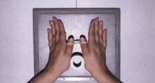
The nogaard method (aka _________ position) is used for early detection of ___________. Describe this view:
Ball Catcher’s
Rheumoatoid arthritis
Bilateral hands (to compare with each other). Hands are oblique 45 degress with palms up.
To avoid excessive MCP joint overlap in an oblique hand, the hand should be angled no more than _____ degrees.
45
What is an alternative to a fan-lateral hand to evaluate a fracture or foreign body.
Lateral in Extension and Flexion
Why is it important to support the fingers so that they remain parallel to the IR?
Prevent foreshortening of phalanges
Prevent obscuring of IP joints
What method is best used to evaluate a Bennett fracture?
AP axial of thumb
CR is angled 15 degrees proximally (towards wrist)
Describe the Brewerton method:
AP axial of the hand. Supinate hand with fingers touching IR. Flex hand 65 degrees. CR 15 degress promixally toward ulna (3rd MCP joint).
Why should the hand be slightly arched / fingers bent for a PA wrist?
Reduce OID of carpals
A PA axial projection to visualize the scaphoid bone requires _________ deviation of the wrist and a _________ degree tube angle directed proximally.
Ulnar
10-15
The Gaynor-Hart method is primarily used to evaluate:
Carpal Tunnel Syndrome
CR 25-30 degrees proximally to long axis od hand
Which oblique will separate the radial head, neck, and tuberosity from the ulna?
Lateral oblique (external rotation)
The coyle method will visualize the _________ & _________ when the patient has their elbow flexed 90 degrees and the CR is angled ________ - degrees medially (towards the shoulder). This can be done with the patient plying down or sitting beside the table.
Radial head
Capitulum
45
The coyle method for the coranoid process is a __________ projection with the elbow 80-degrees. The CR is 45-degrees directed ______ from the shoulder.
Mediolateral (axial)
Away
A partial flexion elbow requires 2 projections to obtain a 'full' AP. Describe each:
AP - forearm parallel to IR
AP - humerus parallel to IR
To visualize the entire circumference of the radial head, 4 projections must be obtained:
All projections are in lateromedial with arm flexed 90 degrees. Epicondyles perpendicular to IR
Supinate hand
Hand in true lateral
Pronate hand
Internally rotate hand
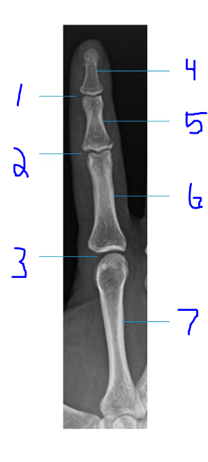
Label
DIP joint
PIP joint
MCP joint
Distal phalanx
Middle phalanx
Proxmial phalax
Metacarpal
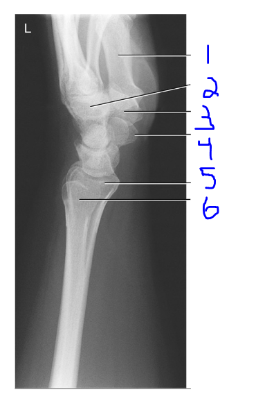
Label
Thumb
Capitate
Trapezium
Scaphoid
Radius
Ulna
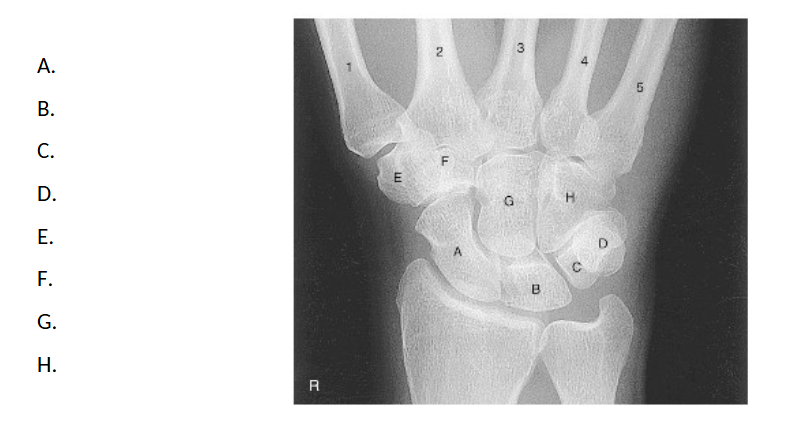
Label
A. Scaphoid
B. Lunate
C. Triquetrum
D. Pisiform
E. Trapezium
F. Trapezoid
G. Capitate
H. Hamate
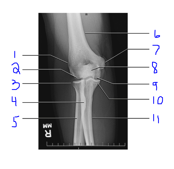
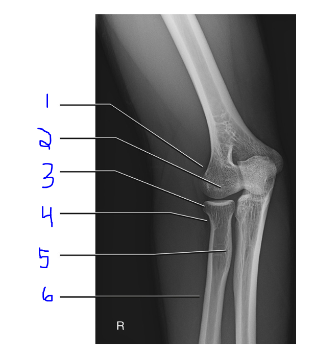
Label
Lateral Epicondyle
Capitulum
Radial Head
Radial Tubercle
Radius
Humerus
Medial Epicondyle
Olecranon Process
Trochlea
Coronoid Process
Ulna
Label
Lateral Epicondyle
Capitulum
Radial Head
Radial Neck
Radial Tubercle
Radial Shaft
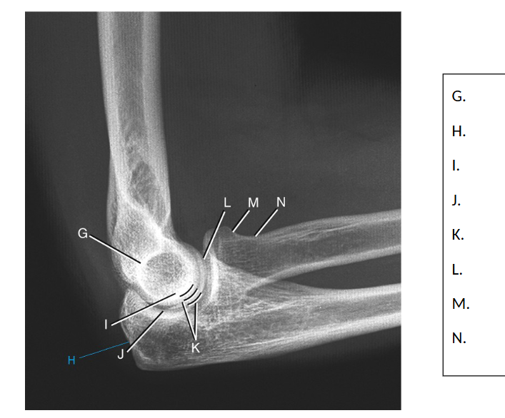
Label
G. Superimposed epicondyles of humerus
H. Olecranon Process
I. Trochlear sulcus
J. Trochlear notch
K. Double outer ridges of capitulum and tochlea
L. Coronoid process of ulna
M. Radial Head
N. Radial Neck
What projection may be done in replacement of a PA Digit?
PA Hand
What projection may be done in replacement of an oblique thumb?
PA Hand
Oblique Hand: Image Criteria
Midshafts of metacarpals should not overlap
Oblique Wrist: Image Criteria
Distal radius, ulna, carpals, and at least to mid metacarpals.
Trapezium and Scaphoid should be well visualized
Lateral Wrist: Image Criteria
Distal radius, ulna, carpals, and at least to mid metacarpals.
Ulnar head superimposed over distal radius
AP Elbow: Image Criteria
Distal humerus, elbow joint space, and proximal radius and ulna are visible
Bilateral epicondyles seen in profile and radial head, neck, and tuberosity separated or only slightly superimposed by ulna
Lateral Elbow: Image Criteria
One-half of radial head should be superimposed by the coronoid process, and olecranon process should be visualized in profile
Superimposition of the humeral epicondyles occurs
Medial Oblique Elbow: Image Criteria
Visualize coronoid process of the ulna in profile
Radial head and neck should be superimposed
Medial epicondyle and trochlea should appear elongated
Olecranon process should appear in olecranon fossa and trochlear notch partially open and visualized
Lateral Oblique Elbow: Image Criteria
Visualize radial head, neck, and tuberosity, free of superimposition by the ulna
Lateral epicondyle and capitulum should appear elongated and in profile
PA thumb results in:
a lot of OID. Not a common projection
PA Stress Thumb (Folio Method)
Hands side by side, rotated laterally 45 degrees, thumbs in PA, wrap rubber bands around distal thumb
Skier’s Thumb (hyperextension and ulnar collateral ligament)
Acute Flexion Elbow Projections
Same patient position but different CR angles
AP - CR perpendicular to distal humerus
PA - CR perpendicular to proximal forearm
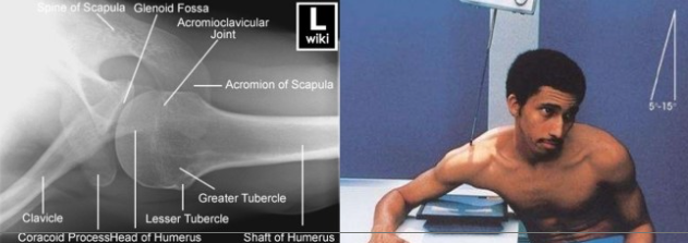
On the proximal humerus, the anterior process directly below the anatomic neck is the __________. The larger process on the lateral aspect is the __________
Lesser tubercle
Greater tubercle
Which muscles attach to the greater tubercle of the humerus?
Pectoralis major and Supraspinatus muscles
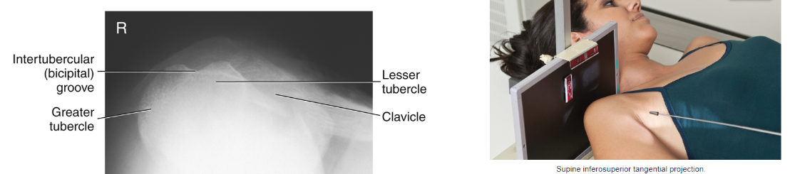
The deep groove between the lesser and greater tubercle is called
Intertubercular or bicipital groove
What rotation of the shoulder allows for a true AP projection of the proximal humerus?
External
The __________ tubercle is located anteriorly and the greater tubercle is __________ in a true AP projection.
Lesser
Laterally
The upper margin of the scapula is located at the level of the __________ posterior rib
2nd
The lower margin of the scapula is located at the level of __________
7th posterior rib (T7)
The bilateral SC joints form the:
Jugular notch
What differences are there in clavicles between a male and a female?
Female - shorter and less curved than male clavicles
Male - thicker and more curve
Axilla refers to:
Armpit
Anatomy that articulates to form the Acromioclavicular (AC) joint:
Lateral/acromial end of the clavicle with the acromion of the scapula
Anatomy that articulates to form the Sternoclavicular (SC) joint:
Medial/sternal end of the clavicle with the manubrium
Anatomy that articulates to form the Scapulohumeral (glenohumeral/shoulder) joint:
Humeral head with the glenoid cavity of the scapula
The wing of the scapular can also be referred to as the:
ala
The anterior surface of the scapula is called __________ surface.
Costal
A thick, beaklike process on the scapula that project anteriorly beneath the clavicle is called the __________. It is located __________ inches interior to the lateral portion of the AC joint.
Coracoid process
2 inches
A long, curved process that extends laterally over the head of the humerus:
Acromion
The 3 joints of the shoulder are classified as:
Synovial
The 3 joints of the shoulder have this type of mobility:
Diarthrodial
List the movement type of each joint:
a. scapulohumeral/glenohumeral:
b. sternoclavicular:
c. acromioclavicular:
Ball and socket
Plane/gliding
Plane/gliding
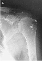
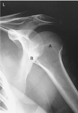
Position and Projection
Pos - External rotation
Proj - AP
A - Greater tubercle
B - Lesser tubercle
Position and Projection
Pos - Internal rotation
Proj - Lateral
A - Greater tubercle
B - Lesser tubercle
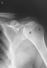
Position and Projection
Pos - Neutral rotation
Proj - Oblique
A - Greater tubercle
B - Lesser tubercle
95% of shoulder dislocations are:
Anterior
To demonstrate a profile view of the glenoid fossa, the patient is AP recumbent and oblique 45 degrees towards the:
Affected side (IR)
If a patient has surgically implanted hardware, it is best practice to set a __________ technique.
Manual
AC joints require that both joints are visualized together, this is called the __________ method. A(n) __________ projection is taken with and without __________. If a fracture is suspected, these views can/cannot be obtained.
Pearson
AP
Weights
Can
A true AP of the clavicle will visualize:
Clavicular body
AC joint
SC joint
Acromion
The lesser tubercle will be in profile and the epicondyles will be superimposed on this projection of the humerus:
Internal rotation (Mediolateral)
Why might an orthostatic breathing technique be used on a transthoracic lateral humerus?
Allows best visualization of humerus by blurring out ribs and lung structures.
An AP scapula requires the arm to be abducted 90 degrees to the body, with the elbow flexed and hand __________. It is best practice to do a __________ breathing technique.
Supinated
Orthostatic
If only 2 views were obtained to rule out a fracture of the shoulder, what are the best views?
AP
Scap Y
The Clark’s method of the shoulder utilizes this projection __________. It can be used to replace the axial shoulder, Lawrence method.
Superoinferior Axial
The Fisk method of the shoulder is best used to evaluate the:
Intertubercular sulcus/groove
The Lawrence method (exaggerated external rotation) is best used to demonstrate a:
Hill-Sachs Defect
Which projection will reduce OID of the humerus? mediolateral/latermedial
Mediolateral
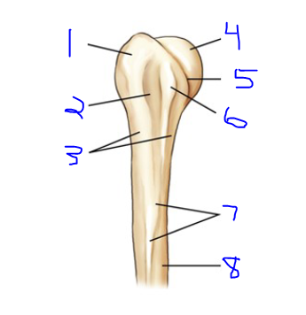
Label
Greater Tubercle
Intertubercular Groove
Surgical Neck
Head
Anatomical Neck
Lesser Tubercle
Deltoid Tuberosity
Body
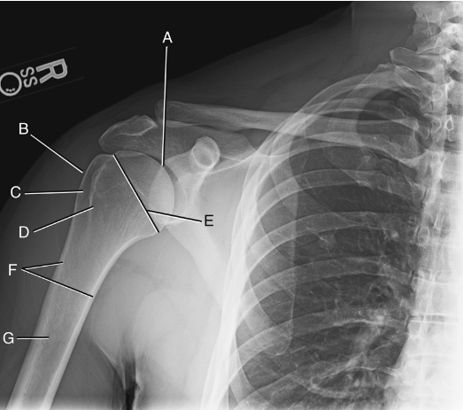
Label
A. Head of Humerus
B. Greater Tubercle
C. Intertubercular Sulcus
D. Lesser Tubercle
E. Anatomical Neck
F. Surgical Neck
G. Body
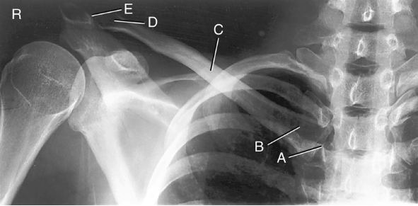
Label
A. Sternoclavicular Joint
B. Sternal Extremity
C. Body
D. Acromial Extremity
E. Acromioclavicular Joint
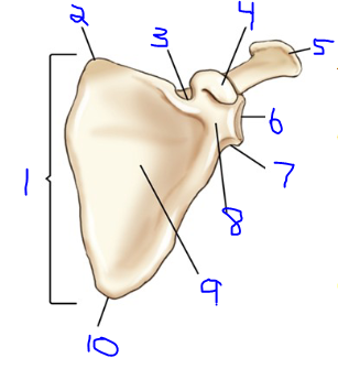
Label
Body
Superior Angle
Subscapular Notch
Coracoid Process
Acromion
Glenoid Cavity
Lateral Angle
Neck
Costal Surface
Inferior Angle
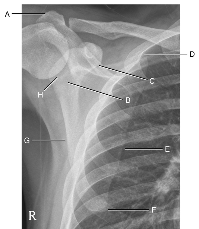
Label
A. Acromion
B. Neck of Scapula
C. Suprascapular Notch
D. Superior Angle
E. Medial Border
F. Inferior Angle
G. Lateral Border
H. Glenoid Cavity