6.6- karyotypes & 6.7 haploid gametes
 Call Kai
 Learn
Learn Practice Test
 Spaced Repetition
 Match
Match Flashcards
 Knowt Play
1/25

There's no tags or description
Looks like no tags are added yet.
Name | Mastery | Learn | Test | Matching | Spaced | Call with Kai | Chat |
|---|
No analytics yet
Send a link to your students to track their progress
26 Terms
A karyotype
•Examination of a prepared slide of stained cells undergoing mitosis revels a jumbled cluster or chromosomes that differ in size and shape.
•Photographic images of the late prophase or metaphase chromosomes can be cut and arranged into matched ordered pairs to create a karyotype.

Karyotypes
•A karyotype is a picture or image of the complete diploid number of chromosomes for a cell.
•Karyotyping is used to:
•determine changes in chromosome number (loss or gain)
•changes in chromosome structure (duplication, inversion or deletion)
Staining of chromosomes
•Regions of a chromosome have more G-C pairs than A-T pairs.
•When stained the G-C rich regions show up lighter.
•This produces a distinct banding pattern.
•Banding patterns can help determine homologous pairs
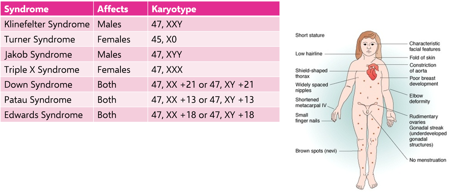
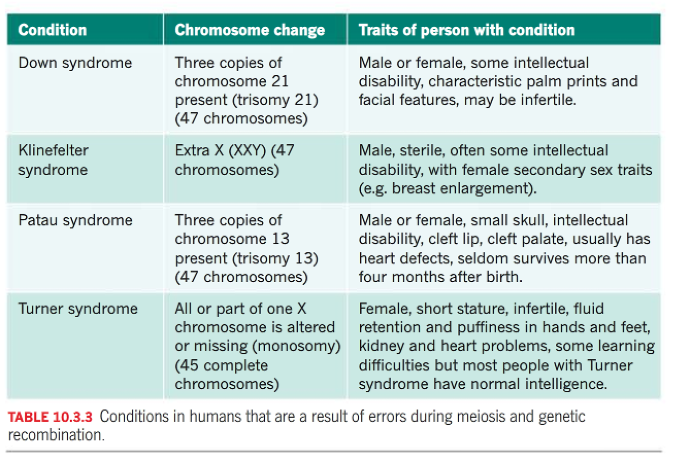
When things go wrong: aneuploidy
•Aneuploidy is the presence of an abnormal number of chromosomes in a cell.
•For example: a human cell having 45 or 47 chromosomes instead of the usual 46.
•
types
•Trisomy: the diploid organism has one extra chromosome (2n+1)
•Monosomy: the diploid organism has one less chromosome (2n-1)
effects
Chromosomal rearrangements
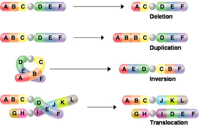
During meiosis changes in chromosome structure can also occur:
•Deletion: segment of the chromosome is lost, can then become attached to a sister chromatid resulting in a duplication
•Duplication: segment of a chromosome is repeated
•Inversion: chromosomal segment reattaches to the original chromosome, but in the reverse orientation
•Translocation: chromosomal segment joins a non-homologous chromosome
What is meiosis?
Meiosis is a complex process that occurs only specialised organs of sexually reproducing animals and plants.
It results in the production of haploid gametes: the sex cells: sperm and eggs (ova).
chromosomes.
more details
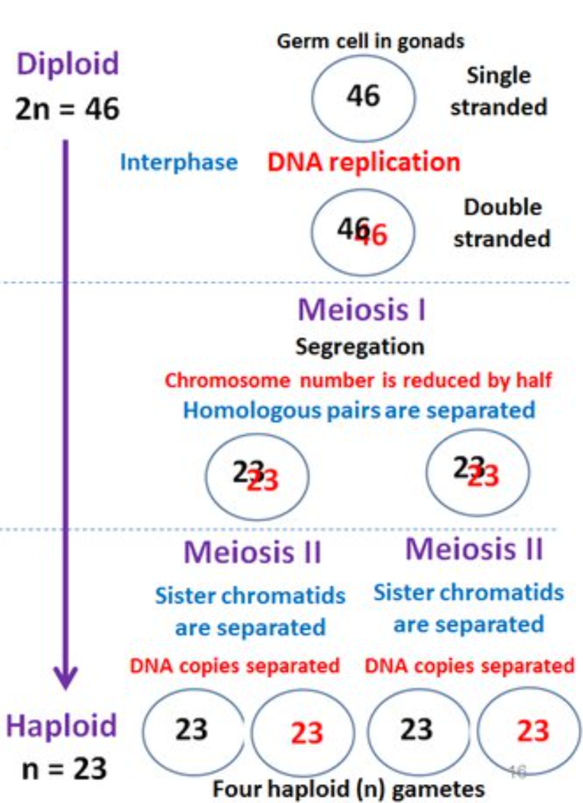
Two divisions of the nucleus take place.
In the first division, each chromosome of a pair separates and goes to each end of the cell.
In the second division, the chromatids of each chromosome separate from each other.
Four gametes are produced – each carrying half the original number of chromosomes.
process of meiosis
Meiosis 1: Interphase
G1 - Cell grows
S - DNA is duplicated
G2 – Normal cell function
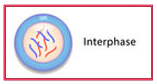
*Synapsis, chiasmata and crossing over
Synapsis: homologous chromosomes lie side by side.
Chiasmata: the point of contact between the homologous pair of chromosomes.
Crossing over: exchange of genetic material, essentially swapping of genes at the same loci.
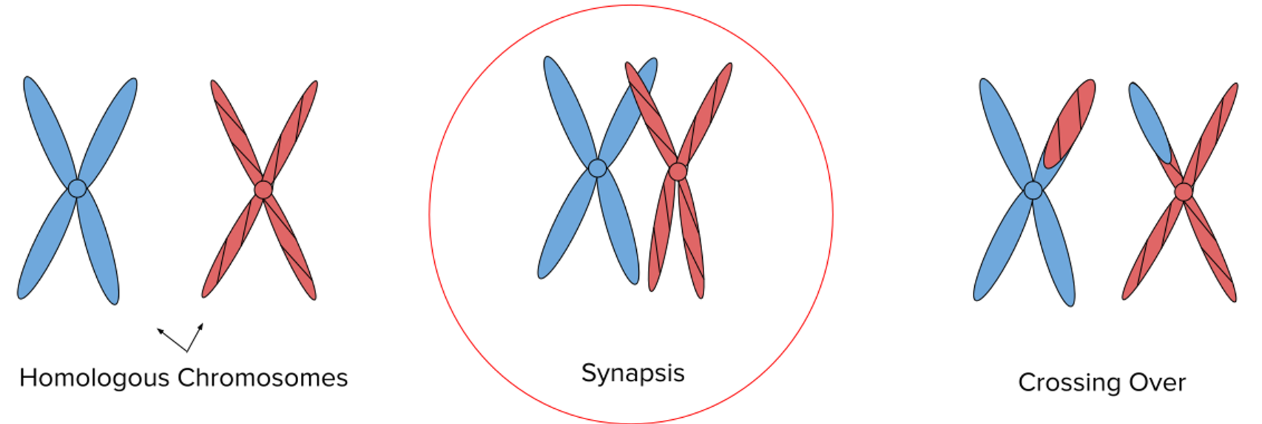
Crossing over leads to recombination…
Crossing over occurs when homologous chromosomes exchange DNA segments during prophase I of meiosis, producing chromosomes with new combinations of alleles. This process is called genetic recombination and increases genetic variation in offspring.
Meiosis 1: Prophase & metaphase 1
prophase 1:
Chromosomes condense and thicken, they’re each composed of two chromatids joined at the centromere.
Homologous chromosomes lie side by side.
*Synapsis, chiasmata and crossing over occur in late prophase.
Metaphase 1
Spindle fibres attach to the centromeres.
Chromosome pairs line up along the equator.
Chromatids become apparent.
The nuclear membrane breaks down.
Meiosis 1: Anaphase 1 & Telophase 1 (and Cytokinesis 1)
Anaphase 1
The spindle fibres pull one of each chromosome pair (maternal and paternal) to opposite poles of the cell.
The separation of each homologous pair occurs independently of the other pairs.
Cell membrane begins to pinch in.
Telophase (cytokinesis 1)
Spindle fibres break down.
Nuclear membrane reforms.
Cytoplasm divides.
Results in two haploid daughter cells.
after meiosis 1
Meiosis I is now complete.
Meiosis II resembles mitosis (except the resulting cells are haploid).
Meiosis 2: Prophase II & Metaphase II
Prophase 2
Nuclear membrane breaks down.
A new meiotic spindle is created.
Metaphase 2
Chromosomes line up in single file along the equator.
Spindle fibres attach to the centromere of each chromosome.
Meiosis 2: Anaphase II & Telophase II (and Cytokinesis II)
Anaphase 2
Centromeres split separating the two sister chromatids.
The now single-strand chromosomes move to opposite ends of the cell.
Telophase II (and Cytokinesis II)
Spindle fibres break down.
Nuclear membrane reforms.
Cytoplasm divides.
Results in four* haploid daughter cells.
Four* haploid daughter cells?
Males make four haploid gametes (sperm) through meiosis.
Females make one large egg cell (ovum) and three smaller ones that degenerate.
This is due to uneven cytoplasm division in meiosis…
compare

Meiosis and Mendel’s Laws
An examination of the process of meiosis reveals two key observations that affect patterns of inheritance in all eukaryotic organisms:
1. The law of segregation
2. The law of independent assortment
The law of segregation
There are two alleles that control each characteristic.
During meiosis, these two alleles segregate - one factor appearing in every gamete.
The offspring can only inherit one allele from each parent.
The alleles recombine at fertilisation.

The law of independent assortment
When the pair of alleles segregate, they do so independently of other pairs of alleles.
They are distributed into gametes independently of other pairs of alleles.
The inheritance of one allele is not dependent on the inheritance of another.

When things go wrong: non-disjunction
•Nondisjunction is the failure of the chromosomes to separate during anaphase I or anaphase 2.
This produces daughter cells with abnormal numbers of chromosomes.

aneuploidy
•Fertilisation occurring after non-disjunction can result in aneuploidy.
•Aneuploidy is the presence of an abnormal number of chromosomes in a cell.
•For example: a human cell having 45 or 47 chromosomes instead of the usual 46.
•
Common aneuploidy examples: